FANCG is an integral scaffold of nuclear Fanconi anemia repair machinery, with additional contextual interaction and mitochondrial evidence. This guide distinguishes actual human experiments, conserved mouse evidence, current ontology, database models and assay-specific uncertainty. It complements the curation journal.
Human FANCG supports the organization of the Fanconi anemia repair complex. Human A/G/F coassociation and complementation-dependent assembly in PMID:11063725(https://pubmed.ncbi.nlm.nih.gov/11063725/), together with the FANCG-mediated A–F bridge described in the accessible PMID:12649160 abstract and human FANCF deletion/association experiments in PMID:15262960(https://pubmed.ncbi.nlm.nih.gov/15262960/), support an integral structural role. The latter study's Figures 4–6 and Table I distinguish graded loss of association from retained complementation for some constructs. Negative pairwise F/G binding in the earlier reticulocyte preparation is retained alongside positive cellular assembly; it does not erase the latter evidence.
Current GO:0030674 explicitly directs integral protein-complex scaffolds to GO:0140378, protein complex scaffold activity. The more specialized GO:0160072, ubiquitin ligase complex scaffold activity, requires its own positioning mechanism; repair-complex membership alone does not establish that narrower activity. FANCL and UBE2T perform ubiquitin transfer. FANCG is not assigned their enzymatic activity.
PMID:22343915(https://pmc.ncbi.nlm.nih.gov/articles/PMC3321854/) supplies direct target localization: Figure 5H includes FANCG in chromatin fractions with H2A control. Actual Figure 5 pixels, main Results/Methods and supplemental fractionation text were inspected. This is stronger than inferring an individual band from complex membership. Nuclear and chromatin localization do not by themselves demonstrate isolated damaged-DNA binding. The specific TAS damaged-DNA-binding assertion from PMID:9806548(https://pubmed.ncbi.nlm.nih.gov/9806548/) remains unresolved because the operative full text was not recovered; its positive complementation abstract is preserved, and no absence of DNA binding is asserted.
Human complex evidence from PMID:17396147(https://pubmed.ncbi.nlm.nih.gov/17396147/), PMID:20347428(https://pubmed.ncbi.nlm.nih.gov/20347428/), PMID:22266823(https://pubmed.ncbi.nlm.nih.gov/22266823/), PMID:22343915(https://pubmed.ncbi.nlm.nih.gov/22343915/) and PMID:22705371(https://pubmed.ncbi.nlm.nih.gov/22705371/) is interpreted at the measured component and complex level. Negative FAAP20–FANCG or FAAP100–FANCG pairwise tests do not contradict core membership. Most FANCM/MHF occurring outside the core is not a denial of the G-containing complex. Xenopus ID2 depletion/add-back in PMID:19965384(https://pubmed.ncbi.nlm.nih.gov/19965384/) establishes pathway steps, not an isolated human FANCG catalytic experiment. The integrated description and core supersede the older absolute no-DNA-binding statement, adaptor terminology and fixed eight-subunit/FANCM–FAAP24 framing.
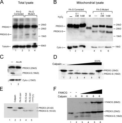
PMID:17060495(https://pubmed.ncbi.nlm.nih.gov/17060495/) provides target-specific FANCG–PRDX3 interaction, mitochondrial imaging/fractionation and partial morphology rescue. Actual Figures 2, 3 and 4 were inspected. Figure 2G retains similar localization of G546R despite reduced PRDX3 interaction; Figure 2H uses stably corrected FA-G cells rather than untransfected endogenous wild type. Figure 3F adds translated FANCG to translated PRDX3 and rat calpain II and shows protection against cleavage. This supports enzyme binding and a contextual protective role, without resolving substrate shielding versus direct calpain inhibition. Lysate peroxidase activity is not purified FANCG peroxidase chemistry. Mitochondrial location and organization remain positive non-core annotations, not complete morphology rescue or universal mitochondrial residence. Supplementary Figure S1 was not inspected.
PMID:12861027(https://pubmed.ncbi.nlm.nih.gov/12861027/) directly tests chicken DT40 FANCG deletion, SCneo recombination and rescue. Chicken cDNA rescues whereas human cDNA does not complement in that host. This is positive conserved-function evidence; failed heterologous rescue is not evidence that human FANCG lost homologous-recombination function. Normal RAD51 foci/SCE and locus-dependent targeting results are retained. Actual rendered main Methods/Results/Discussion were read; no figure pixels were inspected.
PMID:18212739(https://pubmed.ncbi.nlm.nih.gov/18212739/) contains human/hamster FANCG-dependent BRCA2–FANCD2–XRCC3 coassociation and human FANCG S7A tests, in addition to chicken genetic experiments. The historical suggestion that both HR-supporting papers are only chicken studies is therefore superseded. Selected original author-uploaded passages and co-IP/DNase Methods were read; a complete article export and pixels were not recovered. Human association is relevant indirect mechanistic support, not a newly measured human HR reporter endpoint. The inherited human ISS proposal remains UNDECIDED at this precise species/endpoint boundary. Retaining ISS from conservation plus human complex evidence remains a defensible alternative for final synthesis; no rule requiring direct human assays for every GO inference is imposed. Complete current same-role FANCA/FANCC/FANCF queries lack the exact HR term, but that bounded absence is caution rather than a universal curation convention. HR and ICL repair are not ancestors of one another in the saved typed ontology, and no FANCG entry was found in the pinned GO-CAM index.
PMID:9256465(https://pubmed.ncbi.nlm.nih.gov/9256465/) tests human XRCC9/FANCG complementation in hamster UV40, with partial resistance rescue across genotoxins and correction of chromosomal aberrations, while SCE remains unchanged. Broad DNA repair is retained rather than narrowing away the experimentally supported breadth.
Each current partner context was assessed separately. HuRI, PMID:32296183 contributes six FANCG pairs in 18 literal stage records; NeuroNet, PMID:32814053 contributes 21 current partner contexts. Global validation rates are not individual-pair validation. NeuroNet's A/G mammalian luminescence record is distinguished from the other yeast-screen records. DMWD residues 316–649, KIF1B isoform-2 residues 1192–1770 and the ATXN3 N-region with unresolved endpoint remain explicit. The extra IntAct ccsb_9919/Entrez1959 entry is not silently assigned to NDUFV2 or another current GOA partner. Historical HuRI isoform labels differing from current TPRX1/TCEANC/CCHCR1 identifiers are preserved without guessed clone equivalence.
The screen-only SUOX, TPRX1, KLF11, KIF1B, HSPB1, VIM, CYP3A4, GFAP, RAB5A, NOS3, ATXN3, PRPS1 and PLD1 associations remain positive non-core protein binding. Their earlier partner-class replacements are withdrawn conservatively: identifying the partner class does not add a demonstrated FANCG mechanism. Most proposed binding definitions are compatible with the measured association and do not require a neuronal role; lack of a neuronal assay is therefore not a refutation of the interaction. The KIF1B definition additionally specifies stoichiometry, which the fragment screen does not measure. Neither controlled screening nor the paper's aggregation theme is evidence that an individual interaction is false. NDUFV2 and the other unreplaced partners remain subject to the same evidence-based non-core treatment under the project instruction.
The spectrin SH3 and Tax coregulator interpretations preserve the explicit evidence in PMID:19102630(https://pubmed.ncbi.nlm.nih.gov/19102630/) and PMID:22458338(https://pubmed.ncbi.nlm.nih.gov/22458338/). The former is abstract-limited but explicitly reports domain/motif mapping and mutagenesis; the latter includes actual human-cell reporter/knockdown context. Neither establishes FANCG enzymology.
The positive FANCA/FANCG records in PMID:16189514(https://pubmed.ncbi.nlm.nih.gov/16189514/), BioPlex 2.0, BioPlex 3.0, PMID:31467278(https://pubmed.ncbi.nlm.nih.gov/31467278/), PMID:37398436(https://pubmed.ncbi.nlm.nih.gov/37398436/) and PMID:40205054(https://pubmed.ncbi.nlm.nih.gov/40205054/) corroborate independently established assembly biology. The Rual Table S2 Y2H record has empty target co-AP fields. Explicit BioPlex directed files establish G-bait/A-prey, whereas an undirected NDEx edge does not establish IP direction. The FANCA-3 interaction feature in the reference-pair assay is residues 1–297 and is not equated with a full natural product. The generic older BioPlex v2 filename is not treated as proof of the 2017 release identity. Public interaction sources are HuRI, BioPlex, IntAct and the paper-linked supplemental/network resources.
The following source-specific readings were performed separately from the target-study assessments above. Their narrower text or image scope is preserved rather than retroactively expanded.
The displayed human UniProt protein O15287 is 622 residues, matching NP_004620.1. The record has no alternative-products comment. Separate RefSeq/Ensembl models, tagged constructs, antibody antigens, and the older nuclear/cytoplasmic “major/minor form” wording do not establish interchangeable natural splice products. This assessment preserves the inherited source objects and quotations. The independently fetched current source version supplies the exact adopted partner contexts; its mapping to the historical positions is recorded explicitly.
The actual PANTHER v19 PTHR15254 tree places human PTN002495735 beneath PTN001433280. Its ungapped MSA sequence exactly matches the displayed 622-residue O15287 sequence. Two positive IBDs on that path support DNA-damage response and FA nuclear-complex membership, respectively; no path NOT was found. The complex IBD uses human O15287 experimental evidence. The human protein appearing in its own WITH is valid evidence of experimental grounding, not circularity.
The DNA-damage-response donor is reviewed mouse Q9EQR6, MGI:1926471. Current official donor GOA traces the IMP evidence to PMID:11719385 and PMID:11823446. The donor rows' acts_upstream_of_or_within qualifier is retained in the evidence record rather than silently equated with a catalytic claim.
For Yang et al., Blood 98:3435–3440, PMID:11719385, publisher access failed. Selected actual original text was recovered from the author-upload transcription. The read scope was Methods on targeting exons 2–9, splenocyte chromosome assays, marrow colonies and Fancd2 immunoblotting; Results on crosslink and radiation responses; and the Figure 5 caption. In the saved page these are lines 373–483, 508–712, 921–999. The mouse experiments establish crosslink sensitivity and modest radiation responses in defined preparations, with defective damage-induced Fancd2 monoubiquitination. Human PD7/PD20 controls and a nonspecific approximately 150-kDa antibody band are distinguished in the caption. No figure pixels or complete original PDF were inspected.
For Koomen et al., Human Molecular Genetics 11:273–281, PMID:11823446, the complete verified abstract was read; original Methods and figure pixels remained inaccessible. It reports MMC-sensitive Fancg-null MEFs and marrow, but no UV/X-ray/MMS sensitivity in those MEFs. This positive, preparation-specific result is retained alongside the separately read radiation findings in the 2001 study. Neither source supports universal sensitivity under all conditions. The demonstrated response breadth is why the broad DNA-damage-response annotation is retained rather than reduced entirely to ICL repair.
The complete institutional original of PMID:11063725 was available. Selected Results, Methods and Figures 5–6 captions were independently read: human lymphoblast FANCG immunoprecipitation and marker-controlled fractionation demonstrate nuclear and cytoplasmic pools, with an A–F–G complex predominantly nuclear. The pairwise translation experiments are distinguished from cellular co-complex association. No new pixels were viewed for this paper.
PMID:22343915, PMC3321854 was read at selected main Methods, Results and Figure 1/5 caption scope. Human HeLa nuclear purification and reciprocal FANCG immunoprecipitation support assembly membership; Figure 5H and its Results explicitly include FANCG in damage-associated chromatin fractions. No new main-figure pixels or supplement reading was performed for this database assessment. FANCA–FAAP20 binding and other partners' activities are not assigned to FANCG.
The broad nucleus annotation includes these supported pools and the HPA nuclear-speck signal. It is not replaced wholesale by nucleoplasm, because the latter excludes chromosomes. The cytoplasm annotation is also retained: PMID:17060495(https://pubmed.ncbi.nlm.nih.gov/17060495/) uses human HA-FANCG HeLa imaging/fractionation and corrected lymphoblast preparations to examine mitochondrial association. The actual selected Results and construct/fractionation Methods were read; no new pixels or supplements were inspected. This prevents equating every nonnuclear FANCG pool with soluble cytosol. Its cell-cycle localization statement cites earlier work and is not presented as a newly inspected cell-cycle experiment.
The PMID:22266823 main was read for the Introduction and selected FAAP20/core Results. Its inherited quotations provide pathway context; its direct in-vitro FAAP20 association is with FANCA, not FANCG under those conditions. PMID:19965384(https://pmc.ncbi.nlm.nih.gov/articles/PMC2909596/) was independently read for Xenopus egg-extract ID2 depletion/add-back and cisplatin-ICL repair, incision and bypass experiments. These are strong pathway observations, not purified human FANCG chemistry. For PMID:9256465(https://pubmed.ncbi.nlm.nih.gov/9256465/), actual access was the complete cached abstract and selected Discussion, including human XRCC9 complementation of CHO UV40 and the radiation-growth/secondary-mutation cautions. No complete operative Methods/Results reading is claimed.
The eleven assigned Reactome events were read through their actual target-specific input, output and catalyst links. Ten repair events reach human FANCG entity 419532, O15287 residues 1–622, with its own nucleoplasm compartment. They cover core assembly, ID2/UBE2T recruitment, ID2 monoubiquitination, nuclease recruitment, unhooking, POLN recruitment, translesion synthesis, deubiquitination, ATR recruitment and ATR phosphorylation.
These are curated pathway contexts, not ten independent microscopy experiments. Membership in a composite catalyst does not give FANCG the chemistry of FANCL/UBE2T, the nucleases, POLN, USP1 or ATR. The retained nucleoplasm annotations concern FANCG's own modeled compartment, independently corroborated by human nuclear-assembly experiments.
The PKR-response event R-HSA-9835411 instead reaches FANCG entity 420788, residues 1–622, with its own cytosol compartment. The event emphasizes FANCC-dependent PKR binding/inhibition. The complete PMID:15299030 abstract reports functional involvement of FANCA/FANCC/FANCG in human bone-marrow/FA-cell PKR responses; full operative Methods were not accessed. This remains a positive contextual location without claiming purified FANCG–PKR binding or intrinsic inhibitory chemistry.
The inherited GO_REF:0000052 title quotation identifies the annotation method; it does not provide FANCG-specific evidence. The HPA FANCG subcellular record assigns supported nuclear-speck localization with HPA006695 in HeLa, REH and U2OS. Its 138-residue antigen exactly matches O15287 residues 123–260. Actual selected HeLa and selected U2OS pixels were viewed and show nuclear puncta. REH pixels were not read. This is positive localization, with antibody-specificity and functional-site limits: no repair activity within those puncta or independent colocalization quantification was demonstrated in this assessment.
The official IPR039684 FANCG family maps to ICL repair and FA nuclear-complex membership. The actual target match covers residues 6–621. This is a FANCG-family assertion, not a transfer from any TPR-domain protein. The official UniProt SL-0191 and SL-0086 mappings establish the nucleus/cytoplasm terms, with human experiments used as separate corroboration.
The current official ARBA00026271 response, modified 2026-05-04, outputs GO:0006974 with 213 alternative condition sets. Exact historical application to this target was not reconstructed. In particular, the CATH superfamily 1.25.40.10 assignment does not prove the narrower rule FunFam 1.25.40.10:FF:000750. The annotation is retained on independently demonstrated FANCG damage-response biology, without claiming a verified current matching clause or a false biological rule defect.
The core molecular function is protein complex scaffold activity, GO:0140378. The existing complex-level ubiquitin protein ligase activity, GO:0061630 is represented only by contributes_to_molecular_function, the schema slot for an accessory or structural subunit's contribution to an assembled complex. Human assembly evidence and the Fancg-dependent monoubiquitination phenotype in PMID:11719385 support this distinction. The transfer reaction belongs to FANCL/UBE2T; neither an autonomous FANCG catalytic reaction nor the particular ligase–substrate-adaptor positioning of GO:0160072 is asserted. The inherited core quotations remain at their original positions and provide historical interaction/pathway context; the old eight-subunit quotation is not an assertion of invariant modern composition.
ICL repair is the specific biological-process facet. Its typed ancestors include DNA damage response, so the latter is not redundantly listed in the same core. Broad damage-response annotations and preparation-specific radiation results remain positive. Nucleus and chromatin are retained as functional locations. PMID:22343915 Figure 5H and its Results directly detect damage-associated FANCG chromatin loading; broad nucleus also retains the other nuclear pools. Nucleoplasm excludes chromosomes. Positive nuclear-speck detection does not establish repair at those puncta, and mitochondrial/PKR-associated contexts are not forced into the nuclear core.
The historical authored scaffold proposal at position 23 retains GO:0140378 and its authored NEW provenance, but is retired as redundant with the active imported binding-row replacements, including the same PMID:10627486 context. Its original IPI source and two quotation associations remain exact; the scaffold function remains active. The historical human HR proposal at position 48 retains its original ISS source, quotations and UNDECIDED review but is marked retired: this withdraws our specific unresolved proposal from active assertions. It is neither GO Consortium withdrawal nor loss of a current GOA annotation, and it does not negate the positive chicken and human/hamster evidence. Damaged-DNA binding remains a separate unresolved source-specific annotation and research question, without an absolute claim that FANCG cannot bind DNA.
All 74 current imported contexts are represented exactly once. Old positions are retained and expanded partners append; the FANCA context remains at the old NeuroNet position so its assembly-related support is attached appropriately. The old grouped assertions and both accepted scientific assessments remain archived in the private lineage record. No inferred partner, isoform remapping or new machine-source text was introduced.
Reference identity, findings and inherited quotations are preserved. Six overlapping reference assessments retain the historical note once and both complete current scopes. The broader actual Methods/Results or figure access in the target-study assessment does not expand the separate database assessment: this matters particularly for PMID:9256465 and the Figure 5/mitochondrial sources. PMID:22266823 has MEDIUM relevance because the specific inspected FAAP20 interaction is with FANCA rather than FANCG; its historical pathway quotations remain available without becoming a new G-specific Rev1 assay.
Explicit availability flags from the accepted database assessment are checked against the immutable selected normal metadata and can fill previously unspecified fields. Other inherited field absences remain unchanged. External availability and actual selected reading/pixels are recorded separately. In particular, PMID:15262960 has an abstract-only normal despite an accessible complete author original. No normal-cache metadata or publication text was rewritten.
The normal-source union contains 53 genuine records, with exact hash agreement on overlapping paths. The two mouse donor normals were genuinely acquired in the accepted database work; PMID:15262960 is a genuine protected FANCF normal reuse. No new publication acquisition was required for this integration. All inherited publication quotations remain exact, and new supporting citations are bare reference identifiers. Notes distinguish historical scoped work from this final integrated proposal; DRAFT is retained pending canonical validation and unresolved/advisory disposition.
The campaign curation instruction explicitly requires preserving inherited quotations and adding no new publication quotations. The three excerpts below predate this follow-up. They remain byte-identical at their inherited paths; none is being presented as claim-level support merely because it occupies a legacy supporting_text field. The row reasons identify the distinction. The current decisions rest on the following independently inspected evidence.
| Annotation context | Limit of inherited excerpt | Evidence used for the decision |
|---|---|---|
| Chromatin, PMID:22343915 | The abstract excerpt describes FAAP20/FANCA and does not establish FANCG localization. | Original article, Results on damage-induced chromatin loading and Figure 5H: FANCG is detected in HeLa chromatin fractions under untreated/MMC/HU conditions, with H2A loading control. Original Figure 5 image, panel H, was inspected. |
| Tax interaction, PMID:22458338 | The title identifies the paper and is not an experimental result. | Original article, Results/Figure 1 for pairwise Y2H retesting, Figures 2–3 for Tax-dependent reporter experiments, and the Methods subsection on SPG21/FANCG knockdown. The same text is in the unchanged full-text cache, especially lines 109–115 and 123–129 and the named Methods subsection. Pair-specific MAPPIT support is not assumed. |
| Nuclear speck, GO_REF:0000052 | The method title is not FANCG-specific localization evidence. | FANCG HPA record, antibody HPA006695, and the HeLa and U2OS images linked above. These support a nuclear-punctate pool, not repair activity inside the speckles. |
The PMID:22343915 normal cache remains abstract-only. Original Results, Methods, caption and image access are separate from that cache and do not alter its availability metadata. The Figure 5 image inspected here has SHA256 0227b7a9ceea5db069a1128167139cb3de72cd95db4d7b23689e90b5ffff4558.
Likewise, the PRDX3 interpretation used the original PMID:17060495 full text and Figure 3 image, not only its abstract cache. Figure 3F tests protection of translated PRDX3 against calpain-II cleavage after adding translated FANCG. It does not distinguish substrate shielding from calpain inhibition. That inspected image has SHA256 4bbbbb8b72b5e4d51d828d73011d04e0d71592922abc188cf158fcc509b93b66. Supplementary Figure S1 remains uninspected.