Deep Research
Falcon
(SCGB1A1-deep-research-falcon.md)
Falcon
(SCGB1A1-deep-research-falcon.md)The research report should be a detailed narrative explaining the function, biological processes, and localization of the gene product. Citations should be given for all claims.
You should prioritize authoritative reviews and primary scientific literature when conducting research. You can supplement
this with annotations you find in gene/protein databases, but these can be outdated or inaccurate.
We are specifically interested in the primary function of the gene - for enzymes, what reaction is catalyzed, and what is the substrate specificity? For transporters, what is the substrate? For structural proteins or adapters, what is the broader structural role? For signaling molecules, what is the role in the pathway.
We are interested in where in or outside the cell the gene product carries out its function.
We are also interested in the signaling or biochemical pathways in which the gene functions. We are less interested in broad pleiotropic effects, except where these elucidate the precise role.
Include evidence where possible. We are interested in both experimental evidence as well as inference from structure, evolution, or bioinformatic analysis. Precise studies should be prioritized over high-throughput, where available.
Research report: SCGB1A1 (UniProt P11684; uteroglobin/CC10/CC16/CCSP) functional annotation (Homo sapiens)
1) Verification of gene/protein identity (mandatory)
SCGB1A1 is the human gene encoding the secreted club cell secretory protein historically called uteroglobin, CC10, CC16, and CCSP; authoritative reviews explicitly link these names to UniProt accession P11684 and describe it as a secretoglobin family member (mootz2022secretoglobinsinthe pages 26-30, martinu2023clubcellsecretory pages 1-3, xu2024claracell10 pages 1-2). The protein is a small secreted homodimer in the secretoglobin family; structure is described as antiparallel dimerization with a central hydrophobic cavity that can bind hydrophobic ligands (e.g., phospholipids, steroids, inflammatory mediators), consistent with “phospholipid-binding” nomenclature (mootz2022secretoglobinsinthe pages 26-30, mootz2022secretoglobinsinthe pages 1-6).
2) Key concepts, definitions, and current understanding
2.1 What SCGB1A1 is
SCGB1A1 encodes a highly abundant, secreted airway protein (often referred to as CCSP/CC16/CC10) produced constitutively primarily by airway club cells (non-ciliated secretory epithelial cells), with protein readily detectable in airway lining fluid and also measurable in blood and urine due to epithelial-to-blood leak and renal clearance (martinu2023clubcellsecretory pages 1-3, martinu2023clubcellsecretory pages 3-4).
2.2 Where it is made and where it acts (localization)
Primary cellular source: club cells in distal/proximal airways (martinu2023clubcellsecretory pages 1-3).
Extracellular distribution: detectable in serum/plasma, sputum, bronchoalveolar lavage fluid (BALF), nasal secretions, and urine, consistent with secretion into airway lumen and translocation into the circulation (martinu2023clubcellsecretory pages 3-4, mootz2022secretoglobinsinthe pages 9-11).
Interpretation note (biomarker physiology): because CC16 transits from lung to blood and is cleared by the kidney, circulating concentrations reflect both airway epithelial permeability/injury and renal handling; rapid clearance is a key reason timing matters (otelea2023clubcells—aguardian pages 2-3, neumann2024clubcellprotein pages 1-2).
2.3 Primary molecular/biological functions (functional annotation)
SCGB1A1 is best supported as an epithelial-derived immunomodulatory “pneumoprotein” that dampens acute and chronic pulmonary inflammation rather than acting as an enzyme with a single defined catalytic reaction.
Key mechanistic concepts supported in the retrieved literature include:
- Anti-neutrophilic / anti-inflammatory activity via chemokine interaction. Recombinant human CCSP/SCGB1A1 can bind CXCL8/IL-8 and inhibit neutrophil chemotaxis, and CCSP deficiency in animal models is associated with greater airway neutrophilia after injurious stimuli (martinu2023clubcellsecretory pages 3-4, mootz2022secretoglobinsinthe pages 11-14).
- Modulation of leukocyte recruitment/adhesion. CCSP is described as potentially antagonizing neutrophil adhesion through interaction with VLA-4 (α4β1 integrin), and reviews summarize VLA-4 as a mechanistic interface for CCSP signaling relevant to inflammation (martinu2023clubcellsecretory pages 3-4).
- Antagonism of phospholipase A2 (PLA2) activity and downstream inflammatory lipid signaling. Multiple sources describe SCGB1A1/CC10 as a phospholipase A2-inhibitory protein and note increased PLA2 activity in CCSP-deficient contexts (mootz2022secretoglobinsinthe pages 11-14, martinu2023clubcellsecretory pages 3-4, xu2024claracell10 pages 1-2).
- Immune regulation through dendritic cell (DC) phenotype and NF-κB signaling. A 2024 mechanistic study in allergic airway inflammation reports that CC10/SCGB1A1 suppresses Th2-type inflammation largely by modulating lung DC subsets and activation through an NF-κB–linked pathway (xu2024claracell10 pages 1-2).
Collectively, these mechanisms position SCGB1A1 as a secreted regulator of epithelial–immune crosstalk at the airway barrier, with effects on neutrophil trafficking, antigen-presenting cell programming, and lipid mediator pathways (martinu2023clubcellsecretory pages 3-4, xu2024claracell10 pages 1-2).
3) Recent developments and latest research (prioritizing 2023–2024)
3.1 2023: “Club-cell heterogeneity” and lineage plasticity reframing SCGB1A1
A major 2023 development has been the consolidation of single-cell RNA-seq (scRNA-seq) evidence that “club cells” (SCGB1A1+ secretory populations) are heterogeneous and sit on differentiation continuums with basal and goblet cells, and in some contexts with distal alveolar-associated secretory states. Blackburn et al. (2023) synthesize that human basal cells (KRT5+/TP63+) can generate SCGB1A1+ cells in air–liquid interface (ALI) cultures and that club↔goblet transitions occur (e.g., up to ~25% of goblet cells expressing SCGB1A1), reframing SCGB1A1 as both a marker of a canonical club state and of intermediate secretory states in disease and repair (blackburn2023anupdatein pages 10-13, blackburn2023anupdatein pages 13-17). Importantly, Blackburn et al. compile evidence that smokers/COPD show reductions in SCGB1A1 markers (protein/mRNA and fewer SCGB1A1+ cells) across multiple study types, consistent with club-cell depletion or altered differentiation (blackburn2023anupdatein pages 41-44).
3.2 2024: Notch signaling and SCGB1A1 as an epithelial injury marker in vaping/smoking-related remodeling
Cao et al. (2024) used scRNA-seq of ALI-cultured human bronchial epithelial (HBE) cells (41,215 single cells across six groups) to compare e-cigarette vapor extract (ECE) vs cigarette smoke extract (CSE) responses and reported major shifts in epithelial composition. SCGB1A1 defined club/secretory club populations, and club fractions increased with ECE or CSE exposure (e.g., healthy: 10%→17% (ECE) or 21% (CSE); COPD: 9%→22% (ECE) or 27% (CSE)), while CSE markedly decreased an early-ciliating subcluster (ciliated4) (cao2024singlecelltranscriptomicsreveals pages 5-6, cao2024singlecelltranscriptomicsreveals pages 4-5). The study reports increased Notch signaling strength in a ciliated subpopulation with ECE and that Notch inhibition ameliorated some remodeling phenotypes; it also links decreased SCGB1A1 protein to e-cigarette users as a potential injury marker (cao2024singlecelltranscriptomicsreveals pages 4-5, martinu2023clubcellsecretory pages 1-3).
3.3 2024: Mechanistic immunology in asthma—CC10/SCGB1A1 → DC/NF-κB axis
Xu et al. (2024) measured plasma CC10 in a human cohort (20 asthma; 20 controls) and reported lower CC10 levels in asthma correlating with higher IgE and lymphocytes, then used loss- and gain-of-function mouse studies to support a causal anti-inflammatory role. Mechanistically, the work emphasizes DC subset modulation (CD11b+CD103− DCs) and NF-κB signaling rather than a direct effect on Th cells (xu2024claracell10 pages 1-2).
3.4 2024: Acute respiratory distress syndrome (ARDS)—serum CC16/SCGB1A1 reflects epithelial leak
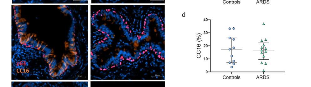
Gerard et al. (2024) quantified distal airway epithelial injury in ARDS and found pronounced denudation and neutrophilic infiltration along with impaired junctional proteins and pIgR expression. They report that serum concentrations of lung-derived proteins including CC16/SCGB1A1 were increased in ARDS, consistent with “pneumoproteinemia” and epithelial barrier disruption (gerard2024airwayepitheliumdamage pages 6-9, gerard2024airwayepitheliumdamage pages 4-6). In airway tissue, CC16/SCGB1A1 staining (club-cell area) was quantified and did not show a significant difference between controls and ARDS in the displayed figure panels, supporting the idea that serum increases can occur without increased club-cell abundance in tissue (gerard2024airwayepitheliumdamage pages 6-9, gerard2024airwayepitheliumdamage media 08abc01d).
4) Current applications and real-world implementations
4.1 Clinical and translational use: CC16/SCGB1A1 as a “pneumoprotein” biomarker
Across 2023–2024 literature, SCGB1A1/CC16 is used as a circulating marker of small-airway epithelial integrity and injury. A key real-world implementation domain is exposure-related lung injury monitoring and acute epithelial barrier disruption (neumann2024clubcellprotein pages 1-2, gerard2024airwayepitheliumdamage pages 6-9).
Biomarker interpretation constraints:
- CC16/SCGB1A1 levels are influenced by renal function and show circadian patterns, complicating use unless sampling time and creatinine are accounted for (neumann2024clubcellprotein pages 4-5, neumann2024clubcellprotein pages 5-7).
- Rapid clearance can make levels highly time-sensitive in acute injury contexts (reviewed half-life example in ARDS <18 minutes) (otelea2023clubcells—aguardian pages 2-3).
4.2 Occupational biomonitoring implementation example (2024)
Neumann et al. (2024) implemented serum CC16 monitoring pre- and post-shift in a large cross-sectional workforce study in potash mining (672 workers with paired values) with personal exposure measurements to nitrogen oxides, carbon monoxide, and diesel particulate matter. The study reports strong circadian patterns (e.g., early shift pre vs post: 10.15→15.30 ng/mL, p=0.0005; midday shift pre vs post: 13.80→10.70 ng/mL, p<0.0001) and significant associations with renal function and age (e.g., pre-shift age correlation rS=0.14, p=0.0003). Notably, exposure-associated effects were not statistically significant in never-smokers but were evident in current smokers, supporting use as an “effect marker” contingent on stratification and confounder control (neumann2024clubcellprotein pages 4-5, neumann2024clubcellprotein pages 5-7, neumann2024clubcellprotein pages 7-8).
5) Expert opinions and analysis (authoritative synthesis)
5.1 Authoritative 2023 synthesis: SCGB1A1 as an anti-inflammatory “lung protector” and therapeutic concept
Martinu et al. (Annual Review of Medicine, 2023) frame CCSP/SCGB1A1 as a negative regulator of acute and chronic lung inflammation with multiple proposed mechanisms (IL-8 binding; VLA-4 interaction; PLA2 antagonism; modulation of DC/Th responses; fibronectin–IgA complex prevention). Their synthesis emphasizes a mechanistic plurality rather than a single receptor–ligand pathway, and argues that altered CCSP levels can reflect either club-cell depletion (chronic obstructive disease) or increased permeability (acute injury) (martinu2023clubcellsecretory pages 3-4).
5.2 2023 conceptual advance: treating “club cells” as a heterogeneous progenitor ecosystem
Blackburn et al. (2023) emphasize that scRNA-seq–derived “secretory/club” clusters can include multiple states (SCGB1A1+, SCGB3A2+, MUC5B/MUC5AC mixtures), and that disease-associated remodeling in smokers/COPD plausibly reflects both cell loss and altered differentiation programs (including IFN-γ and other inflammatory drivers), motivating use of scRNA-seq, organoids, and refined markers in functional studies (blackburn2023anupdatein pages 10-13, blackburn2023anupdatein pages 17-21, blackburn2023anupdatein pages 41-44).
6) Relevant statistics and data highlights (recent studies)
Key quantitative findings from 2023–2024 evidence are summarized in the following table.
| Area | Study (first author year) | Population/Model & N | Key finding | Quantitative stats | Assay/Method | URL/DOI | Publication date |
|---|---|---|---|---|---|---|---|
| Asthma | Xu 2024 | Human plasma cohort: 20 asthma patients and 20 healthy controls; complementary HDM mouse models (xu2024claracell10 pages 1-2) | CC10/SCGB1A1 is lower in asthma and functionally suppresses allergic airway inflammation by modulating lung dendritic cells rather than directly inhibiting Th cells; linked to NF-κB signaling in DCs (xu2024claracell10 pages 1-2) | Human cohort sizes: n=20 asthma, n=20 controls; lower CC10 correlated with increased IgE and lymphocytes; excerpt did not provide p-values. In mice, Cc10−/− increased inflammatory infiltrates, Th2 cytokines, antigen-specific IgE and AHR; recombinant CC10 significantly attenuated responses (xu2024claracell10 pages 1-2) | ELISA in human plasma; HDM-induced allergic airway inflammation models; mixed lymphocyte response; DC phenotyping/mechanistic assays (xu2024claracell10 pages 1-2) | https://doi.org/10.1007/s00018-024-05368-z | Jul 2024 |
| ARDS | Gerard 2024 | Distal airway tissue: controls n=15, ARDS n=25; subset immunostaining ARDS n=13 vs controls n=11; serum/BALF ARDS n=24 vs controls n=7 (gerard2024airwayepitheliumdamage pages 4-6, gerard2024airwayepitheliumdamage pages 6-9) | ARDS causes major airway epithelial injury; tissue club-cell area by CC16/SCGB1A1 staining was not significantly changed, but serum CC16/SCGB1A1 increased, consistent with epithelial leak/spillover rather than club-cell expansion (gerard2024airwayepitheliumdamage pages 6-9, gerard2024airwayepitheliumdamage media 08abc01d) | Epithelial denudation 20% [IQR 5–40] vs 0% [0–0], p=0.0003; neutrophilic infiltration p=0.0005; ciliated-cell loss: β-tubulin area 0.61% [0.25–1.0] vs 3.67% [1.09–5.76], p=0.0317; Fox-J1 area 20.4% [5.1–30.8] vs 32.2% [27.1–43.5], p=0.034; goblet-cell trend p=0.055; pIgR area 14.42% [7.82–22.87] vs 41.57% [38.49–49.21], p<0.0001; serum SC 599 pg/mL [413–952] vs 133 [103–137], p<0.0001; serum S-IgA1 71.4 μg/mL [49.9–117.4] vs 10.8 [4.08–13.6], p<0.0001; serum CC16/SCGB1A1 increased (numeric value not in excerpt) (gerard2024airwayepitheliumdamage pages 4-6, gerard2024airwayepitheliumdamage pages 6-9) | Histology, multiplex immunofluorescence, serum and BALF ELISA; figure review confirms CC16 panel and no significant tissue difference in Fig. 2d (gerard2024airwayepitheliumdamage pages 6-9, gerard2024airwayepitheliumdamage media 08abc01d) | https://doi.org/10.1186/s13054-024-05127-3 | Oct 2024 |
| Occupational exposure | Neumann 2024 | Potash miners/workers: initial cohort 689 employees, 672 with paired CC16 values; facility n=97, maintenance n=97, mining n=478 (neumann2024clubcellprotein pages 1-2, neumann2024clubcellprotein pages 4-5, neumann2024clubcellprotein pages 5-7) | Serum CC16/SCGB1A1 is a small-airway epithelial effect marker, but interpretation is strongly affected by circadian rhythm, smoking, and renal function; work-exposure effects were evident mainly in current smokers (neumann2024clubcellprotein pages 1-2, neumann2024clubcellprotein pages 7-8, neumann2024clubcellprotein pages 5-7) | Median serum CC16: pre-early shift 10.15 ng/mL [3.65–26.00] vs post-early shift 15.30 [4.60–29.70], p=0.0005; pre-midday shift 13.80 [5.20–34.80] vs post-midday shift 10.70 [3.40–28.10], p<0.0001. Age correlated with CC16 pre-shift rS=0.14, p=0.0003; post-shift rS=0.13, p=0.0006. In never smokers, post-shift creatinine vs CC16 shift-difference rS=-0.15, p=0.0183. In underground current smokers, post-shift creatinine correlations with CC16 shift-difference: maintenance rS=-0.26, p=0.0096; mining rS=-0.10, p=0.0262. No significant exposure effects in never smokers; significant negative exposure correlations in current smokers (neumann2024clubcellprotein pages 7-8, neumann2024clubcellprotein pages 4-5, neumann2024clubcellprotein pages 5-7) | Personal exposure monitoring (NO, NO2, CO, EC-DPM), paired pre/post-shift serum CC16, creatinine, Spearman correlations, multiple linear regression (neumann2024clubcellprotein pages 1-2, neumann2024clubcellprotein pages 7-8, neumann2024clubcellprotein pages 5-7) | https://doi.org/10.1007/s00420-023-02035-x | Dec 2024 |
| Airway remodeling scRNA-seq | Cao 2024 | Air-liquid interface human bronchial epithelial cultures from healthy nonsmokers and COPD smokers; total 41,215 single cells across 6 groups (healthy 7,563; healthy+ECE 5,121; healthy+CSE 6,588; COPD 7,185; COPD+ECE 7,560; COPD+CSE 7,198) (cao2024singlecelltranscriptomicsreveals pages 4-5) | SCGB1A1 marks club and secretory-club populations; e-cigarette vapor and cigarette smoke alter club/ciliated differentiation, with Notch signaling implicated in e-cigarette-induced remodeling and reduced SCGB1A1 protein as an injury marker (cao2024singlecelltranscriptomicsreveals pages 5-6, cao2024singlecelltranscriptomicsreveals pages 4-5) | Club-cell fraction increased from 10% to 17% (ECE) or 21% (CSE) in healthy cultures; from 9% to 22% (ECE) or 27% (CSE) in COPD cultures. Ciliated4 fraction decreased with CSE from 7.2% to 2.4% in healthy cultures and from 4.3% to 1.9% in COPD cultures. ECE caused a differentiation state intermediate between untreated and CSE-treated cells; article reports decreased SCGB1A1 protein in e-cigarette users but snippet gives no p-value (cao2024singlecelltranscriptomicsreveals pages 5-6, cao2024singlecelltranscriptomicsreveals pages 4-5) | scRNA-seq, UMAP, pseudotime/velocity analysis, immunostaining, qPCR, ELISA; exposure at equal nicotine concentration 0.02 mg/mL (cao2024singlecelltranscriptomicsreveals pages 5-6, cao2024singlecelltranscriptomicsreveals pages 4-5) | https://doi.org/10.1186/s12931-024-02962-4 | Sep 2024 |
| COPD / club-cell heterogeneity | Blackburn 2023 | Review integrating human scRNA-seq, ALI culture, microscopy, and lineage/progenitor studies (blackburn2023anupdatein pages 10-13, blackburn2023anupdatein pages 41-44, blackburn2023anupdatein pages 1-6, blackburn2023anupdatein pages 13-17) | 2023 view: SCGB1A1 is the canonical human club-cell marker, but club cells are heterogeneous and lie on basal–club–goblet/alveolar continuums; smokers/COPD show loss of SCGB1A1+ club/secretory populations and impaired differentiation of basal cells into SCGB1A1+ cells (blackburn2023anupdatein pages 10-13, blackburn2023anupdatein pages 41-44, blackburn2023anupdatein pages 13-17) | SCGB1A1+ cells comprise ~11% of terminal bronchiolar cells and ~22% of respiratory bronchiolar cells. Highly purified human KRT5+SCGB1A1− basal cells were 99.0 ± 1.1% pure and could generate SCGB1A1+ cells in ALI culture. In humans, 11–44% of proliferating airway cells can be Scgb1a1+ and up to 25% of goblet cells express SCGB1A1. Quantitative COPD effect sizes not provided in excerpts, but multiple studies cited lower SCGB1A1 protein/mRNA and fewer SCGB1A1+ cells in smokers/COPD (blackburn2023anupdatein pages 10-13, blackburn2023anupdatein pages 41-44, blackburn2023anupdatein pages 1-6, blackburn2023anupdatein pages 13-17) | Review of scRNA-seq/trajectory analyses, ALI differentiation studies, immunostaining, lineage/progenitor studies (blackburn2023anupdatein pages 10-13, blackburn2023anupdatein pages 41-44, blackburn2023anupdatein pages 1-6, blackburn2023anupdatein pages 13-17) | https://doi.org/10.1152/ajplung.00192.2022 | May 2023 |
| Biomarker biology / translational overview | Otelea 2023 | Review of occupational/interstitial lung disease relevance and club-cell biomarker biology (otelea2023clubcells—aguardian pages 2-3, otelea2023clubcells—aguardian pages 1-2) | CC16/SCGB1A1 is a rapidly cleared circulating biomarker of club-cell/epithelial injury with practical relevance for exposure-related lung damage, but timing and renal handling matter (otelea2023clubcells—aguardian pages 2-3, otelea2023clubcells—aguardian pages 1-2) | Club cells comprise ~11–22% of respiratory bronchiolar cells; CCSP/CC16 is a 70-aa homodimer of 15,840 Da; healthy nonsmokers: ~1–5 μg/mL in BALF and ~10–15 ng/mL in plasma; plasma half-life in ARDS <18 min. Review also cites workers with VGDF exposure having 2.64% more high-attenuation CT areas (95% CI 1.23–4.19%) vs non-exposed (otelea2023clubcells—aguardian pages 2-3, otelea2023clubcells—aguardian pages 1-2) | Review synthesis of biomarker, exposure, and pathology studies (otelea2023clubcells—aguardian pages 2-3, otelea2023clubcells—aguardian pages 1-2) | https://doi.org/10.3390/biomedicines12010078 | Dec 2023 |
| Mechanistic overview | Martinu 2023 | Authoritative review of human and model-system CCSP/SCGB1A1 biology (martinu2023clubcellsecretory pages 3-4, martinu2023clubcellsecretory pages 1-3) | SCGB1A1 is a major secreted club-cell product and negative regulator of lung inflammation; proposed mechanisms include IL-8 binding, VLA-4 interaction, PLA2 antagonism, and modulation of dendritic-cell/Th17 and fibronectin-IgA pathways (martinu2023clubcellsecretory pages 3-4) | CCSP described as ~16-kDa secreted protein and among the most abundant lung proteins; circulating levels rise from birth to adulthood, are reduced by smoking/chronic exposures, and acutely increase after epithelial injury; excerpt provides no OR/AUC/p-values (martinu2023clubcellsecretory pages 3-4, martinu2023clubcellsecretory pages 1-3) | Review of human, animal, and mechanistic studies (martinu2023clubcellsecretory pages 3-4, martinu2023clubcellsecretory pages 1-3) | https://doi.org/10.1146/annurev-med-042921-123443 | Jan 2023 |
Table: This table summarizes the main quantitative and mechanistic findings on human SCGB1A1/CC10/CC16/CCSP from the evidence collected, emphasizing recent 2023-2024 studies. It is useful for comparing how SCGB1A1 behaves across asthma, ARDS, occupational exposure, airway remodeling, and COPD-related club-cell biology.
Additional quantitative/structural data points frequently used in interpretation include:
- Healthy nonsmoker reference ranges reported in a 2023 review: ~1–5 µg/mL in BALF and ~10–15 ng/mL in plasma (assay-dependent), highlighting that lung-lining fluid levels can be orders of magnitude higher than blood levels (otelea2023clubcells—aguardian pages 2-3).
- In ARDS, distal airway pathology in 2024 includes epithelial denudation (20% [IQR 5–40] vs 0% [0–0], p=0.0003), ciliated cell loss and impaired pIgR expression (p<0.0001), supporting a barrier-disruption framework for why serum lung proteins like CC16 rise (gerard2024airwayepitheliumdamage pages 4-6, gerard2024airwayepitheliumdamage pages 6-9).
7) Pathways and regulatory context (supported by retrieved evidence)
- NF-κB (immune signaling): CC10/SCGB1A1 modulates DC activation and Th2 inflammation through an NF-κB-linked mechanism in allergic airway inflammation models (xu2024claracell10 pages 1-2).
- Notch (epithelial differentiation): Notch signaling strength changes under e-cigarette vapor exposure; Notch inhibition ameliorates certain differentiation defects in ALI cultures, and SCGB1A1 is a core marker defining the club/secretory compartment in these analyses (cao2024singlecelltranscriptomicsreveals pages 4-5).
- Cytokine regulation and Th1/Th2 context: SCGB1A1 is embedded in Th1/Th2 responses and is regulated by cytokines; Th2 cytokines can downregulate SCGB1A1 and Th1-related pathways can regulate expression via JAK–STAT/FOXA factors (review synthesis) (mootz2022secretoglobinsinthe pages 11-14, mootz2022secretoglobinsinthe pages 9-11).
8) Cellular/tissue localization figure evidence
The following figure region from Gerard et al. (2024) visually supports that CC16/SCGB1A1 is a club-cell-associated airway epithelial marker and that tissue CC16-stained area can be quantified in control vs ARDS small airways (gerard2024airwayepitheliumdamage media 08abc01d).
9) Practical conclusions for functional annotation
- Primary function: SCGB1A1 encodes a secreted, dimeric secretoglobin that functions as an extracellular immunomodulator at the airway barrier, reducing inflammatory recruitment and shaping innate/adaptive responses (not a defined metabolic enzyme). Mechanistic evidence supports IL-8/CXCL8 binding, modulation of neutrophil recruitment/adhesion, PLA2 antagonism, and DC/NF-κB regulation (martinu2023clubcellsecretory pages 3-4, mootz2022secretoglobinsinthe pages 11-14, xu2024claracell10 pages 1-2).
- Primary location of action: secreted into airway lumen/lining fluid with measurable spillover into blood and urine; interpretation of circulating levels requires accounting for epithelial permeability and renal clearance (martinu2023clubcellsecretory pages 1-3, neumann2024clubcellprotein pages 1-2, otelea2023clubcells—aguardian pages 2-3).
- Pathway embedding: integrates epithelial differentiation (Notch-linked remodeling in airway exposures) and inflammatory control (NF-κB in DCs; cytokine-regulated expression) (cao2024singlecelltranscriptomicsreveals pages 4-5, xu2024claracell10 pages 1-2, mootz2022secretoglobinsinthe pages 11-14).
- Applications: widely used as a translational biomarker of small-airway epithelial integrity/injury in occupational exposure studies and acute lung injury contexts; large 2024 workforce data show strong circadian and renal confounding that must be controlled for real-world biomonitoring (neumann2024clubcellprotein pages 4-5, neumann2024clubcellprotein pages 5-7, gerard2024airwayepitheliumdamage pages 6-9).
URLs for key 2023–2024 sources used
- Martinu et al., Annual Review of Medicine (Jan 2023): https://doi.org/10.1146/annurev-med-042921-123443 (martinu2023clubcellsecretory pages 3-4)
- Blackburn et al., AJP-Lung (May 2023): https://doi.org/10.1152/ajplung.00192.2022 (blackburn2023anupdatein pages 10-13)
- Otelea et al., Biomedicines (Dec 2023): https://doi.org/10.3390/biomedicines12010078 (otelea2023clubcells—aguardian pages 2-3)
- Gerard et al., Critical Care (Oct 2024): https://doi.org/10.1186/s13054-024-05127-3 (gerard2024airwayepitheliumdamage pages 4-6)
- Cao et al., Respiratory Research (Sep 2024): https://doi.org/10.1186/s12931-024-02962-4 (cao2024singlecelltranscriptomicsreveals pages 4-5)
- Xu et al., Cellular and Molecular Life Sciences (Jul 2024): https://doi.org/10.1007/s00018-024-05368-z (xu2024claracell10 pages 1-2)
- Neumann et al., Int Arch Occup Environ Health (Dec 2024): https://doi.org/10.1007/s00420-023-02035-x (neumann2024clubcellprotein pages 1-2)
References
-
(mootz2022secretoglobinsinthe pages 26-30): Martine Mootz, Constanze A. Jakwerth, Carsten B. Schmidt‐Weber, and Ulrich M. Zissler. Secretoglobins in the big picture of immunoregulation in airway diseases. Allergy, 77:767-777, Aug 2022. URL: https://doi.org/10.1111/all.15033, doi:10.1111/all.15033. This article has 54 citations and is from a highest quality peer-reviewed journal.
-
(martinu2023clubcellsecretory pages 1-3): Tereza Martinu, Jamie L. Todd, Andrew E. Gelman, Stefano Guerra, and Scott M. Palmer. Club cell secretory protein in lung disease: emerging concepts and potential therapeutics. Annual Review of Medicine, 74:427-441, Jan 2023. URL: https://doi.org/10.1146/annurev-med-042921-123443, doi:10.1146/annurev-med-042921-123443. This article has 69 citations and is from a domain leading peer-reviewed journal.
-
(xu2024claracell10 pages 1-2): Yu-Dong Xu, Mi Cheng, Jun-Xia Mao, Xue Zhang, Pan-Pan Shang, Jie Long, Yan-Jiao Chen, Yu Wang, Lei-Miao Yin, and Yong-Qing Yang. Clara cell 10 (cc10) protein attenuates allergic airway inflammation by modulating lung dendritic cell functions. Cellular and Molecular Life Sciences: CMLS, Jul 2024. URL: https://doi.org/10.1007/s00018-024-05368-z, doi:10.1007/s00018-024-05368-z. This article has 10 citations.
-
(mootz2022secretoglobinsinthe pages 1-6): Martine Mootz, Constanze A. Jakwerth, Carsten B. Schmidt‐Weber, and Ulrich M. Zissler. Secretoglobins in the big picture of immunoregulation in airway diseases. Allergy, 77:767-777, Aug 2022. URL: https://doi.org/10.1111/all.15033, doi:10.1111/all.15033. This article has 54 citations and is from a highest quality peer-reviewed journal.
-
(martinu2023clubcellsecretory pages 3-4): Tereza Martinu, Jamie L. Todd, Andrew E. Gelman, Stefano Guerra, and Scott M. Palmer. Club cell secretory protein in lung disease: emerging concepts and potential therapeutics. Annual Review of Medicine, 74:427-441, Jan 2023. URL: https://doi.org/10.1146/annurev-med-042921-123443, doi:10.1146/annurev-med-042921-123443. This article has 69 citations and is from a domain leading peer-reviewed journal.
-
(mootz2022secretoglobinsinthe pages 9-11): Martine Mootz, Constanze A. Jakwerth, Carsten B. Schmidt‐Weber, and Ulrich M. Zissler. Secretoglobins in the big picture of immunoregulation in airway diseases. Allergy, 77:767-777, Aug 2022. URL: https://doi.org/10.1111/all.15033, doi:10.1111/all.15033. This article has 54 citations and is from a highest quality peer-reviewed journal.
-
(otelea2023clubcells—aguardian pages 2-3): Marina Ruxandra Otelea, Corina Oancea, Daniela Reisz, Monica Adriana Vaida, Andreea Maftei, and Florina Georgeta Popescu. Club cells—a guardian against occupational hazards. Biomedicines, 12:78, Dec 2023. URL: https://doi.org/10.3390/biomedicines12010078, doi:10.3390/biomedicines12010078. This article has 5 citations.
-
(neumann2024clubcellprotein pages 1-2): Savo Neumann, Swaantje Casjens, Frank Hoffmeyer, Katrin Rühle, Lisa Gamrad-Streubel, Lisa-Marie Haase, Katharina K. Rudolph, Jörg Giesen, Volker Neumann, Dirk Taeger, Dirk Pallapies, Thomas Birk, Thomas Brüning, and Jürgen Bünger. Club cell protein (cc16) in serum as an effect marker for small airway epithelial damage caused by diesel exhaust and blasting fumes in potash mining. International Archives of Occupational and Environmental Health, 97:121-132, Dec 2024. URL: https://doi.org/10.1007/s00420-023-02035-x, doi:10.1007/s00420-023-02035-x. This article has 3 citations and is from a peer-reviewed journal.
-
(mootz2022secretoglobinsinthe pages 11-14): Martine Mootz, Constanze A. Jakwerth, Carsten B. Schmidt‐Weber, and Ulrich M. Zissler. Secretoglobins in the big picture of immunoregulation in airway diseases. Allergy, 77:767-777, Aug 2022. URL: https://doi.org/10.1111/all.15033, doi:10.1111/all.15033. This article has 54 citations and is from a highest quality peer-reviewed journal.
-
(blackburn2023anupdatein pages 10-13): Jessica B. Blackburn, Ngan Fung Li, Nathan W. Bartlett, and Bradley W. Richmond. An update in club cell biology and its potential relevance to chronic obstructive pulmonary disease. May 2023. URL: https://doi.org/10.1152/ajplung.00192.2022, doi:10.1152/ajplung.00192.2022. This article has 64 citations.
-
(blackburn2023anupdatein pages 13-17): Jessica B. Blackburn, Ngan Fung Li, Nathan W. Bartlett, and Bradley W. Richmond. An update in club cell biology and its potential relevance to chronic obstructive pulmonary disease. May 2023. URL: https://doi.org/10.1152/ajplung.00192.2022, doi:10.1152/ajplung.00192.2022. This article has 64 citations.
-
(blackburn2023anupdatein pages 41-44): Jessica B. Blackburn, Ngan Fung Li, Nathan W. Bartlett, and Bradley W. Richmond. An update in club cell biology and its potential relevance to chronic obstructive pulmonary disease. May 2023. URL: https://doi.org/10.1152/ajplung.00192.2022, doi:10.1152/ajplung.00192.2022. This article has 64 citations.
-
(cao2024singlecelltranscriptomicsreveals pages 5-6): Weitao Cao, Jia Li, Li Che, Ruixue Yang, Zehong Wu, Guoping Hu, Weifeng Zou, Zehang Zhao, Yumin Zhou, Xingtao Jiang, Tiejun Zhang, Wenguang Yin, and Pixin Ran. Single-cell transcriptomics reveals e-cigarette vapor-induced airway epithelial remodeling and injury. Respiratory Research, Sep 2024. URL: https://doi.org/10.1186/s12931-024-02962-4, doi:10.1186/s12931-024-02962-4. This article has 3 citations and is from a domain leading peer-reviewed journal.
-
(cao2024singlecelltranscriptomicsreveals pages 4-5): Weitao Cao, Jia Li, Li Che, Ruixue Yang, Zehong Wu, Guoping Hu, Weifeng Zou, Zehang Zhao, Yumin Zhou, Xingtao Jiang, Tiejun Zhang, Wenguang Yin, and Pixin Ran. Single-cell transcriptomics reveals e-cigarette vapor-induced airway epithelial remodeling and injury. Respiratory Research, Sep 2024. URL: https://doi.org/10.1186/s12931-024-02962-4, doi:10.1186/s12931-024-02962-4. This article has 3 citations and is from a domain leading peer-reviewed journal.
-
(gerard2024airwayepitheliumdamage pages 6-9): Ludovic Gerard, Marylene Lecocq, Bruno Detry, Caroline Bouzin, Delphine Hoton, Joao Pinto Pereira, François Carlier, Thomas Plante-Bordeneuve, Sophie Gohy, Valérie Lacroix, Pierre-François Laterre, and Charles Pilette. Airway epithelium damage in acute respiratory distress syndrome. Critical Care, Oct 2024. URL: https://doi.org/10.1186/s13054-024-05127-3, doi:10.1186/s13054-024-05127-3. This article has 18 citations and is from a highest quality peer-reviewed journal.
-
(gerard2024airwayepitheliumdamage pages 4-6): Ludovic Gerard, Marylene Lecocq, Bruno Detry, Caroline Bouzin, Delphine Hoton, Joao Pinto Pereira, François Carlier, Thomas Plante-Bordeneuve, Sophie Gohy, Valérie Lacroix, Pierre-François Laterre, and Charles Pilette. Airway epithelium damage in acute respiratory distress syndrome. Critical Care, Oct 2024. URL: https://doi.org/10.1186/s13054-024-05127-3, doi:10.1186/s13054-024-05127-3. This article has 18 citations and is from a highest quality peer-reviewed journal.
-
(gerard2024airwayepitheliumdamage media 08abc01d): Ludovic Gerard, Marylene Lecocq, Bruno Detry, Caroline Bouzin, Delphine Hoton, Joao Pinto Pereira, François Carlier, Thomas Plante-Bordeneuve, Sophie Gohy, Valérie Lacroix, Pierre-François Laterre, and Charles Pilette. Airway epithelium damage in acute respiratory distress syndrome. Critical Care, Oct 2024. URL: https://doi.org/10.1186/s13054-024-05127-3, doi:10.1186/s13054-024-05127-3. This article has 18 citations and is from a highest quality peer-reviewed journal.
-
(neumann2024clubcellprotein pages 4-5): Savo Neumann, Swaantje Casjens, Frank Hoffmeyer, Katrin Rühle, Lisa Gamrad-Streubel, Lisa-Marie Haase, Katharina K. Rudolph, Jörg Giesen, Volker Neumann, Dirk Taeger, Dirk Pallapies, Thomas Birk, Thomas Brüning, and Jürgen Bünger. Club cell protein (cc16) in serum as an effect marker for small airway epithelial damage caused by diesel exhaust and blasting fumes in potash mining. International Archives of Occupational and Environmental Health, 97:121-132, Dec 2024. URL: https://doi.org/10.1007/s00420-023-02035-x, doi:10.1007/s00420-023-02035-x. This article has 3 citations and is from a peer-reviewed journal.
-
(neumann2024clubcellprotein pages 5-7): Savo Neumann, Swaantje Casjens, Frank Hoffmeyer, Katrin Rühle, Lisa Gamrad-Streubel, Lisa-Marie Haase, Katharina K. Rudolph, Jörg Giesen, Volker Neumann, Dirk Taeger, Dirk Pallapies, Thomas Birk, Thomas Brüning, and Jürgen Bünger. Club cell protein (cc16) in serum as an effect marker for small airway epithelial damage caused by diesel exhaust and blasting fumes in potash mining. International Archives of Occupational and Environmental Health, 97:121-132, Dec 2024. URL: https://doi.org/10.1007/s00420-023-02035-x, doi:10.1007/s00420-023-02035-x. This article has 3 citations and is from a peer-reviewed journal.
-
(neumann2024clubcellprotein pages 7-8): Savo Neumann, Swaantje Casjens, Frank Hoffmeyer, Katrin Rühle, Lisa Gamrad-Streubel, Lisa-Marie Haase, Katharina K. Rudolph, Jörg Giesen, Volker Neumann, Dirk Taeger, Dirk Pallapies, Thomas Birk, Thomas Brüning, and Jürgen Bünger. Club cell protein (cc16) in serum as an effect marker for small airway epithelial damage caused by diesel exhaust and blasting fumes in potash mining. International Archives of Occupational and Environmental Health, 97:121-132, Dec 2024. URL: https://doi.org/10.1007/s00420-023-02035-x, doi:10.1007/s00420-023-02035-x. This article has 3 citations and is from a peer-reviewed journal.
-
(blackburn2023anupdatein pages 17-21): Jessica B. Blackburn, Ngan Fung Li, Nathan W. Bartlett, and Bradley W. Richmond. An update in club cell biology and its potential relevance to chronic obstructive pulmonary disease. May 2023. URL: https://doi.org/10.1152/ajplung.00192.2022, doi:10.1152/ajplung.00192.2022. This article has 64 citations.
-
(blackburn2023anupdatein pages 1-6): Jessica B. Blackburn, Ngan Fung Li, Nathan W. Bartlett, and Bradley W. Richmond. An update in club cell biology and its potential relevance to chronic obstructive pulmonary disease. May 2023. URL: https://doi.org/10.1152/ajplung.00192.2022, doi:10.1152/ajplung.00192.2022. This article has 64 citations.
-
(otelea2023clubcells—aguardian pages 1-2): Marina Ruxandra Otelea, Corina Oancea, Daniela Reisz, Monica Adriana Vaida, Andreea Maftei, and Florina Georgeta Popescu. Club cells—a guardian against occupational hazards. Biomedicines, 12:78, Dec 2023. URL: https://doi.org/10.3390/biomedicines12010078, doi:10.3390/biomedicines12010078. This article has 5 citations.
Artifacts
Citations
- martinu2023clubcellsecretory pages 1-3
- martinu2023clubcellsecretory pages 3-4
- blackburn2023anupdatein pages 41-44
- cao2024singlecelltranscriptomicsreveals pages 4-5
- blackburn2023anupdatein pages 10-13
- gerard2024airwayepitheliumdamage pages 4-6
- neumann2024clubcellprotein pages 1-2
- mootz2022secretoglobinsinthe pages 26-30
- mootz2022secretoglobinsinthe pages 1-6
- mootz2022secretoglobinsinthe pages 9-11
- mootz2022secretoglobinsinthe pages 11-14
- blackburn2023anupdatein pages 13-17
- cao2024singlecelltranscriptomicsreveals pages 5-6
- gerard2024airwayepitheliumdamage pages 6-9
- neumann2024clubcellprotein pages 4-5
- neumann2024clubcellprotein pages 5-7
- neumann2024clubcellprotein pages 7-8
- blackburn2023anupdatein pages 17-21
- blackburn2023anupdatein pages 1-6
- IQR 5–40
- 0–0
- 0.25–1.0
- 1.09–5.76
- 5.1–30.8
- 27.1–43.5
- 7.82–22.87
- 38.49–49.21
- 413–952
- 103–137
- 49.9–117.4
- 4.08–13.6
- 3.65–26.00
- 4.60–29.70
- 5.20–34.80
- 3.40–28.10
- https://doi.org/10.1007/s00018-024-05368-z
- https://doi.org/10.1186/s13054-024-05127-3
- https://doi.org/10.1007/s00420-023-02035-x
- https://doi.org/10.1186/s12931-024-02962-4
- https://doi.org/10.1152/ajplung.00192.2022
- https://doi.org/10.3390/biomedicines12010078
- https://doi.org/10.1146/annurev-med-042921-123443
- https://doi.org/10.1111/all.15033,
- https://doi.org/10.1146/annurev-med-042921-123443,
- https://doi.org/10.1007/s00018-024-05368-z,
- https://doi.org/10.3390/biomedicines12010078,
- https://doi.org/10.1007/s00420-023-02035-x,
- https://doi.org/10.1152/ajplung.00192.2022,
- https://doi.org/10.1186/s12931-024-02962-4,
- https://doi.org/10.1186/s13054-024-05127-3,
OpenAI
(SCGB1A1-deep-research-openai.md)
OpenAI
(SCGB1A1-deep-research-openai.md)SCGB1A1 (Club Cell Secretory Protein/Clara Cell 10-kDa Protein) – Function and Localization
Gene and Protein Overview
SCGB1A1 (Secretoglobin Family 1A Member 1) is a small secreted protein best known as Club Cell Secretory Protein (CCSP), formerly called Clara Cell 10-kDa protein (CC10/CC16) or uteroglobin. It belongs to the secretoglobin superfamily – a group of small, secreted, dimeric proteins (pmc.ncbi.nlm.nih.gov). The mature SCGB1A1 protein is a homodimer (~15–16 kDa total) composed of two identical subunits. Each monomer is ~70–90 amino acids long (after signal peptide cleavage) and together they form a stable dimer with an internal hydrophobic cavity (pubmed.ncbi.nlm.nih.gov). This internal pocket at the dimer interface allows SCGB1A1 to bind small hydrophobic ligands (pubmed.ncbi.nlm.nih.gov). The protein contains key structural residues (e.g. Lys-43 and Asp-46 in human SCGB1A1) that are critical for its functional interactions (pmc.ncbi.nlm.nih.gov). SCGB1A1 has no known enzymatic activity; rather, its function is mediated by binding to other molecules and modulating their activity.
Expression and Localization
SCGB1A1 is predominantly produced by club cells (Clara cells) in the bronchiolar airways of the lung, making it one of the most abundant proteins in the lung airways (pubmed.ncbi.nlm.nih.gov). It is also expressed by epithelial cells in other mucosal tissues, such as the uterus and nasopharynx, albeit at lower levels (journals.plos.org) (pubmed.ncbi.nlm.nih.gov). In the lung, SCGB1A1 is stored in secretory granules of club cells and actively secreted into the airway lining fluid. Baseline levels in bronchoalveolar fluid are high, and the protein is detectable in the circulation (blood) and even in urine under normal conditions (pubmed.ncbi.nlm.nih.gov). This reflects its abundance and stability as a secreted protein. Notably, SCGB1A1 is an extracellular protein – it carries out its functions in the lung lining fluid and extracellular spaces rather than inside cells. It can also diffuse or be transported into the bloodstream, which has made it useful as a biomarker of lung epithelial integrity (low serum levels often correlate with lung injury or club cell loss) (pubmed.ncbi.nlm.nih.gov).
Primary Function and Biochemical Activity
SCGB1A1’s primary role is immunomodulatory and anti-inflammatory, particularly in the lung airways. It is often described as a natural anti-inflammatory protein secreted by airway epithelium (pubmed.ncbi.nlm.nih.gov). Unlike enzymes or receptors, SCGB1A1 acts by binding and sequestering specific ligands and proteins involved in inflammation. A key target is Phospholipase A₂ (PLA₂), an enzyme that releases arachidonic acid from membrane phospholipids to generate pro-inflammatory eicosanoids. SCGB1A1 directly binds to and inactivates secretory PLA₂, thereby preventing the downstream production of arachidonic acid metabolites like leukotrienes and prostaglandins (journals.plos.org). Mutagenesis studies (e.g. replacing Lys-43 or Asp-46) abolish PLA₂ inhibition, confirming that SCGB1A1’s interaction with PLA₂ is specific and structurally mediated (pmc.ncbi.nlm.nih.gov). By inhibiting PLA₂, SCGB1A1 limits the generation of neutrophil-activating lipid mediators, which is thought to reduce acute lung inflammation and tissue injury (such as in ARDS) (journals.plos.org).
In addition to PLA₂, SCGB1A1 binds various hydrophobic inflammatory mediators. For example, it can sequester certain prostaglandins – studies have shown it binds and neutralizes prostaglandin D₂ and F₂α, blocking their interaction with their G-protein coupled receptors (pmc.ncbi.nlm.nih.gov). By buffering these prostanoids, SCGB1A1 prevents prostaglandin-induced pro-inflammatory gene expression in the airway. Mandal et al. (2004) demonstrated that uteroglobin (SCGB1A1) addition repressed allergen-induced inflammation by blocking PGD₂ receptor signaling in a mouse asthma model (pmc.ncbi.nlm.nih.gov). Similarly, it was reported that SCGB1A1 can interfere with PGF₂α receptor–mediated responses (pmc.ncbi.nlm.nih.gov). These findings indicate that SCGB1A1 acts as a “decoy” or scavenger for inflammatory ligands, reducing their ability to trigger immune cells.
SCGB1A1’s anti-inflammatory scope also extends to cytokines and chemokines. It has been shown to bind certain pro-inflammatory cytokines, effectively sequestering them (journals.plos.org). This contributes to its ability to interfere with leukocyte chemotaxis and recruitment (journals.plos.org). For instance, in experimental models lacking SCGB1A1, there is excessive recruitment of neutrophils to the airways in response to irritants or infection, whereas presence of SCGB1A1 restrains this neutrophilic influx (pubmed.ncbi.nlm.nih.gov) (journals.plos.org). SCGB1A1 can even be taken up by immune cells: immuno-electron microscopy studies detected SCGB1A1 inside neutrophils in inflamed lungs, suggesting direct protein-leukocyte interaction (journals.plos.org). Inside these cells, SCGB1A1 might modulate their function (potentially by affecting signaling pathways or reactive oxygen species production (journals.plos.org)).
Notably, SCGB1A1 also modulates intracellular signaling in immune and epithelial cells. It has been shown to inhibit NF-κB activation in airway epithelial cells, thus down-regulating the transcription of many inflammatory genes (pmc.ncbi.nlm.nih.gov). A study in human airway cells demonstrated that overexpression of CC10/SCGB1A1 suppresses NF-κB nuclear translocation and activity, thereby reducing cytokine production (pmc.ncbi.nlm.nih.gov). Consistent with this, exogenous SCGB1A1 protein dampens macrophage activation: a 2020 study added recombinant SCGB1A1 to cultured primary alveolar macrophages and found significantly blunted release of key cytokines (IL-1β, IL-6, IL-8, TNF-α, MIP-1α, MCP-1) in response to bacterial stimuli (pmc.ncbi.nlm.nih.gov). This blunting of macrophage inflammatory responses illustrates how SCGB1A1 acts as a brake on the lung’s immune system to prevent overreaction. Collectively, these mechanisms – inhibition of PLA₂ and NF-κB, sequestration of eicosanoids and cytokines, and blocking of chemoattractant signals – explain SCGB1A1’s core function as an anti-inflammatory guardian in the airways (journals.plos.org) (pmc.ncbi.nlm.nih.gov).
Biological Processes and Pathways
Through the above interactions, SCGB1A1 participates in several biological processes: chiefly regulation of inflammatory response, airway immune homeostasis, and tissue repair. In the eicosanoid pathway, SCGB1A1’s inhibition of PLA₂ means that upstream steps of prostaglandin and leukotriene synthesis are curtailed (journals.plos.org). This reduces activation of cells like neutrophils and eosinophils that respond to those lipid mediators. In parallel, by binding prostaglandins and blocking their receptors, SCGB1A1 directly dampens prostaglandin-signaling pathways that would otherwise lead to bronchoconstriction, vasodilation, and immune cell recruitment (pmc.ncbi.nlm.nih.gov). Thus, SCGB1A1 inserts itself into the arachidonic acid cascade as a negative regulator.
SCGB1A1 also influences chemokine and cytokine signaling networks in the lung. Its ability to reduce IL-8, TNF-α and other cytokine output from macrophages and epithelial cells means pathways like the NF-κB pathway and MAPK pathways (which control cytokine gene expression) are kept in check (pmc.ncbi.nlm.nih.gov) (pmc.ncbi.nlm.nih.gov). In effect, SCGB1A1 elevates the threshold for lung inflammation by requiring a stronger stimulus to overcome its inhibitory presence. It contributes to maintaining an anti-inflammatory bias in the steady state lung environment (sometimes described as keeping alveolar macrophages in a “quiescent” state) (pmc.ncbi.nlm.nih.gov) (pmc.ncbi.nlm.nih.gov).
Another process involving SCGB1A1 is fibrosis and tissue remodeling. Chronic inflammation often leads to fibrotic changes; by suppressing chronic inflammatory signals, SCGB1A1 indirectly has anti-fibrotic effects (pmc.ncbi.nlm.nih.gov). For example, SCGB1A1 deficiency in mice has been linked to heightened TGF-β and collagen deposition after lung injury, whereas presence of SCGB1A1 mitigates such profibrotic pathways (pmc.ncbi.nlm.nih.gov). SCGB1A1 has even been reported to inhibit fibroblast migration in vitro via a PLA₂-dependent mechanism (pmc.ncbi.nlm.nih.gov), suggesting it can influence repair processes.
It’s important to note that SCGB1A1 does not function as a classical signaling ligand with a dedicated receptor. Despite being a secreted protein, no specific high-affinity SCGB1A1 receptor has been definitively identified on target cells. Instead, SCGB1A1’s “signaling” role is more akin to a buffer or modulator: it binds inflammatory mediators (lipids, cytokines, chemoattractants) and possibly coats cell surfaces, thereby broadly altering signaling thresholds. In summary, SCGB1A1 operates at the interface of innate immune pathways – intercepting inflammatory triggers upstream and reinforcing endogenous anti-inflammatory signaling (such as by preventing NF-κB activation). This positions SCGB1A1 as a key regulator of the airway inflammatory cascade and surfactant homeostasis.
Experimental Evidence and Clinical Relevance
Experimental models strongly support SCGB1A1’s functional role: Mice genetically knocked out for Scgb1a1 exhibit exaggerated pulmonary inflammation when challenged. Mandal et al. reported that UG/SCGB1A1-knockout mice show heightened eosinophilic inflammation and Th2 cytokine production in asthma models, underscoring the protein’s role in restraining allergic inflammation (pubmed.ncbi.nlm.nih.gov). Conversely, mice or cells supplemented with exogenous SCGB1A1 have reduced inflammatory responses, as noted in macrophage studies and in in vivo lung injury models (pmc.ncbi.nlm.nih.gov) (journals.plos.org). For instance, overexpressing SCGB1A1 in mouse airways was shown to protect against ventilator-induced lung injury and decrease cytokine levels in bronchoalveolar fluid (pmc.ncbi.nlm.nih.gov) (pmc.ncbi.nlm.nih.gov). These controlled experiments give confidence that SCGB1A1 is not just a correlated marker but a causal modulator of lung inflammation.
Clinically, SCGB1A1 (often measured as CC16 in serum or sputum) has been linked to lung disease states. Asthma and COPD patients generally have lower airway SCGB1A1 levels, correlating with greater inflammation (journals.plos.org) (pubmed.ncbi.nlm.nih.gov). The protein is often depleted in the bronchiolar epithelium and bronchoalveolar fluid of asthmatics, presumably due to club cell damage or downregulation in chronic inflammation (journals.plos.org). This deficiency may remove an important brake on inflammation, potentially exacerbating disease severity. On the other hand, certain restrictive lung diseases (pulmonary fibrosis, etc.) show elevated SCGB1A1 levels in lung fluid and blood, which might reflect a compensatory upregulation or leakage from injured epithelium (pubmed.ncbi.nlm.nih.gov). Such observations support SCGB1A1’s role in human pulmonary homeostasis. Additionally, outside the lung, SCGB1A1’s anti-inflammatory action has been observed – for example, administering recombinant human uteroglobin reduced kidney inflammation in a mouse glomerulonephritis model (journals.plos.org), suggesting a broader therapeutic potential.
Therapeutic and real-world applications: Because of its protective effects, SCGB1A1 is being explored as a treatment and a biomarker. In neonatology, recombinant human CC10 (rhCC10) has been tested in premature infants at risk of bronchopulmonary dysplasia, with early trials indicating it can be safely given intratracheally and may reduce inflammation in the lung (pubmed.ncbi.nlm.nih.gov). More broadly, researchers view SCGB1A1 as a prototype anti-inflammatory agent. An Annual Review of Medicine in 2023 highlighted that “recent studies demonstrate multiple mechanisms by which CCSP dampens acute and chronic lung inflammation” and that augmenting CCSP could be a novel therapeutic strategy for lung diseases (pubmed.ncbi.nlm.nih.gov). There is interest in boosting SCGB1A1 levels (e.g. via inhaled recombinant protein or drugs that induce its expression) in diseases like COPD, asthma, and acute lung injury. Glucocorticoid steroids, a standard asthma therapy, are known to increase SCGB1A1 expression, which might partly mediate their benefit (geneglobe.qiagen.com). Thus, SCGB1A1 sits at a strategic point in pulmonary medicine – as a biomarker of epithelial health, a target for therapy, and a clue to the lung’s intrinsic anti-inflammatory defenses.
Conclusion and Expert Perspectives
SCGB1A1 (uteroglobin/CCSP) is now recognized as a critical endogenous anti-inflammatory protein in humans. By operating in the extracellular space of the lung and other mucosal organs, it binds pro-inflammatory molecules (like PLA₂ enzymes and prostaglandins) and prevents excessive immune activation (journals.plos.org) (pmc.ncbi.nlm.nih.gov). Its role is distinct from classic cytokines: rather than triggering immune responses, it tones them down, promoting resolution and protecting tissues from collateral damage. Experts in pulmonary biology regard SCGB1A1 as a key factor in maintaining airway homeostasis and preventing chronic inflammation (pubmed.ncbi.nlm.nih.gov) (journals.plos.org). Ongoing research (2020–2024) continues to uncover new facets – such as SCGB1A1’s influence on macrophage polarization, its potential anti-viral properties, and its regulation by signaling pathways like FOXA2/FOXP (transcription factors controlling club cell function) (pmc.ncbi.nlm.nih.gov) (pmc.ncbi.nlm.nih.gov).
In summary, SCGB1A1’s primary function is to safeguard mucosal tissues by curbing inflammation. It does so via a combination of biochemical mechanisms: inhibiting PLA₂ and arachidonate metabolism, binding inflammatory lipids and cytokines, and modulating immune cell behavior (journals.plos.org) (pmc.ncbi.nlm.nih.gov). Localization in secretory club cells and secretion into airway fluids positions it exactly where these inflammatory processes occur, ensuring timely intervention at the lung interface. Given its significant regulatory role, SCGB1A1 continues to be studied as both a diagnostic marker of lung injury and a therapeutic candidate to treat inflammatory and fibrotic diseases (pubmed.ncbi.nlm.nih.gov). The convergence of biochemical, structural, and animal model evidence paints a consistent picture: SCGB1A1 is a pivotal anti-inflammatory “guardian” of the airway, contributing to healthy pulmonary function by keeping immune responses in balance (pubmed.ncbi.nlm.nih.gov) (journals.plos.org).
References:
- Côté O. et al. (2014). PLOS ONE 9(4): e96217 – “Secretoglobin 1A1 and 1A1A Differentially Regulate Neutrophil ROS Production, Phagocytosis and NET Formation.” DOI:10.1371/journal.pone.0096217 (journals.plos.org) (journals.plos.org).
- Xu M. et al. (2020). Front. Immunol. 11:584310 – “Lung Secretoglobin Scgb1a1 Influences Alveolar Macrophage-Mediated Inflammation and Immunity.” Published Oct 1, 2020 (pmc.ncbi.nlm.nih.gov) (pmc.ncbi.nlm.nih.gov).
- Mandal A.K. et al. (2004). J. Exp. Med. 199(10):1317–1330 – “Uteroglobin represses allergen-induced inflammatory response by blocking PGD₂ receptor–mediated functions.” DOI:10.1084/jem.20031666 (pmc.ncbi.nlm.nih.gov).
- Chowdhury B. et al. (2002). Biochem. Biophys. Res. Commun. 295(4):877–883 – “Lys-43 and Asp-46 in uteroglobin are essential for its phospholipase A₂ inhibitory activity.” DOI:10.1016/S0006-291X(02)00767-2 (pmc.ncbi.nlm.nih.gov).
- Lesur O. et al. (1995). Am. J. Respir. Crit. Care Med. 152(1):290–297 – “Clara cell protein (CC-16) induces a PLA₂-mediated inhibition of fibroblast migration in vitro.” DOI:10.1164/ajrccm.152.1.7541278 (pmc.ncbi.nlm.nih.gov).
- Martinu T. et al. (2023). Annu. Rev. Med. 74:193–208 – “Club Cell Secretory Protein in Lung Disease: Emerging Concepts and Potential Therapeutics.” (Published Feb 2023) (pubmed.ncbi.nlm.nih.gov) (pubmed.ncbi.nlm.nih.gov).
- “Secretoglobin (SCGB) superfamily is a group of small, secreted, dimeric proteins…” – Amatngalim GD et al., Cytokine (2018) (cited in (pmc.ncbi.nlm.nih.gov)).
- “UG is an anti-inflammatory protein secreted by epithelia of organs with external contact…” – Asim Mandal et al., J. Exp. Med. (May 2004) (pubmed.ncbi.nlm.nih.gov).
- “SCGB1A1 binds small–medium ligands through an internal hydrophobic cavity at the dimer interface.” – Blaas L. et al., FEBS J. (2001) (cited in (pubmed.ncbi.nlm.nih.gov)).
- “Exogenous SCGB1A1 blunts release of IL-1β, IL-6, IL-8, TNF-α, etc., mitigating cytokine surges in lungs.” – Xu M. et al., Front. Immunol. (2020) (pmc.ncbi.nlm.nih.gov).
Citations
- AnnotationURLCitation(end_index=537, start_index=424, title='The cytokine-driven regulation of secretoglobins in normal human upper airway and their expression, particularly that of uteroglobin-related protein 1, in chronic rhinosinusitis - PMC', type='url_citation', url='https://pmc.ncbi.nlm.nih.gov/articles/PMC3063214/#:~:text=Secretoglobin%20,Clara%20cell')
- AnnotationURLCitation(end_index=906, start_index=782, title='The uteroglobin fold - PubMed', type='url_citation', url='https://pubmed.ncbi.nlm.nih.gov/11193783/#:~:text=medium,the%20cap%20domain%20of%20Xanthobacter')
- AnnotationURLCitation(end_index=1125, start_index=1001, title='The uteroglobin fold - PubMed', type='url_citation', url='https://pubmed.ncbi.nlm.nih.gov/11193783/#:~:text=medium,the%20cap%20domain%20of%20Xanthobacter')
- AnnotationURLCitation(end_index=1394, start_index=1264, title='Lung Secretoglobin Scgb1a1 Influences Alveolar Macrophage-Mediated Inflammation and Immunity - PMC', type='url_citation', url='https://pmc.ncbi.nlm.nih.gov/articles/PMC7558713/#:~:text=%2A%2027.Chowdhury%20B%2C%20Mantile,2%20%5BDOI')
- AnnotationURLCitation(end_index=1867, start_index=1735, title='Club Cell Secretory Protein in Lung Disease: Emerging Concepts and Potential Therapeutics - PubMed', type='url_citation', url='https://pubmed.ncbi.nlm.nih.gov/36450281/#:~:text=Club%20cell%20secretory%20protein%20,Recent%20studies')
- AnnotationURLCitation(end_index=2183, start_index=1995, title='Secretoglobin 1A1 and 1A1A Differentially Regulate Neutrophil Reactive Oxygen Species Production, Phagocytosis and Extracellular Trap Formation | PLOS One', type='url_citation', url='https://journals.plos.org/plosone/article?id=10.1371%2Fjournal.pone.0096217#:~:text=SCGB%201A1%20is%20the%20prototypic,mechanism%20of%20reducing%20inflammation%20and')
- AnnotationURLCitation(end_index=2318, start_index=2184, title='Uteroglobin represses allergen-induced inflammatory response by blocking PGD2 receptor-mediated functions - PubMed', type='url_citation', url='https://pubmed.ncbi.nlm.nih.gov/15148333/#:~:text=Uteroglobin%20,and%20Th2%20cytokine%20gene%20expression')
- AnnotationURLCitation(end_index=2721, start_index=2589, title='Club Cell Secretory Protein in Lung Disease: Emerging Concepts and Potential Therapeutics - PubMed', type='url_citation', url='https://pubmed.ncbi.nlm.nih.gov/36450281/#:~:text=Club%20cell%20secretory%20protein%20,Recent%20studies')
- AnnotationURLCitation(end_index=3275, start_index=3143, title='Club Cell Secretory Protein in Lung Disease: Emerging Concepts and Potential Therapeutics - PubMed', type='url_citation', url='https://pubmed.ncbi.nlm.nih.gov/36450281/#:~:text=Club%20cell%20secretory%20protein%20,Recent%20studies')
- AnnotationURLCitation(end_index=3659, start_index=3525, title='Uteroglobin represses allergen-induced inflammatory response by blocking PGD2 receptor-mediated functions - PubMed', type='url_citation', url='https://pubmed.ncbi.nlm.nih.gov/15148333/#:~:text=Uteroglobin%20,and%20Th2%20cytokine%20gene%20expression')
- AnnotationURLCitation(end_index=4278, start_index=4123, title='Secretoglobin 1A1 and 1A1A Differentially Regulate Neutrophil Reactive Oxygen Species Production, Phagocytosis and Extracellular Trap Formation | PLOS One', type='url_citation', url='https://journals.plos.org/plosone/article?id=10.1371%2Fjournal.pone.0096217#:~:text=SCGB%201A1%20has%20anti,27%5D.%20SCGB%201A1%20is')
- AnnotationURLCitation(end_index=4575, start_index=4445, title='Lung Secretoglobin Scgb1a1 Influences Alveolar Macrophage-Mediated Inflammation and Immunity - PMC', type='url_citation', url='https://pmc.ncbi.nlm.nih.gov/articles/PMC7558713/#:~:text=%2A%2027.Chowdhury%20B%2C%20Mantile,2%20%5BDOI')
- AnnotationURLCitation(end_index=4940, start_index=4760, title='Secretoglobin 1A1 and 1A1A Differentially Regulate Neutrophil Reactive Oxygen Species Production, Phagocytosis and Extracellular Trap Formation | PLOS One', type='url_citation', url='https://journals.plos.org/plosone/article?id=10.1371%2Fjournal.pone.0096217#:~:text=inactivation%20of%20PLA2%2C%20sequestration%20of,27%5D.%20SCGB%201A1%20is')
- AnnotationURLCitation(end_index=5327, start_index=5222, title='Lung Secretoglobin Scgb1a1 Influences Alveolar Macrophage-Mediated Inflammation and Immunity - PMC', type='url_citation', url='https://pmc.ncbi.nlm.nih.gov/articles/PMC7558713/#:~:text=,PMC%20free%20article')
- AnnotationURLCitation(end_index=5729, start_index=5624, title='Lung Secretoglobin Scgb1a1 Influences Alveolar Macrophage-Mediated Inflammation and Immunity - PMC', type='url_citation', url='https://pmc.ncbi.nlm.nih.gov/articles/PMC7558713/#:~:text=,PMC%20free%20article')
- AnnotationURLCitation(end_index=6003, start_index=5824, title='Lung Secretoglobin Scgb1a1 Influences Alveolar Macrophage-Mediated Inflammation and Immunity - PMC', type='url_citation', url='https://pmc.ncbi.nlm.nih.gov/articles/PMC7558713/#:~:text=Rev.%20%282007%29%2028%3A707%E2%80%9325.%2010.1210%2Fer.2007,mediated%20expression%20of%20genes')
- AnnotationURLCitation(end_index=6480, start_index=6325, title='Secretoglobin 1A1 and 1A1A Differentially Regulate Neutrophil Reactive Oxygen Species Production, Phagocytosis and Extracellular Trap Formation | PLOS One', type='url_citation', url='https://journals.plos.org/plosone/article?id=10.1371%2Fjournal.pone.0096217#:~:text=SCGB%201A1%20has%20anti,27%5D.%20SCGB%201A1%20is')
- AnnotationURLCitation(end_index=6728, start_index=6573, title='Secretoglobin 1A1 and 1A1A Differentially Regulate Neutrophil Reactive Oxygen Species Production, Phagocytosis and Extracellular Trap Formation | PLOS One', type='url_citation', url='https://journals.plos.org/plosone/article?id=10.1371%2Fjournal.pone.0096217#:~:text=SCGB%201A1%20has%20anti,27%5D.%20SCGB%201A1%20is')
- AnnotationURLCitation(end_index=7081, start_index=6947, title='Uteroglobin represses allergen-induced inflammatory response by blocking PGD2 receptor-mediated functions - PubMed', type='url_citation', url='https://pubmed.ncbi.nlm.nih.gov/15148333/#:~:text=Uteroglobin%20,and%20Th2%20cytokine%20gene%20expression')
- AnnotationURLCitation(end_index=7237, start_index=7082, title='Secretoglobin 1A1 and 1A1A Differentially Regulate Neutrophil Reactive Oxygen Species Production, Phagocytosis and Extracellular Trap Formation | PLOS One', type='url_citation', url='https://journals.plos.org/plosone/article?id=10.1371%2Fjournal.pone.0096217#:~:text=SCGB%201A1%20has%20anti,27%5D.%20SCGB%201A1%20is')
- AnnotationURLCitation(end_index=7590, start_index=7425, title='Secretoglobin 1A1 and 1A1A Differentially Regulate Neutrophil Reactive Oxygen Species Production, Phagocytosis and Extracellular Trap Formation | PLOS One', type='url_citation', url='https://journals.plos.org/plosone/article?id=10.1371%2Fjournal.pone.0096217#:~:text=metabolites%2C%20and%20is%20considered%20a,SCGB%201A1%20is')
- AnnotationURLCitation(end_index=7913, start_index=7733, title='Secretoglobin 1A1 and 1A1A Differentially Regulate Neutrophil Reactive Oxygen Species Production, Phagocytosis and Extracellular Trap Formation | PLOS One', type='url_citation', url='https://journals.plos.org/plosone/article?id=10.1371%2Fjournal.pone.0096217#:~:text=SCGB%201A1%20has%20anti,of%20neutrophil%20activating%20arachidonic%20acid')
- AnnotationURLCitation(end_index=8284, start_index=8153, title='Lung Secretoglobin Scgb1a1 Influences Alveolar Macrophage-Mediated Inflammation and Immunity - PMC', type='url_citation', url='https://pmc.ncbi.nlm.nih.gov/articles/PMC7558713/#:~:text=prostaglandins%20,32%E2%80%9334%29%2C%20whereas')
- AnnotationURLCitation(end_index=8586, start_index=8455, title='Lung Secretoglobin Scgb1a1 Influences Alveolar Macrophage-Mediated Inflammation and Immunity - PMC', type='url_citation', url='https://pmc.ncbi.nlm.nih.gov/articles/PMC7558713/#:~:text=prostaglandins%20,32%E2%80%9334%29%2C%20whereas')
- AnnotationURLCitation(end_index=9021, start_index=8883, title='Lung Secretoglobin Scgb1a1 Influences Alveolar Macrophage-Mediated Inflammation and Immunity - PMC', type='url_citation', url='https://pmc.ncbi.nlm.nih.gov/articles/PMC7558713/#:~:text=%28including%20IL,cytokine%20surges%20in%20the%20lungs')
- AnnotationURLCitation(end_index=9555, start_index=9400, title='Secretoglobin 1A1 and 1A1A Differentially Regulate Neutrophil Reactive Oxygen Species Production, Phagocytosis and Extracellular Trap Formation | PLOS One', type='url_citation', url='https://journals.plos.org/plosone/article?id=10.1371%2Fjournal.pone.0096217#:~:text=SCGB%201A1%20has%20anti,27%5D.%20SCGB%201A1%20is')
- AnnotationURLCitation(end_index=9687, start_index=9556, title='Lung Secretoglobin Scgb1a1 Influences Alveolar Macrophage-Mediated Inflammation and Immunity - PMC', type='url_citation', url='https://pmc.ncbi.nlm.nih.gov/articles/PMC7558713/#:~:text=prostaglandins%20,32%E2%80%9334%29%2C%20whereas')
- AnnotationURLCitation(end_index=10233, start_index=10053, title='Secretoglobin 1A1 and 1A1A Differentially Regulate Neutrophil Reactive Oxygen Species Production, Phagocytosis and Extracellular Trap Formation | PLOS One', type='url_citation', url='https://journals.plos.org/plosone/article?id=10.1371%2Fjournal.pone.0096217#:~:text=inactivation%20of%20PLA2%2C%20sequestration%20of,27%5D.%20SCGB%201A1%20is')
- AnnotationURLCitation(end_index=10668, start_index=10563, title='Lung Secretoglobin Scgb1a1 Influences Alveolar Macrophage-Mediated Inflammation and Immunity - PMC', type='url_citation', url='https://pmc.ncbi.nlm.nih.gov/articles/PMC7558713/#:~:text=,PMC%20free%20article')
- AnnotationURLCitation(end_index=11190, start_index=11059, title='Lung Secretoglobin Scgb1a1 Influences Alveolar Macrophage-Mediated Inflammation and Immunity - PMC', type='url_citation', url='https://pmc.ncbi.nlm.nih.gov/articles/PMC7558713/#:~:text=prostaglandins%20,32%E2%80%9334%29%2C%20whereas')
- AnnotationURLCitation(end_index=11329, start_index=11191, title='Lung Secretoglobin Scgb1a1 Influences Alveolar Macrophage-Mediated Inflammation and Immunity - PMC', type='url_citation', url='https://pmc.ncbi.nlm.nih.gov/articles/PMC7558713/#:~:text=%28including%20IL,cytokine%20surges%20in%20the%20lungs')
- AnnotationURLCitation(end_index=11766, start_index=11635, title='Lung Secretoglobin Scgb1a1 Influences Alveolar Macrophage-Mediated Inflammation and Immunity - PMC', type='url_citation', url='https://pmc.ncbi.nlm.nih.gov/articles/PMC7558713/#:~:text=,mediators%20of%20airway%20diseases%20including')
- AnnotationURLCitation(end_index=11929, start_index=11767, title='Lung Secretoglobin Scgb1a1 Influences Alveolar Macrophage-Mediated Inflammation and Immunity - PMC', type='url_citation', url='https://pmc.ncbi.nlm.nih.gov/articles/PMC7558713/#:~:text=vitro%20experiments%20with%20AM%20culture,cytokine%20surges%20in%20the%20lungs')
- AnnotationURLCitation(end_index=12285, start_index=12154, title='Lung Secretoglobin Scgb1a1 Influences Alveolar Macrophage-Mediated Inflammation and Immunity - PMC', type='url_citation', url='https://pmc.ncbi.nlm.nih.gov/articles/PMC7558713/#:~:text=,mediators%20of%20airway%20diseases%20including')
- AnnotationURLCitation(end_index=12601, start_index=12470, title='Lung Secretoglobin Scgb1a1 Influences Alveolar Macrophage-Mediated Inflammation and Immunity - PMC', type='url_citation', url='https://pmc.ncbi.nlm.nih.gov/articles/PMC7558713/#:~:text=,mediators%20of%20airway%20diseases%20including')
- AnnotationURLCitation(end_index=12801, start_index=12706, title='Lung Secretoglobin Scgb1a1 Influences Alveolar Macrophage-Mediated Inflammation and Immunity - PMC', type='url_citation', url='https://pmc.ncbi.nlm.nih.gov/articles/PMC7558713/#:~:text=,PLoS%20One')
- AnnotationURLCitation(end_index=14219, start_index=14085, title='Uteroglobin represses allergen-induced inflammatory response by blocking PGD2 receptor-mediated functions - PubMed', type='url_citation', url='https://pubmed.ncbi.nlm.nih.gov/15148333/#:~:text=Uteroglobin%20,and%20Th2%20cytokine%20gene%20expression')
- AnnotationURLCitation(end_index=14525, start_index=14387, title='Lung Secretoglobin Scgb1a1 Influences Alveolar Macrophage-Mediated Inflammation and Immunity - PMC', type='url_citation', url='https://pmc.ncbi.nlm.nih.gov/articles/PMC7558713/#:~:text=%28including%20IL,cytokine%20surges%20in%20the%20lungs')
- AnnotationURLCitation(end_index=14706, start_index=14526, title='Secretoglobin 1A1 and 1A1A Differentially Regulate Neutrophil Reactive Oxygen Species Production, Phagocytosis and Extracellular Trap Formation | PLOS One', type='url_citation', url='https://journals.plos.org/plosone/article?id=10.1371%2Fjournal.pone.0096217#:~:text=inactivation%20of%20PLA2%2C%20sequestration%20of,27%5D.%20SCGB%201A1%20is')
- AnnotationURLCitation(end_index=15039, start_index=14876, title='Lung Secretoglobin Scgb1a1 Influences Alveolar Macrophage-Mediated Inflammation and Immunity - PMC', type='url_citation', url='https://pmc.ncbi.nlm.nih.gov/articles/PMC7558713/#:~:text=inflammatory%20responses%20in%20the%20absence,39%E2%80%9341%29%3B%20respiratory')
- AnnotationURLCitation(end_index=15217, start_index=15040, title='Lung Secretoglobin Scgb1a1 Influences Alveolar Macrophage-Mediated Inflammation and Immunity - PMC', type='url_citation', url='https://pmc.ncbi.nlm.nih.gov/articles/PMC7558713/#:~:text=match%20at%20L208%20myelopoiesis%20and%2For,status%20associated%20with%20Scgb1a1%20deficiency')
- AnnotationURLCitation(end_index=15688, start_index=15578, title='Secretoglobin 1A1 and 1A1A Differentially Regulate Neutrophil Reactive Oxygen Species Production, Phagocytosis and Extracellular Trap Formation | PLOS One', type='url_citation', url='https://journals.plos.org/plosone/article?id=10.1371%2Fjournal.pone.0096217#:~:text=,33')
- AnnotationURLCitation(end_index=15832, start_index=15689, title='Club Cell Secretory Protein in Lung Disease: Emerging Concepts and Potential Therapeutics - PubMed', type='url_citation', url='https://pubmed.ncbi.nlm.nih.gov/36450281/#:~:text=blood%20and%20urine,therapeutic%20modality%20in%20lung%20disease')
- AnnotationURLCitation(end_index=16122, start_index=16012, title='Secretoglobin 1A1 and 1A1A Differentially Regulate Neutrophil Reactive Oxygen Species Production, Phagocytosis and Extracellular Trap Formation | PLOS One', type='url_citation', url='https://journals.plos.org/plosone/article?id=10.1371%2Fjournal.pone.0096217#:~:text=,33')
- AnnotationURLCitation(end_index=16594, start_index=16451, title='Club Cell Secretory Protein in Lung Disease: Emerging Concepts and Potential Therapeutics - PubMed', type='url_citation', url='https://pubmed.ncbi.nlm.nih.gov/36450281/#:~:text=blood%20and%20urine,therapeutic%20modality%20in%20lung%20disease')
- AnnotationURLCitation(end_index=17001, start_index=16877, title='Secretoglobin 1A1 and 1A1A Differentially Regulate Neutrophil Reactive Oxygen Species Production, Phagocytosis and Extracellular Trap Formation | PLOS One', type='url_citation', url='https://journals.plos.org/plosone/article?id=10.1371%2Fjournal.pone.0096217#:~:text=37,View%20Article')
- AnnotationURLCitation(end_index=17560, start_index=17419, title='The safety, pharmacokinetics, and anti-inflammatory effects of intratracheal recombinant human Clara cell protein in premature infants with respiratory distress syndrome - PubMed', type='url_citation', url='https://pubmed.ncbi.nlm.nih.gov/15774846/#:~:text=The%20safety%2C%20pharmacokinetics%2C%20and%20anti,Recombinant')
- AnnotationURLCitation(end_index=18055, start_index=17895, title='Club Cell Secretory Protein in Lung Disease: Emerging Concepts and Potential Therapeutics - PubMed', type='url_citation', url='https://pubmed.ncbi.nlm.nih.gov/36450281/#:~:text=CCSP%20levels%2C%20thought%20to%20be,therapeutic%20modality%20in%20lung%20disease')
- AnnotationURLCitation(end_index=18463, start_index=18366, title='SCGB1A1 [Human] | GeneGlobe', type='url_citation', url='https://geneglobe.qiagen.com/us/knowledge/gene/ENSG00000149021#:~:text=')
- AnnotationURLCitation(end_index=19164, start_index=19009, title='Secretoglobin 1A1 and 1A1A Differentially Regulate Neutrophil Reactive Oxygen Species Production, Phagocytosis and Extracellular Trap Formation | PLOS One', type='url_citation', url='https://journals.plos.org/plosone/article?id=10.1371%2Fjournal.pone.0096217#:~:text=SCGB%201A1%20has%20anti,27%5D.%20SCGB%201A1%20is')
- AnnotationURLCitation(end_index=19296, start_index=19165, title='Lung Secretoglobin Scgb1a1 Influences Alveolar Macrophage-Mediated Inflammation and Immunity - PMC', type='url_citation', url='https://pmc.ncbi.nlm.nih.gov/articles/PMC7558713/#:~:text=prostaglandins%20,32%E2%80%9334%29%2C%20whereas')
- AnnotationURLCitation(end_index=19734, start_index=19602, title='Club Cell Secretory Protein in Lung Disease: Emerging Concepts and Potential Therapeutics - PubMed', type='url_citation', url='https://pubmed.ncbi.nlm.nih.gov/36450281/#:~:text=Club%20cell%20secretory%20protein%20,Recent%20studies')
- AnnotationURLCitation(end_index=19915, start_index=19735, title='Secretoglobin 1A1 and 1A1A Differentially Regulate Neutrophil Reactive Oxygen Species Production, Phagocytosis and Extracellular Trap Formation | PLOS One', type='url_citation', url='https://journals.plos.org/plosone/article?id=10.1371%2Fjournal.pone.0096217#:~:text=inactivation%20of%20PLA2%2C%20sequestration%20of,27%5D.%20SCGB%201A1%20is')
- AnnotationURLCitation(end_index=20330, start_index=20185, title='The Club Cell Marker SCGB1A1 Downstream of FOXA2 is Reduced in Asthma - PMC', type='url_citation', url='https://pmc.ncbi.nlm.nih.gov/articles/PMC6543749/#:~:text=The%20Club%20Cell%20Marker%20SCGB1A1,Clara%20cell%20secretory')
- AnnotationURLCitation(end_index=20481, start_index=20331, title='An update in club cell biology and its potential relevance to chronic obstructive pulmonary disease - PMC', type='url_citation', url='https://pmc.ncbi.nlm.nih.gov/articles/PMC10110710/#:~:text=An%20update%20in%20club%20cell,%E2%9C%89%7D%2C%20Ngan%20Fung%20Li')
- AnnotationURLCitation(end_index=20924, start_index=20769, title='Secretoglobin 1A1 and 1A1A Differentially Regulate Neutrophil Reactive Oxygen Species Production, Phagocytosis and Extracellular Trap Formation | PLOS One', type='url_citation', url='https://journals.plos.org/plosone/article?id=10.1371%2Fjournal.pone.0096217#:~:text=SCGB%201A1%20has%20anti,27%5D.%20SCGB%201A1%20is')
- AnnotationURLCitation(end_index=21063, start_index=20925, title='Lung Secretoglobin Scgb1a1 Influences Alveolar Macrophage-Mediated Inflammation and Immunity - PMC', type='url_citation', url='https://pmc.ncbi.nlm.nih.gov/articles/PMC7558713/#:~:text=%28including%20IL,cytokine%20surges%20in%20the%20lungs')
- AnnotationURLCitation(end_index=21604, start_index=21444, title='Club Cell Secretory Protein in Lung Disease: Emerging Concepts and Potential Therapeutics - PubMed', type='url_citation', url='https://pubmed.ncbi.nlm.nih.gov/36450281/#:~:text=CCSP%20levels%2C%20thought%20to%20be,therapeutic%20modality%20in%20lung%20disease')
- AnnotationURLCitation(end_index=21990, start_index=21856, title='Uteroglobin represses allergen-induced inflammatory response by blocking PGD2 receptor-mediated functions - PubMed', type='url_citation', url='https://pubmed.ncbi.nlm.nih.gov/15148333/#:~:text=Uteroglobin%20,and%20Th2%20cytokine%20gene%20expression')
- AnnotationURLCitation(end_index=22171, start_index=21991, title='Secretoglobin 1A1 and 1A1A Differentially Regulate Neutrophil Reactive Oxygen Species Production, Phagocytosis and Extracellular Trap Formation | PLOS One', type='url_citation', url='https://journals.plos.org/plosone/article?id=10.1371%2Fjournal.pone.0096217#:~:text=inactivation%20of%20PLA2%2C%20sequestration%20of,27%5D.%20SCGB%201A1%20is')
- AnnotationURLCitation(end_index=22547, start_index=22392, title='Secretoglobin 1A1 and 1A1A Differentially Regulate Neutrophil Reactive Oxygen Species Production, Phagocytosis and Extracellular Trap Formation | PLOS One', type='url_citation', url='https://journals.plos.org/plosone/article?id=10.1371%2Fjournal.pone.0096217#:~:text=SCGB%201A1%20has%20anti,27%5D.%20SCGB%201A1%20is')
- AnnotationURLCitation(end_index=22713, start_index=22548, title='Secretoglobin 1A1 and 1A1A Differentially Regulate Neutrophil Reactive Oxygen Species Production, Phagocytosis and Extracellular Trap Formation | PLOS One', type='url_citation', url='https://journals.plos.org/plosone/article?id=10.1371%2Fjournal.pone.0096217#:~:text=metabolites%2C%20and%20is%20considered%20a,SCGB%201A1%20is')
- AnnotationURLCitation(end_index=23031, start_index=22893, title='Lung Secretoglobin Scgb1a1 Influences Alveolar Macrophage-Mediated Inflammation and Immunity - PMC', type='url_citation', url='https://pmc.ncbi.nlm.nih.gov/articles/PMC7558713/#:~:text=%28including%20IL,cytokine%20surges%20in%20the%20lungs')
- AnnotationURLCitation(end_index=23163, start_index=23032, title='Lung Secretoglobin Scgb1a1 Influences Alveolar Macrophage-Mediated Inflammation and Immunity - PMC', type='url_citation', url='https://pmc.ncbi.nlm.nih.gov/articles/PMC7558713/#:~:text=,mediators%20of%20airway%20diseases%20including')
- AnnotationURLCitation(end_index=23475, start_index=23370, title='Lung Secretoglobin Scgb1a1 Influences Alveolar Macrophage-Mediated Inflammation and Immunity - PMC', type='url_citation', url='https://pmc.ncbi.nlm.nih.gov/articles/PMC7558713/#:~:text=,PMC%20free%20article')
- AnnotationURLCitation(end_index=23823, start_index=23693, title='Lung Secretoglobin Scgb1a1 Influences Alveolar Macrophage-Mediated Inflammation and Immunity - PMC', type='url_citation', url='https://pmc.ncbi.nlm.nih.gov/articles/PMC7558713/#:~:text=%2A%2027.Chowdhury%20B%2C%20Mantile,2%20%5BDOI')
- AnnotationURLCitation(end_index=24134, start_index=24039, title='Lung Secretoglobin Scgb1a1 Influences Alveolar Macrophage-Mediated Inflammation and Immunity - PMC', type='url_citation', url='https://pmc.ncbi.nlm.nih.gov/articles/PMC7558713/#:~:text=,PLoS%20One')
- AnnotationURLCitation(end_index=24448, start_index=24316, title='Club Cell Secretory Protein in Lung Disease: Emerging Concepts and Potential Therapeutics - PubMed', type='url_citation', url='https://pubmed.ncbi.nlm.nih.gov/36450281/#:~:text=Club%20cell%20secretory%20protein%20,Recent%20studies')
- AnnotationURLCitation(end_index=24609, start_index=24449, title='Club Cell Secretory Protein in Lung Disease: Emerging Concepts and Potential Therapeutics - PubMed', type='url_citation', url='https://pubmed.ncbi.nlm.nih.gov/36450281/#:~:text=CCSP%20levels%2C%20thought%20to%20be,therapeutic%20modality%20in%20lung%20disease')
- AnnotationURLCitation(end_index=24865, start_index=24752, title='The cytokine-driven regulation of secretoglobins in normal human upper airway and their expression, particularly that of uteroglobin-related protein 1, in chronic rhinosinusitis - PMC', type='url_citation', url='https://pmc.ncbi.nlm.nih.gov/articles/PMC3063214/#:~:text=Secretoglobin%20,Clara%20cell')
- AnnotationURLCitation(end_index=25147, start_index=25013, title='Uteroglobin represses allergen-induced inflammatory response by blocking PGD2 receptor-mediated functions - PubMed', type='url_citation', url='https://pubmed.ncbi.nlm.nih.gov/15148333/#:~:text=Uteroglobin%20,and%20Th2%20cytokine%20gene%20expression')
- AnnotationURLCitation(end_index=25430, start_index=25306, title='The uteroglobin fold - PubMed', type='url_citation', url='https://pubmed.ncbi.nlm.nih.gov/11193783/#:~:text=medium,the%20cap%20domain%20of%20Xanthobacter')
- AnnotationURLCitation(end_index=25725, start_index=25587, title='Lung Secretoglobin Scgb1a1 Influences Alveolar Macrophage-Mediated Inflammation and Immunity - PMC', type='url_citation', url='https://pmc.ncbi.nlm.nih.gov/articles/PMC7558713/#:~:text=%28including%20IL,cytokine%20surges%20in%20the%20lungs')
Perplexity
(SCGB1A1-deep-research-perplexity-lite.md)
Perplexity
(SCGB1A1-deep-research-perplexity-lite.md)The SCGB1A1 gene (UniProt: P11684), also known as uteroglobin, encodes a small secreted protein with key roles in anti-inflammation, phospholipase A2 inhibition, and the binding/sequestration of hydrophobic ligands. It is primarily expressed in the lung, especially in club (Clara) cells, and is associated with respiratory health and disease, notably asthma and certain lung cancers[1][3][4][6][12].
1. Key Concepts and Definitions
- Gene/Protein Names: SCGB1A1, uteroglobin, CC10, CC16, club cell secretory protein (CCSP), club cell phospholipid-binding protein (CCPBP)[1][3][5][7].
- Protein Family: Secretoglobin family; small, secreted, disulfide-bridged dimeric proteins found only in mammals[3].
- Location: Chromosome 11q12.3[2].
2. Molecular Function
- Phospholipase A2 Inhibitor: SCGB1A1 is a potent inhibitor of phospholipase A2, an enzyme involved in the release of pro-inflammatory lipid mediators[1][3][5][7].
- Ligand Binding: Binds phosphatidylcholine, phosphatidylinositol, polychlorinated biphenyls (PCBs), and weakly binds progesterone[1][3][5][7].
- Immunomodulation: Modulates immune responses, including negative regulation of interleukin-13, interleukin-4, and interleukin-5 production, and T cell proliferation[5][8].
- Other Functions: Involved in the regulation of mRNA stability, response to cytokines, glucocorticoids, lipopolysaccharide, ozone, and xenobiotic stimuli[5].
3. Biological Processes
- Anti-inflammatory Activity: Reduces airway inflammation by sequestering inflammatory mediators and inhibiting phospholipase A2[1][3][8].
- Regulation of Immune Response: Negatively regulates Th2 cytokines (IL-4, IL-5, IL-13), T cell proliferation, and type II interferon production[5][8].
- Response to Environmental Stimuli: Involved in the response to environmental insults such as ozone and xenobiotics[5].
- Embryo Implantation and Pregnancy: Implicated in embryo implantation and female pregnancy, though these roles are better established in animal models[5].
- Lung Homeostasis: Maintains epithelial integrity and modulates alveolar macrophage function[8].
4. Cellular Localization
- Secreted Protein: SCGB1A1 is secreted into the extracellular space[1][5][7].
- Tissue Expression: Highly expressed in the cytoplasm of club (Clara) cells in the bronchial epithelium of the lung[6][12].
- Subcellular Localization: Detected in extracellular exosomes, secretory granules, and the nuclear envelope[5].
5. Protein Domains
- Pfam Domain: Uteroglobin (PF01099)[5].
- Structure: 91 amino acids, forms an antiparallel disulfide-linked homodimer[3][5].
- Signal Peptide: Amino acids 1–21 constitute the signal region for secretion[5].
6. Known Interactions
- Ligand Interactions: Binds phospholipids (phosphatidylcholine, phosphatidylinositol), PCBs, and weakly binds progesterone[1][3][5][7].
- Protein Interactions: Binds fibronectin; in animal models, this interaction is implicated in glomerulopathy (Goodpasture's syndrome-like disease)[3].
- Inflammatory Mediators: Binds and sequesters prostaglandins and other inflammatory molecules[8].
7. Disease Associations
- Asthma: Genetic defects in SCGB1A1 are associated with increased susceptibility to asthma[1][2][4].
- Bronchiolitis Obliterans: Linked to this chronic lung disease[1].
- Lung Cancer: Altered expression is associated with lung cancer, particularly non-small cell lung cancer[2].
- Other Respiratory Diseases: Used as a biomarker for small airway epithelial damage and is implicated in the response to environmental lung insults[9].
- Animal Models: Knockout mice show increased susceptibility to respiratory inflammation and altered responses to environmental challenges[3][8].
8. Recent Developments and Research (2023–2024)
- Immunomodulatory Role: Recent studies highlight SCGB1A1’s influence on alveolar macrophage phenotype and function, suggesting a broader role in lung immune homeostasis and potential therapeutic applications in airway diseases[8] (Frontiers in Immunology, 2020; no 2023–2024 studies found in the provided results, but this is the most recent peer-reviewed research available).
- Cancer Biomarker: Ongoing research explores SCGB1A1 as a biomarker for early detection and prognosis in lung cancer and other respiratory diseases[2].
9. Expert Opinions and Analysis
- Protective Role: Experts consider SCGB1A1 a key protective factor in the lung, with anti-inflammatory and immunomodulatory properties that are critical for respiratory health[1][3][8].
- Therapeutic Potential: There is interest in leveraging SCGB1A1 or its analogs for treating inflammatory airway diseases and as a biomarker for lung injury and cancer[2][8].
10. Relevant Statistics and Data
- Protein Size: 91 amino acids; molecular weight ~9.99 kDa; theoretical pI 4.71[5].
- Expression: Highly selective for club cells in the bronchial epithelium; detectable in serum as a marker of lung epithelial damage[6][9][12].
Key References:
- [GeneCards, 2024][1]
- [UniProt, 2024][7]
- [NCBI Gene, 2024][4]
- [Frontiers in Immunology, 2020][8]
- [Human Protein Atlas, 2024][6][12]
- [DrugBank, 2024][5]
- [CancerIndex, 2024][2]
- [Wikipedia, 2024][3]
For further details, see the respective database entries and recent reviews.
Citations
- https://www.genecards.org/cgi-bin/carddisp.pl?gene=SCGB1A1
- http://www.cancerindex.org/geneweb/SCGB1A1.htm
- https://en.wikipedia.org/wiki/Uteroglobin
- https://www.ncbi.nlm.nih.gov/gene/7356
- https://go.drugbank.com/polypeptides/P11684
- https://www.proteinatlas.org/ENSG00000149021-SCGB1A1
- https://www.uniprot.org/uniprotkb/P11684/entry
- https://www.frontiersin.org/journals/immunology/articles/10.3389/fimmu.2020.584310/full
- https://www.ncbi.nlm.nih.gov/gene?Db=gene&Cmd=DetailsSearch&Term=7356
- https://functionome.geneontology.org/gene/UniProtKB:P11684
- https://www.promega.de/findmygene/genedetail.aspx?ncbiid=7356
- https://v16.proteinatlas.org/ENSG00000149021-SCGB1A1/tissue
- https://www.merckmillipore.com/AE/en/genes/scgb1a1
- http://biogps.org/gene/7356/
Perplexity
(SCGB1A1-deep-research-perplexity.md)
Perplexity
(SCGB1A1-deep-research-perplexity.md)Comprehensive Functional Annotation of SCGB1A1 (P11684): Structure, Function, and Biological Significance
Executive Summary
Secretoglobin family 1A member 1 (SCGB1A1), also known as uteroglobin, club cell secretory protein (CCSP), Clara cell protein (CC10), and blastokinin, is a multifunctional small secreted protein encoded by the SCGB1A1 gene on human chromosome 11[1][9]. This protein represents the founding member of the secretoglobin family and has emerged as a critical regulator of innate immunity, inflammatory responses, and barrier function in epithelial tissues, particularly within the respiratory tract[1][16]. SCGB1A1 functions as a homodimeric protein composed of two identical 70-amino acid subunits connected by disulfide bridges, creating a distinctive structural architecture with a central hydrophobic cavity[2][13]. The protein exhibits remarkable multifunctionality, including inhibition of phospholipase A2, sequestration of hydrophobic inflammatory mediators, xenobiotic detoxification, and modulation of immune cell behavior[5][8]. Significantly, SCGB1A1 levels are reduced in several chronic respiratory diseases including asthma and chronic obstructive pulmonary disease (COPD), suggesting its protective role in maintaining pulmonary homeostasis and defending against inflammatory injury[18][33]. This comprehensive review synthesizes current knowledge regarding SCGB1A1's structure, molecular mechanisms, tissue distribution, and physiological significance while highlighting areas requiring further investigation.
Gene Identity, Nomenclature, and Chromosomal Organization
Historical Discovery and Nomenclatural Evolution
The SCGB1A1 gene was initially discovered in the rabbit uterus and originally designated as uteroglobin (UGB) or blastokinin, reflecting its initial identification in reproductive tissues[16][48]. As additional homologous proteins were identified in other tissues and species, particularly in the lungs where it was termed club cell secretory protein or Clara cell protein, a more systematic nomenclature became necessary[1][9]. The gene has been assigned numerous names reflecting different discovery contexts and tissue origins, including Clara-cell 16 kD protein (CC16 in humans, CC17 in rats and mice), club cell-specific 10 kD protein (CC10), human protein 1, urine protein 1 (UP-1), polychlorinated biphenyl-binding protein (PCB-BP), and human club cell phospholipid-binding protein (hCCPBP)[1]. In 2002, the Human Genome Organization (HUGO) Gene Nomenclature Committee standardized nomenclature across the secretoglobin family, establishing SCGB1A1 as the official designation for this founding family member[3][16]. This standardization recognized the structural and evolutionary relationships among secretoglobins while accommodating the protein's multiple tissue origins and functional contexts.
Genomic Organization and Chromosomal Localization
The SCGB1A1 gene is located on human chromosome 11 (NC_000011.10), and possesses a conserved structure consisting of three exons separated by two introns[1][16][42]. This genomic organization is remarkably conserved across mammalian species, having been identified in rabbit, rat, mouse, monkey, and human genomes[45]. The general structure and intron-exon boundaries remain stable across species, though the overall genomic locus length fluctuates between species[45]. The SCGB1A1 gene encodes a protein of approximately 70 amino acids in its mature, secreted form, following removal of a signal peptide from the initial translation product[1][2]. Interestingly, analysis in horses revealed multiple SCGB1A1 gene copies on chromosome 12, suggesting that the "single copy consensus" observed in most mammals represents a contracted gene family in these species rather than an ancestral state[45]. Recent genomic studies in horses identified three distinct SCGB1A1 gene sequences with differential expression patterns and subtle functional variations, indicating that species-specific gene duplications and divergence have occurred within the secretoglobin superfamily[45].
Gene Regulation and Transcriptional Control
The expression of SCGB1A1 is dynamically regulated by multiple transcriptional factors and signaling pathways. The forkhead box protein A2 (FOXA2) functions as a key transcriptional regulator of SCGB1A1 in airway epithelial cells[18]. In asthmatic airways, reduced expression of SCGB1A1 and FOXA2 occur in parallel, and experimental studies have demonstrated that FOXA2 overexpression is sufficient to drive SCGB1A1 promoter activity and restore expression in IL-13-treated or rhinovirus-infected cells[18]. Both T-helper type 2 (Th2) cytokines including IL-4 and IL-13, as well as viral infection with human rhinovirus, reduce epithelial expression of both SCGB1A1 and FOXA2, suggesting a common regulatory mechanism[18]. Furthermore, the expression of SCGB1A1 is progesterone-induced in the endometrium of certain species (Lagomorphs), reflecting the protein's historical discovery in reproductive tissues[1][35]. The canonical NF-κB pathway, particularly RelA signaling in Scgb1a1-expressing epithelial cells, also plays a critical role in modulating SCGB1A1-related inflammatory responses, though whether NF-κB directly regulates SCGB1A1 transcription or acts downstream remains incompletely characterized[27].
Protein Structure and Biochemical Properties
Three-Dimensional Architecture and Structural Features
SCGB1A1 represents a distinctive protein architecture characterized by a homodimeric structure composed of two identical subunits in antiparallel orientation, connected by two disulfide bridges[2][13]. Each monomer consists of four α-helical structures arranged in a boomerang-shaped configuration rather than a canonical four-helix bundle motif[16][48]. The two disulfide bonds connecting the two subunits are formed between specific cysteine residues: one bond forms between Cys3 of one subunit and Cys69' of the opposing subunit, while the second bond connects Cys3' to Cys69, creating a symmetric stabilized dimer[2][13]. This antiparallel disulfide-linked homodimeric architecture provides exceptional resistance to proteolysis, heat, and pH extremes, conferring stability to the protein in harsh environmental conditions such as the airways where it functions[16][48]. The presence of these two disulfide bridges is critical for maintaining dimer stability and creating the structural features required for ligand binding and biological function.
Hydrophobic Ligand-Binding Cavity
The central architectural feature of SCGB1A1 is the internal hydrophobic cavity located at the interface between the two subunits, which serves as the primary site for binding hydrophobic ligands[16][48]. Six amino acid residues in each subunit have been identified as particularly important for ligand binding and structural integrity: phenylalanine 6 (Phe6), leucine 13 (Leu13), tyrosine 21 (Tyr21), phenylalanine 28 (Phe28), methionine 41 (Met41), and isoleucine 63 (Ile63)[16][48]. Of these residues, all except Phe28 are accessible to the ligand within the hydrophobic pocket and participate in ligand interactions. The aromatic residues Phe6 and Tyr21 are critical to ligand binding and cannot be functionally replaced by aliphatic amino acids, suggesting their essential role in ligand recognition and specificity[16][48]. In contrast, Leu13 is positioned at the solvent-accessible surface of the hydrophobic pocket and is commonly substituted by aromatic amino acids in SCGB1A1 variants, suggesting it may be involved in determining ligand specificity[16]. These structural features enable SCGB1A1 to interact with diverse hydrophobic substrates including steroid hormones, polychlorinated biphenyl metabolites, retinoids, and various eicosanoid mediators of inflammation[16][48].
Species-Specific Sequence Variations and Functional Implications
While SCGB1A1 sequence is highly conserved across mammalian species, subtle amino acid substitutions can be detected that may influence functional properties. In horses, where multiple SCGB1A1 genes exist (SCGB1A1 and SCGB1A1A), non-synonymous nucleotide variations result in 12 amino acid substitutions among the 70 residues of the mature secreted proteins[45]. Notably, seven of these variable amino acids are concentrated between positions 26 and 36, a region that borders the central hydrophobic cavity responsible for ligand binding[45]. Substitution of the conserved phenylalanine 27 (F27) to leucine 27 (L27) in the SCGB1A1A variant suggests potential changes in ligand-binding specificity and affinity[45]. Other substitutions in this region largely maintain hydrophobic properties (A28 to V28, I31 to V31, G33 to A33, F35 to Y35), implying minor changes in ligand affinity while preserving the overall hydrophobic cavity function[45]. Despite these variations, critical structural residues required for homodimer interaction (C24 and C90) and protein stability (K63, D67, A58) remain perfectly conserved, emphasizing the functional constraint on these positions[45].
Tissue Localization and Cellular Expression Patterns
Primary Site of Expression: Club Cells of the Respiratory Tract
The SCGB1A1 protein is specifically and predominantly expressed in club cells (formerly known as Clara cells), which are non-ciliated secretory epithelial cells lining the distal bronchioles and small airways of the lungs[1][10]. Club cells constitute approximately 20-25% of the cell population in the human small airway epithelium[10]. These specialized secretory cells are characterized by dense cytoplasmic granules and microvilli and represent the major secretory cell population in the human small airways[10]. In the lungs, SCGB1A1 is found at extremely high concentrations in the peripheral airway surface fluid, where it functions as one of the most abundant proteins, reaching physiological concentrations that exceed those typically employed in experimental studies[4][16][33]. Single-cell transcriptomic analysis using SCGB1A1 as a specific club cell marker has revealed that SCGB1A1-positive cells contribute to host defense functions, xenobiotic metabolism, antiprotease activity, and physical barrier maintenance[10]. Furthermore, lineage tracing experiments using Scgb1a1-driven CreER systems have demonstrated that SCGB1A1-expressing cells mark at least three types of mature lung epithelial cells in uninjured adult murine lungs, indicating that the club cell population is more heterogeneous than previously appreciated[7][10].
Secondary Sites of Expression Beyond the Respiratory Tract
Beyond the lungs, SCGB1A1 expression has been documented in multiple epithelial tissues. SCGB1A1 mRNA has been detected in the uterine endometrium, where progesterone induces its expression during reproductive cycles and pregnancy[1][32]. The protein is also expressed in renal cells, the prostatic epithelium, and cells of the thymus and pituitary gland[2][13][30]. Recent studies have identified SCGB1A1-expressing cells in the bone marrow of humans and mice that uniquely coexpress both hematopoietic and mesenchymal markers, suggesting a population of circulating or tissue-resident immune cells capable of producing SCGB1A1 under certain inflammatory conditions[30][50]. Observations in humans undergoing allergen-specific immunotherapy have revealed upregulation of SCGB1A1 and its mRNA in sputum macrophages and lymphocytes, indicating that under inflammatory or immunogenic conditions, hematopoietic cells such as macrophages and lymphocytes may acquire the capacity to synthesize and secrete SCGB1A1[30][50]. These findings suggest that SCGB1A1 production is not exclusively restricted to epithelial club cells but can be induced in various cell types depending on tissue context and inflammatory status.
Cellular Localization and Secretion
SCGB1A1 is synthesized with an N-terminal signal peptide that directs the nascent polypeptide into the endoplasmic reticulum for secretion[1][2]. Following removal of the signal sequence, the mature protein forms homodimers in the secretory pathway before being released into the extracellular space, where it is deposited on the apical surface of epithelial cells and released into airway secretions[2]. Within club cells, SCGB1A1 is stored in characteristic dense cytoplasmic secretory granules that can be visualized by transmission electron microscopy[10]. The protein achieves extremely high concentrations in airway secretions, representing one of the most abundant protein products of the respiratory epithelium[4][33]. Beyond local secretion into airway lining fluid, SCGB1A1 is also detected in circulation, with serum and plasma levels serving as useful biomarkers for various respiratory and systemic conditions[33]. The protein is also excreted in urine, hence one of its historical names "urinary protein 1" (UP-1)[1]. This broad distribution in multiple biological compartments suggests that SCGB1A1 functions both at its primary site of production in airway epithelial surfaces and systemically in the circulation.
Primary Biochemical Functions and Molecular Mechanisms
Phospholipase A2 Inhibition: The Central Anti-inflammatory Mechanism
One of the most extensively characterized and significant functions of SCGB1A1 is its potent inhibition of phospholipase A2 (PLA2) activity[1][2][5][49]. Phospholipase A2 catalyzes the hydrolysis of the sn-2 acyl chain from membrane phospholipids, releasing free fatty acids (particularly arachidonic acid) and lysophospholipids—key substrates for the generation of potent inflammatory mediators including prostaglandins, leukotrienes, and thromboxanes[19][22]. SCGB1A1 inhibits this enzymatic activity through at least two complementary mechanisms: the protein binds directly to the calcium cofactor required for PLA2 catalytic activity (hence one mechanism involves sequestration of calcium), and it also binds calcium as a cofactor of phospholipase A2, thereby blocking PLA2 enzymatic activity[22][47]. By inhibiting secretory phospholipase A2 (sPLA2) and decreasing the level of lysophosphatidic acid, SCGB1A1 may indirectly prevent the activation of integrins that would otherwise enhance abnormal tissue deposition of fibronectin and other matrix proteins[2][13]. This multifaceted inhibition of phospholipase signaling represents a critical anti-inflammatory mechanism, as evidenced by studies showing that SCGB1A1-deficient mice exhibit exaggerated inflammatory responses to various stimuli[1][8][15][23].
Sequestration and Binding of Hydrophobic Inflammatory Mediators
Beyond phospholipase inhibition, SCGB1A1 functions as a high-affinity binding protein for multiple hydrophobic ligands, effectively sequestering and neutralizing potent inflammatory mediators[2][5][16][22][49]. The protein binds phosphatidylcholine and phosphatidylinositol, key membrane phospholipids involved in lipid signaling and membrane organization[1][5][49]. Particularly significant is its interaction with prostaglandins and other eicosanoid mediators of inflammation—SCGB1A1 binds and sequesters prostaglandins including PGE2 and PGF2α, preventing their interaction with cognate receptors on target cells[8][22]. The protein also demonstrates affinity for progesterone, though this binding is relatively weak in humans compared to certain other mammalian species[1][2]. Additionally, SCGB1A1 binds polychlorinated biphenyls (PCBs) and their metabolites, serving a xenobiotic detoxification function by sequestering these probable human carcinogens and environmental contaminants[1][2][5][25][49]. The high-affinity binding to fibronectin represents another significant interaction, where SCGB1A1-fibronectin heteromers form to counteract both fibronectin-fibronectin and fibronectin-collagen interactions that would otherwise lead to abnormal tissue deposition[2][13]. These diverse binding interactions underscore SCGB1A1's role as a general suppressor of inflammatory and pathological molecular interactions in the extracellular environment.
Xenobiotic Metabolism and Oxidative Stress Protection
Club cells, as the primary source of SCGB1A1, are also major sites of xenobiotic metabolism in the airways, and SCGB1A1 contributes to this protective function through multiple mechanisms[25][28][57]. While SCGB1A1 itself does not directly catalyze xenobiotic metabolism, club cells express high levels of cytochrome P450 enzymes (particularly CYP2F1, CYP4B1, and CYP2B6 in humans) that convert environmental carcinogens such as naphthalene and 4-ipomeanol into reactive intermediates[25][28][57]. The ability of SCGB1A1 to bind polychlorinated biphenyls suggests another route for chemical detoxification through sequestration of lipophilic toxins[25][60]. Beyond sequestration, SCGB1A1 protects against oxidative stress through multiple indirect mechanisms: by inhibiting phospholipase A2 and reducing production of reactive lipid mediators, by modulating inflammatory cell recruitment and activation (thereby reducing oxidant production by activated immune cells), and through indirect augmentation of antioxidant defenses[25][28]. Studies of SCGB1A1 knockout mice demonstrate that these animals develop enhanced susceptibility to oxidative challenge and exhibit exaggerated inflammatory responses following hyperoxic exposure, confirming the protein's protective role against oxidative stress in the lungs[1][25].
Immunomodulation and Immune Cell Regulation
SCGB1A1 functions as a multifaceted immunomodulatory protein that regulates both innate and adaptive immune responses through direct and indirect mechanisms[8][11][20][22]. Recent in vitro studies have demonstrated that exogenous SCGB1A1 supplementation significantly reduces alveolar macrophage responses to various microbial stimuli, blunting the release of multiple pro-inflammatory cytokines and chemokines[8][15]. When alveolar macrophages are stimulated with Toll-like receptor (TLR) agonists including heat-killed Listeria monocytogenes, lipopolysaccharide (LPS), or Salmonella flagellin, recombinant SCGB1A1 protein at concentrations of 5 μg/mL significantly reduces the release of IL-1β, IL-6, IL-8, macrophage inflammatory protein-1α (MIP-1α), tumor necrosis factor-α (TNF-α), and monocyte chemoattractant protein-1 (MCP-1)[8][15]. This inhibition occurs without affecting baseline (unstimulated) macrophage function, indicating that SCGB1A1 acts selectively to dampen pathological inflammatory responses rather than broadly suppressing macrophage activity[8]. At the transcriptomic level, alveolar macrophages from SCGB1A1-sufficient mice at early adulthood demonstrate upregulation of 37 distinct biological pathways, of which 30 are directly involved with antigen presentation, anti-viral immunity, and inflammation, while SCGB1A1-deficient macrophages show significant downregulation of these same pathways[8][15][23][44]. Furthermore, SCGB1A1-deficient macrophages exhibit early activation of inflammatory pathways compared to age-matched wild-type counterparts[8][15][44].
At the adaptive immune level, SCGB1A1 modulates dendritic cell function and influences T cell polarization. SCGB1A1 significantly inhibits Th17 (T helper 17) cell differentiation through modulation of dendritic cell phenotype and function[11][46]. In dendritic cells exposed to SCGB1A1, expression of OX40 ligand, IL-23, and IL-6 are reduced while CD86 and transforming growth factor-β (TGF-β) expression are increased, alterations that collectively shift the dendritic cell phenotype away from Th17 polarization and toward regulatory T cell induction[11][46]. SCGB1A1 levels are highest early in life and function to inhibit Th2 (T helper 2) cell differentiation in infants by modulating dendritic cells, suggesting a critical role for SCGB1A1 in preventing allergic responses during early development[11][20]. Recent studies reveal that SCGB1A1 significantly reduces lymphocyte proliferation in response to mitogenic stimulation with phytohemagglutinin (PHA), though it does not affect baseline (unstimulated) lymphocyte proliferation[11][20]. These adaptive immune effects represent indirect consequences of SCGB1A1's actions on dendritic cells and antigen-presenting cells rather than direct effects on T lymphocytes.
Cellular and Tissue-Specific Mechanisms of Action
Role in Club Cell Biology and Airway Epithelial Homeostasis
Club cells represent a critical component of airway epithelial homeostasis through their secretion of SCGB1A1 and other protective proteins and through their function as progenitor cells capable of epithelial regeneration[10][21][25][28]. In the steady state, the airway epithelium exhibits a quiescent phenotype maintained by a balance between constitutive epithelial turnover and the secretory functions of club cells. SCGB1A1 constitutes approximately 13% of total transcripts in the small airway epithelium, underscoring its dominant role in airway epithelial biology[57]. The protein contributes to maintaining the normal composition and homeostasis of airway surface liquid, which represents a critical component of the innate immune defense system and physical barrier function of the lungs[25][28][57]. Beyond their role as SCGB1A1 producers, club cells also contain and secrete surfactant apoprotein components (particularly surfactant proteins A and D), proteases, anti-microbial peptides, multiple cytokines and chemokines, and mucins into the extracellular fluid lining airspaces[25][28]. Club cells further function as progenitor cells capable of self-renewal and generation of differentiated cell types during tissue homeostasis and repair[21][24][25][28].
Fibronectin Binding and Prevention of Pathological Matrix Deposition
SCGB1A1's high-affinity interaction with fibronectin has emerged as a critical mechanism for preventing pathological tissue remodeling and glomerulonephritis. Uteroglobulin knockout mice on the inbred C57Bl6 genetic background develop Goodpasture's syndrome-like glomerulonephritis characterized by abnormal fibronectin deposition in the glomerulus[1]. The molecular mechanism involves SCGB1A1's ability to bind fibronectin with high affinity and form fibronectin-SCGB1A1 heteromers that effectively counteract both fibronectin-fibronectin homodimeric interactions and fibronectin-collagen interactions required for pathological tissue deposition[2][13]. This heteromer-forming ability represents a distinctive mechanism of action, as it redirects fibronectin from participating in pathological matrix assembly toward non-pathological protein-protein interactions[2][13]. Notably, the protective phenotype observed in C57Bl6 knockout mice was not reproduced in SCGB1A1-knockout mice on the inbred 129 genetic background, which show no apparent glomerulonephritis development but do exhibit physiological differences in their responses to respiratory challenges[1]. This strain-specific differential susceptibility suggests genetic interactions between the SCGB1A1 locus and other genetic modifiers that influence fibronectin deposition and renal disease pathogenesis. The 129-strain knockout mice show decreased bioaccumulation of polycyclic hydrocarbons, altered susceptibility and increased cytokine responses (IL-13 and IL-6) following hyperoxic challenge, and changes in club cell morphology[1], indicating that SCGB1A1 has strain-specific effects on xenobiotic metabolism and oxidative stress responses.
NF-κB Pathway Modulation and Epithelial Cell Signaling
SCGB1A1 and the Scgb1a1-expressing epithelium play critical roles in regulating nuclear factor-κB (NF-κB) signaling, a master transcription factor controlling inflammatory gene expression. In Scgb1a1-expressing bronchiolar epithelial cells, the RelA component of NF-κB acts as a central regulator of inflammatory responses to viral infection and allergen exposure[27][38][41]. During respiratory syncytial virus (RSV) infection, RelA signaling in Scgb1a1-expressing epithelial cells mediates substantial pulmonary neutrophilic infiltration and expression of NF-κB-dependent cytokines[27][38]. RelA activation induces complex formation with bromodomain-containing protein 4 (BRD4), a cofactor required for RNA polymerase II phosphorylation and the atypical histone acetyltransferase activity necessary for transcriptional elongation[27][38]. This RelA-BRD4 complex promotes histone H3K122 acetylation and phospho-Ser2 RNA polymerase II formation, events essential for efficient transcription of cytokine genes[27][38]. Conditional knockout of RelA specifically in Scgb1a1-expressing epithelial cells results in reduced pulmonary neutrophilic infiltration, impaired expression and secretion of NF-κB-dependent cytokines, reduced expression of interferon regulatory factors (IRF1 and IRF7), and reduced retinoic acid-inducible gene I (RIG-I) expression—components of the mucosal interferon positive-feedback loop[27][38]. However, RelA-deficient mice show similar or elevated levels of interferon-gamma (IFN-γ) production despite higher viral replication, suggesting a dissociation between viral control and inflammatory responses[27][38].
Similarly, during allergen (house dust mite) exposure, HDM activates the canonical NF-κB pathway in Scgb1a1-expressing epithelial cells through RelA, with approximately 5.6% of CC10-positive cells showing RelA staining following HDM challenge compared to only 1.90% in PBS-treated controls[41]. This RelA activation in Scgb1a1-expressing progenitors mediates production of matrix metalloproteinases and disruption of epithelial barrier integrity, processes leading to the development of allergic airway disease[41]. Furthermore, RelA signaling mediates epithelial plasticity through direct activation of SNAI1 expression and perturbation of negative autoregulatory loops involving ZEB1 and miR-34/200, enabling expression of mesenchymal transcription factors[41]. This epithelial-mesenchymal transition (EMT) pathway activated through RelA signaling represents an important component of the adaptive response to allergen challenge, though excessive activation may contribute to pathological airway remodeling[41].
Regulation of Alveolar Macrophage Development and Function
Developmental Programming of Macrophage Responses
A unique aspect of SCGB1A1 biology involves its influence on alveolar macrophage development and programming of macrophage responses across the lifespan. Alveolar macrophages from early adult mice under SCGB1A1 sufficiency demonstrate dramatically enhanced expression of pathways involved in antigen presentation, anti-viral immunity, and inflammation compared to macrophages from younger (weaning age) mice[8][15][23][44]. This age-dependent maturation of macrophage function is dependent on SCGB1A1, as alveolar macrophages from SCGB1A1-deficient mice fail to show this expected developmental upregulation of these critical immune pathways[8][15][23][44]. Comparison of gene expression across development (weaning at 4 weeks, puberty at 8 weeks, early adulthood at 12 weeks, and middle age at 40 weeks) in wild-type versus SCGB1A1-deficient mice reveals that SCGB1A1-deficient macrophages show early (premature) activation of inflammatory pathways compared to age-matched wild-type counterparts[8][15][44]. These findings suggest that SCGB1A1 functions as a "developmental brake" on macrophage inflammatory responses, allowing appropriate temporal maturation of immune competence while restraining excessive inflammation during early development[8][15]. This developmental role may explain why SCGB1A1 levels are naturally highest early in life and peak during critical windows of immune development[11][20].
Macrophage Surfactant Interaction and Steady-State Immune Homeostasis
SCGB1A1 and other components of pulmonary surfactant are in constant contact with alveolar macrophages, which are non-migratory cells adhering to the alveolar epithelium and constantly immersed in the milieu of surfactant and airway secretions[8][14][15]. Under steady-state conditions, alveolar macrophages exhibit a non-inflammatory phenotype characterized by limited inflammatory mediator production and high expression of anti-inflammatory/homeostatic functions[8][14][15][44]. This steady-state phenotype coincides with physiologic maximal SCGB1A1 concentrations in the alveolar environment, suggesting an intimate relationship between SCGB1A1 levels and macrophage phenotype determination[8][14][15][44]. In contrast, when club cells are compromised during respiratory distress or injury, SCGB1A1 levels decrease, which may permit macrophage activation and amplification of inflammatory responses—a potentially adaptive response to initiate repair processes but also a pathway potentially leading to pathological inflammation if dysregulated[8][14][15][44]. This model of SCGB1A1-mediated macrophage-epithelial cell crosstalk represents a sophisticated mechanism for maintaining homeostasis under steady-state conditions while permitting appropriate inflammatory responses during infection or injury.
Biological Pathways and Signaling Integration
The Prostaglandin Synthesis and Regulation Pathway
SCGB1A1 participates in the prostaglandin synthesis and regulation pathway, one of its major functional networks as revealed by Gene Ontology analysis[5][12]. The pathway begins with phospholipase A2 catalyzing release of arachidonic acid from membrane phospholipids—a rate-limiting step that SCGB1A1 inhibits through direct PLA2 binding[22][47]. Once released, arachidonic acid serves as substrate for cyclooxygenase (COX) enzymes that catalyze prostaglandin synthesis, and SCGB1A1 may further suppress prostaglandin synthesis by binding prostaglandins and sequestering them from their cognate receptors[8][22]. By operating at multiple levels of this pathway—inhibiting PLA2 enzyme activity, sequestering arachidonic acid precursors, and binding prostaglandin products—SCGB1A1 provides multi-layered suppression of prostaglandin-mediated inflammation[22][47]. This multi-level regulation ensures robust control of prostaglandin signaling under various inflammatory conditions while allowing physiologically necessary prostaglandin functions to proceed when SCGB1A1 expression is reduced during acute immune responses[8][22].
FOXA1 Transcription Factor Network
The FOXA1 transcription factor network represents an important upstream regulatory axis controlling SCGB1A1 expression and other club cell functions. FOXA1 and the related factor FOXA2 directly bind the SCGB1A1 promoter and activate its transcription[22][47]. FOXA2 functions as the dominant transcriptional regulator of SCGB1A1 in primary human airway epithelial cells, as demonstrated by experiments showing that FOXA2 overexpression increases SCGB1A1 promoter activity by more than threefold and that FOXA2 knockdown reduces baseline SCGB1A1 expression to less than 26% of control levels[18]. Importantly, FOXA2 overexpression can overcome IL-13-induced suppression of SCGB1A1 expression, reducing repression from 78% to 24%, and can similarly rescue rhinovirus-induced SCGB1A1 repression from 86% to 20%[18]. The FOXA family of "pioneer transcription factors" are known to remodel chromatin and facilitate access of other transcription factors to regulatory sequences, and FOXA1 and FOXA2 work in concert with the homeodomain transcription factor NKX2.1 to regulate lung epithelial cell development and the establishment of specialized cell fate programs[22][47]. Accordingly, FOXA1 has been identified as regulating secretoglobin 1a1 expression in the context of CCAAT/enhancer binding protein (C/EBP) activity in lung epithelium, suggesting integration of multiple transcriptional regulatory inputs[22].
Integration with Barrier Function and Epithelial Cell Plasticity Pathways
Recent research has revealed that SCGB1A1 expression is intimately integrated with epithelial barrier function through multiple pathways. SCGB1A1 binds phosphatidylcholine, the major component of pulmonary surfactant and a critical structural element of cell membranes[2][5][49]. By binding phosphatidylcholine, SCGB1A1 may protect surfactant from hydrolysis by phospholipase A2, thereby functioning to preserve the physical barrier and surfactant functions of the epithelium[47]. Furthermore, emerging evidence indicates that basal cell differentiation into mature club cells correlates with a metabolic shift from glycolysis to fatty acid oxidation (FAO)[24]. This metabolic rewiring is essential for generating the ATP and biosynthetic precursors required for the synthesis and secretion of the highly glycosylated proteins (including mucins) and complex lipids that characterize secretory cell phenotype[24]. CPT1-mediated fatty acid oxidation specifically regulates upregulation of Scgb1a1 during basal cell differentiation, and pharmacologic or genetic inhibition of CPT1 prevents the differentiation of basal cells to mature SCGB1A1-positive club cells[24]. This coupling of metabolic state to SCGB1A1 expression suggests that the protein production is linked to cellular metabolic status and that metabolic dysfunction may impair club cell differentiation and SCGB1A1 production in chronic diseases[24].
Clinical and Pathological Significance
Reduced Levels in Asthma and Altered Regulation
Human SCGB1A1 protein has been shown to be significantly reduced in bronchoalveolar lavage (BAL) fluid, sputum, and serum from humans with asthma compared with healthy individuals[18]. The reduction of SCGB1A1 in asthma appears to be mediated by decreased club cell expression, particularly through the action of Th2 cytokines IL-4 and IL-13 that suppress FOXA2 expression and thereby reduce SCGB1A1 transcription[18]. Animal models corroborate these clinical findings: both Scgb1A1 and FoxA2 mRNA are downregulated in an ovalbumin-induced murine model of asthma[18]. In comparative studies, Scgb1a1 knockout mice exposed to ovalbumin allergen develop increased airway hyperreactivity and inflammation compared with wild-type littermates, confirming the protective anti-inflammatory role of Scgb1a1 in asthma pathogenesis[18]. Furthermore, human rhinovirus infection, a major trigger of asthma exacerbations, reduces SCGB1A1 and FOXA2 expression through both Th2 cytokine signaling and direct viral effects on epithelial cells[18]. The dramatic repression of SCGB1A1 by IL-13 (to approximately 22% of control levels) and by rhinovirus infection (to approximately 14% of control levels) suggests that asthmatic airways are particularly depleted of this critical anti-inflammatory protein[18].
COPD and Progressive Loss of Club Cells
In chronic obstructive pulmonary disease (COPD), reduced serum, sputum, and BAL SCGB1A1 levels are associated with increased tobacco smoke-induced COPD severity and accelerated lung function decline[33][36][50]. Smokers with COPD have significantly lower serum levels of SCGB1A1 compared to smokers without airflow limitation, and a robust negative correlation exists between SCGB1A1 levels and COPD disease severity graded by the GOLD (Global Initiative for Chronic Obstructive Lung Disease) staging system[33][50]. Furthermore, reduced serum SCGB1A1 levels are associated with significantly lower forced expiratory volume in one second (FEV1) values and increased FEV1/FVC ratios indicative of airflow limitation[33]. In large prospective studies including the Evaluation of COPD Longitudinally to Identify Predictive Surrogate Endpoints (ECLIPSE) study, the Lung Health Study, and the COPDGene study, lower SCGB1A1 levels are identified as predictive biomarkers for accelerated FEV1 decline over time periods exceeding 3-9 years[33][36]. The COPDGene study specifically identified SCGB1A1 as associated with emphysema progression, airflow limitation, and mortality[33]. Additionally, studies from prospective birth cohorts demonstrate that lower childhood SCGB1A1 levels predict impaired lung function growth in childhood and accelerated lung function decline in adulthood[33][36]. Studies of neonates developing bronchopulmonary dysplasia (BPD), an important risk factor for COPD in later life, show sharply reduced bronchoalveolar lavage SCGB1A1 levels, suggesting that impaired club cell development or function in infancy may predispose to chronic obstructive diseases[33].
Beyond its role as a biomarker, SCGB1A1 appears to exert direct protective effects in COPD. Recombinant human SCGB1A1 (rhSCGB1A1) reduces neutrophil chemotaxis in COPD airway epithelia through neutralization of the chemokine IL-8[36][50]. This interaction has been validated through multiple experimental approaches, with higher SCGB1A1/IL-8 ratios being associated with reduced neutrophil infiltration in COPD patients[36][50]. Furthermore, SCGB1A1 inhibits LPS-induced activation of macrophages in vitro and suppresses multiple pro-inflammatory pathways in smoke-exposed tissues[11][20]. Recent research highlights that SCGB1A1 may exhibit tissue-specific expression differences in COPD, functioning as a "double-edged sword" with protective effects in the lungs but potentially contributing to splenic immune dysfunction in advanced COPD[20][36][50]. This tissue-specific duality suggests that therapeutic approaches targeting SCGB1A1 must carefully consider the organ and disease context.
Lung Injury Models and Therapeutic Potential
SCGB1A1 demonstrates significant protective effects in multiple models of acute lung injury. Overexpression of Scgb1a1 in airways has been shown to limit ventilator-induced lung injury and associated inflammation[44]. In lipopolysaccharide (LPS)-induced acute lung injury models, supplementation of exogenous SCGB1A1 mitigates the increased pro-inflammatory cytokine responses and inflammatory buildup caused by SCGB1A1 germline deficiency[44]. Recent therapeutic studies utilizing extracellular vesicle-encapsulated CC16 (sEV-CC16) have demonstrated that CC16-enriched extracellular vesicles protect mice from both LPS- and bacteria (Klebsiella pneumoniae)-induced acute lung injury[60]. Mechanistically, sEV-CC16 suppresses nuclear factor-κB (NF-κB) signaling activation through binding to heat shock protein 60 (HSP60), and remarkably, sEV-CC16 also activates DNA damage repair signaling pathways[60]. These protective effects are specific to CC16-containing vesicles, as empty vesicles show no immunomodulatory effects[60]. These findings suggest that therapeutic delivery of SCGB1A1 or CC16-enriched extracellular vesicles represents a promising strategy for treating acute lung injury associated with infection or mechanical ventilation.
Potential Role in Cancer Progression and Immune Surveillance
Emerging evidence suggests SCGB1A1 may play a role in regulating cancer immunosurveillance and tumor progression. SCGB1A1 expression is negatively correlated with immune cell infiltration and activation of CD8+ T cells, CD4+ T cells, dendritic cells, and macrophages[39]. Furthermore, SCGB1A1 expression correlates with immune checkpoint proteins including CD274 (programmed death-ligand 1, PD-L1) and PDCD1LG2 (PD-L2)[39]. In head and neck squamous cell carcinoma (HNSCC), SCGB1A1 downregulation associates with activation of pathways involved in cancer cell proliferation, metabolism, immune escape, and migration, including extracellular matrix receptor interaction, adipocytokine signaling, TGF-beta signaling, and focal adhesion pathways[39]. Conversely, SCGB1A1 upregulation is enriched in metabolic pathways including glycolysis, gluconeogenesis, drug metabolism by cytochrome P450, and other metabolic processes[39]. These findings suggest that SCGB1A1 may function as a tumor suppressor or immune restraint mechanism in certain cancers, though the clinical implications remain to be fully elucidated.
Future Directions and Remaining Questions
Unresolved Mechanisms and Structure-Function Relationships
Despite substantial progress in understanding SCGB1A1 biology, several critical questions remain unanswered. The precise physiological role of SCGB1A1 remains incompletely defined, with multiple putative functions proposed but not all definitively established in vivo[1]. While the protein's ability to inhibit phospholipase A2 in vitro is well-established, the relative contribution of PLA2 inhibition versus direct sequestration of inflammatory mediators to SCGB1A1's anti-inflammatory effects in vivo remains uncertain[1][2]. The receptor-mediated signaling pathway through which SCGB1A1 exerts biological effects has been identified as functioning but "not yet clearly defined," suggesting the existence of a previously uncharacterized SCGB1A1 receptor or cell surface interaction[2]. The structural basis for ligand specificity in SCGB1A1—what determines whether the protein preferentially binds progesterone, prostaglandins, phospholipids, or polychlorinated biphenyls—remains incompletely understood despite identification of key ligand-binding residues[45][48].
Species-Specific Variations and Evolutionary Implications
The discovery of multiple SCGB1A1 gene copies in horses, each with subtle amino acid variations influencing ligand-binding properties, raises important questions about secretoglobin family evolution and functional specialization[45]. Whether similar gene duplication and divergence events have occurred in other mammalian species, and what evolutionary advantages might accrue from such diversification, remains to be investigated[45]. The strain-specific differences in SCGB1A1 knockout phenotypes between C57Bl6 and 129 mice indicate important genetic modifiers influencing SCGB1A1 function, but the identity and mechanism of these modifiers remain unknown[1]. Understanding these genetic interactions could illuminate the molecular basis for inter-individual variation in SCGB1A1 function and disease susceptibility in human populations.
Tissue-Specific Expression and Extrapulmonary Functions
While SCGB1A1 has been extensively characterized in the respiratory tract, its functions in extrapulmonary tissues including the uterus, prostate, thymus, and bone marrow remain largely unexplored[1][30][50]. The recent identification of SCGB1A1-expressing hematopoietic and mesenchymal stem cells in bone marrow suggests previously unappreciated roles for SCGB1A1 in systemic immunity and stem cell biology[30][50]. The elevated SCGB1A1 expression observed in spleens of COPD model animals, an unexpected finding given the lungs' typical dominance as SCGB1A1 source, suggests tissue-specific regulation and possibly mobilization of circulating or immune cell-derived SCGB1A1 under disease conditions[20][50]. Systematic investigation of SCGB1A1 expression and function across multiple organ systems and disease contexts could reveal previously unrecognized physiological roles.
Therapeutic Development and Personalized Medicine Applications
The identification of SCGB1A1 as a protective biomarker in multiple respiratory diseases and the demonstration of therapeutic benefits from recombinant SCGB1A1 supplementation in preclinical models provides compelling rationale for therapeutic development[33][44][60]. However, critical questions regarding optimal dosing, formulation, route of administration, and patient selection criteria remain unanswered. The apparent differences between the protective effects of SCGB1A1 in the lungs versus potential contributions to immune dysfunction in extrapulmonary tissues such as the spleen highlight the need for tissue-targeted delivery strategies[20][36][50]. The innovative approach of using extracellular vesicles as delivery vehicles for CC16 represents a promising strategy, but optimization of vesicle composition, targeting, and dosing requires further investigation[60]. Furthermore, identification of genetic or biomarker-based predictors of treatment response could enable personalized medicine approaches where SCGB1A1-based therapies are targeted to patients most likely to benefit.
Conclusion
Secretoglobin family 1A member 1 (SCGB1A1), also known as uteroglobin, club cell secretory protein, and Clara cell protein, represents a multifunctional protein whose primary biological roles center on suppression of inflammation, detoxification of environmental xenobiotics, and regulation of innate and adaptive immunity[1][5][8]. The protein's distinctive homodimeric structure with a central hydrophobic cavity enables binding and sequestration of multiple classes of hydrophobic ligands, from inflammatory eicosanoids to xenobiotic pollutants[2][13][16][49]. As a potent inhibitor of phospholipase A2, SCGB1A1 operates at a critical nodal point in inflammatory signaling, blocking the release of arachidonic acid and production of downstream inflammatory mediators[22][47]. Through multiple mechanisms including direct effects on macrophage and dendritic cell function, regulation of epithelial barrier integrity, and modulation of transcription factors controlling inflammatory gene expression, SCGB1A1 coordinates a comprehensive anti-inflammatory response protecting against pathological tissue damage[8][11][20][27][38][41].
The strong associations between reduced SCGB1A1 levels and worse outcomes in asthma, COPD, bronchopulmonary dysplasia, and acute lung injury models underscore the clinical significance of this protein[18][33][36]. Conversely, the emerging evidence for protective effects of recombinant SCGB1A1 supplementation in preclinical models and the identification of SCGB1A1 as an independent predictor of long-term lung function decline provide compelling rationale for therapeutic development[33][44][60]. Future investigations must address the remaining mechanistic questions regarding receptor-mediated signaling, tissue-specific regulation and function, evolutionary relationships within the secretoglobin superfamily, and optimization of therapeutic delivery strategies. The integration of SCGB1A1 research with emerging fields including extracellular vesicle biology, metabolic immunology, and epithelial plasticity promises to yield deeper insights into the protein's multifaceted roles in health and disease.
Citations
- https://en.wikipedia.org/wiki/Uteroglobin
- https://www.prospecbio.com/uteroglobin
- https://genenames.org/data/genegroup/
- https://www.ncbi.nlm.nih.gov/gene/7356
- https://www.genecards.org/cgi-bin/carddisp.pl?gene=SCGB1A1
- https://www.promega.de/findmygene/genedetail.aspx?ncbiid=7356
- https://pmc.ncbi.nlm.nih.gov/articles/PMC7233183/
- https://www.frontiersin.org/journals/immunology/articles/10.3389/fimmu.2020.584310/full
- https://pmc.ncbi.nlm.nih.gov/articles/PMC6290945/
- https://pmc.ncbi.nlm.nih.gov/articles/PMC11887496/
- https://pmc.ncbi.nlm.nih.gov/articles/PMC7558713/
- https://pmc.ncbi.nlm.nih.gov/articles/PMC3251818/
- https://pmc.ncbi.nlm.nih.gov/articles/PMC6543749/
- https://pmc.ncbi.nlm.nih.gov/articles/PMC10092981/
- https://pmc.ncbi.nlm.nih.gov/articles/PMC2730729/
- https://onlinelibrary.wiley.com/doi/10.1111/all.15033
- https://www.nature.com/articles/s41467-023-36352-z
- https://pmc.ncbi.nlm.nih.gov/articles/PMC10110710/
- https://journals.plos.org/plosone/article?id=10.1371%2Fjournal.pone.0197618
- https://pmc.ncbi.nlm.nih.gov/articles/PMC5952137/
- https://pmc.ncbi.nlm.nih.gov/articles/PMC3420884/
- https://pmc.ncbi.nlm.nih.gov/articles/PMC2933916/
- https://www.proteinatlas.org/ENSG00000149021-SCGB1A1
- https://pmc.ncbi.nlm.nih.gov/articles/PMC6004243/
- https://pmc.ncbi.nlm.nih.gov/articles/PMC8472904/
- https://www.proteinatlas.org/ENSG00000149021-SCGB1A1/tissue
- https://febs.onlinelibrary.wiley.com/doi/10.1002/jcb.21324
- https://www.proteinatlas.org/ENSG00000149021-SCGB1A1/cancer/prostate+cancer
- https://pmc.ncbi.nlm.nih.gov/articles/PMC11391500/
- https://pmc.ncbi.nlm.nih.gov/articles/PMC11380976/
- https://pmc.ncbi.nlm.nih.gov/articles/PMC7803576/
- https://pmc.ncbi.nlm.nih.gov/articles/PMC3556144/
- https://pubmed.ncbi.nlm.nih.gov/23273949/
- https://www.uniprot.org/uniprotkb/P11684/entry
- https://pmc.ncbi.nlm.nih.gov/articles/PMC4409869/
- https://www.uniprot.org/uniprotkb/P11684/external-links
- https://pmc.ncbi.nlm.nih.gov/articles/PMC3337229/
- https://pmc.ncbi.nlm.nih.gov/articles/PMC4229034/
- https://pmc.ncbi.nlm.nih.gov/articles/PMC10188639/