Egfr

UniProt ID: G3V6K6
Organism: Rattus norvegicus
Review Status: IN PROGRESS
📝 Provide Detailed Feedback

Gene Description

Egfr (EGFR/ErbB1) is the rat ortholog of the epidermal growth factor receptor, a single-pass type I transmembrane receptor tyrosine kinase (EC 2.7.10.1). Its extracellular region contains cysteine-rich (furin-like) subdomains that bind EGF-family ligands; ligand binding stabilizes an active conformation, exposes a dimerization arm, and drives receptor dimerization. Dimerization activates the intracellular tyrosine kinase domain (asymmetric kinase-domain interaction), which autophosphorylates C-terminal tail tyrosines, creating SH2/PTB docking sites for adaptors (GRB2, SHC1, GAB1, PLCgamma). This couples EGFR to the canonical RAS-RAF-MEK-ERK (MAPK), PI3K-AKT-mTOR, and PLCgamma/Ca2+ pathways, governing proliferation, survival, motility, and context-dependent transcription. The core function is therefore ligand-activated transmembrane receptor tyrosine kinase signaling at the plasma membrane. Activated EGFR is internalized by clathrin-mediated endocytosis and can continue signaling from early endosomes before recycling or Cbl/Cbl-b ubiquitin-dependent lysosomal degradation; this trafficking shapes signal duration. In native rat inner medullary collecting duct, EGFR is localized to the basolateral plasma membrane and EGF stimulation increases EGFR tyrosine phosphorylation (e.g. Y1091/Y1171) with strong ErbB/PI3K-Akt/MAPK pathway enrichment. EGFR is highly pleiotropic, contributing to many downstream developmental, proliferative, and physiological processes that are not its core biochemical function.

Existing Annotations Review

GO Term Evidence Action Reason
GO:0004714 transmembrane receptor protein tyrosine kinase activity
IBA
GO_REF:0000033
ACCEPT
Summary: This is the core molecular function of EGFR - a ligand-activated transmembrane receptor tyrosine kinase that transmits a signal across the plasma membrane by catalyzing tyrosine autophosphorylation. The IBA inference is phylogenetically sound for the EGF receptor subfamily.
Reason: Core molecular function, well supported by family membership, domain architecture, and rat phosphoproteomic evidence of EGF-induced EGFR tyrosine phosphorylation.
Supporting Evidence:
file:rat/Egfr/Egfr-deep-research-falcon.md
EGFR is a **receptor protein-tyrosine kinase** that increases intrinsic kinase activity upon ligand-induced dimerization and catalyzes autophosphorylation on cytoplasmic tyrosines, generating SH2/PTB docking sites for signaling proteins such as GRB2, GAB1, and PLCγ.
file:rat/Egfr/Egfr-deep-research-falcon.md
EGFR is a single-pass transmembrane receptor with an extracellular ligand-binding region containing cysteine-rich subdomains, a transmembrane helix, and an intracellular tyrosine kinase domain with a C-terminal phosphotyrosine tail for adaptor docking.
GO:0005886 plasma membrane
IBA
GO_REF:0000033
ACCEPT
Summary: EGFR is a single-pass type I plasma membrane receptor where ligand binding occurs outside the cell and kinase signaling occurs inside. This is the core site of its primary function.
Reason: Core localization for a transmembrane receptor; consistent with UniProt cell membrane assignment and rat tissue evidence.
Supporting Evidence:
file:rat/Egfr/Egfr-deep-research-falcon.md
EGFR is primarily a **plasma-membrane** receptor (ligand binding outside the cell; kinase signaling inside), and it is internalized after activation.
GO:0043235 receptor complex
IBA
GO_REF:0000033
ACCEPT
Summary: Activated EGFR functions as a dimer (homodimer or ErbB heterodimer), forming a signaling receptor complex. This is integral to the activation mechanism.
Reason: EGFR signaling requires ligand-induced dimerization into a receptor complex; well supported.
Supporting Evidence:
file:rat/Egfr/Egfr-deep-research-falcon.md
EGFR is activated by binding EGF-family ligands, which stabilizes an active extracellular conformation, exposes a dimerization interface, and promotes receptor dimerization.
GO:0030182 neuron differentiation
IBA
GO_REF:0000033
KEEP AS NON CORE
Summary: EGFR signaling contributes to neuronal/glial differentiation in some contexts, but this is a downstream pleiotropic developmental outcome rather than the core biochemical function of the receptor.
Reason: A downstream developmental process; valid but not part of the core receptor tyrosine kinase function.
GO:0043066 negative regulation of apoptotic process
IBA
GO_REF:0000033
KEEP AS NON CORE
Summary: EGFR-driven PI3K-AKT signaling promotes cell survival, which manifests as negative regulation of apoptosis. This is a downstream consequence of core signaling rather than the core function itself.
Reason: Downstream survival effect of EGFR signaling; pleiotropic, not core.
GO:0043410 positive regulation of MAPK cascade
IBA
GO_REF:0000033
KEEP AS NON CORE
Summary: A defining downstream output of EGFR is activation of the RAS-RAF-MEK-ERK (MAPK) cascade via GRB2/SHC1 recruitment. This is one of the principal signaling consequences of receptor activation, but it is a downstream BP consequence of the core RTK activity rather than the core function itself.
Reason: Direct downstream signaling pathway; well-supported in rat tissue phosphoproteomics, but represents a BP consequence of core RTK activity (which requires RAS/RAF/MEK/ERK, other gene products) rather than the core molecular function. Treated consistently with other downstream BP outcomes (apoptosis, proliferation) in this review.
Supporting Evidence:
file:rat/Egfr/Egfr-deep-research-falcon.md
Autophosphorylation enables recruitment of adaptor/effector proteins (e.g., GRB2, GAB1, PLCγ), coupling EGFR to major signaling routes including **RAS–RAF–MEK–ERK (MAPK)** and **PI3K–AKT–mTOR**, among others.
file:rat/Egfr/Egfr-deep-research-falcon.md
In rat IMCD, EGF increases phosphorylation on EGFR tyrosines (e.g., Y1091/Y1171 in rat phosphoproteomics), consistent with enhanced kinase activity and generation of SH2-binding motifs that recruit adaptors such as Shc1 and Grb2 to couple EGFR to Ras and downstream MAPK signaling.
GO:0007173 epidermal growth factor receptor signaling pathway
IBA
GO_REF:0000033
ACCEPT
Summary: EGFR is the defining receptor of the epidermal growth factor receptor signaling pathway. This is a core biological process for the gene.
Reason: Core biological process - EGFR is the namesake and central node of this pathway.
Supporting Evidence:
file:rat/Egfr/Egfr-deep-research-falcon.md
In native rat IMCD, EGF stimulation produces phosphoproteomic signatures consistent with canonical ErbB signaling (Raf/MEK/ERK; PI3K-Akt; mTOR; endocytosis-associated networks), indicating that rat Egfr is embedded in the conserved EGFR signaling architecture in intact epithelial tissue.
GO:0050679 positive regulation of epithelial cell proliferation
IBA
GO_REF:0000033
KEEP AS NON CORE
Summary: EGFR mitogenic signaling promotes epithelial cell proliferation. This is an important but downstream/pleiotropic consequence of core signaling rather than the core biochemical function.
Reason: Downstream proliferative outcome of EGFR signaling; pleiotropic.
GO:0009925 basal plasma membrane
IBA
GO_REF:0000033
KEEP AS NON CORE
Summary: In polarized epithelia EGFR localizes to the basal/basolateral plasma membrane. This is a context-specific refinement of plasma membrane localization rather than a distinct core function.
Reason: Context-specific (polarized epithelial) localization refinement; supported by rat collecting duct evidence for the basolateral domain.
Supporting Evidence:
file:rat/Egfr/Egfr-deep-research-falcon.md
In native rat IMCD, EGFR is reported at the **basolateral plasma membrane**, consistent with epithelial polarity and paracrine signaling in the collecting duct.
GO:0048408 epidermal growth factor binding
IBA
GO_REF:0000033
ACCEPT
Summary: EGFR binds EGF-family ligands via its extracellular cysteine-rich domains; ligand binding is the obligatory first step of receptor activation. This is a core molecular function.
Reason: Core molecular function - ligand recognition is intrinsic to receptor activation.
Supporting Evidence:
file:rat/Egfr/Egfr-deep-research-falcon.md
EGFR is activated by binding EGF-family ligands, which stabilizes an active extracellular conformation, exposes a dimerization interface, and promotes receptor dimerization.
GO:0000166 nucleotide binding
IEA
GO_REF:0000104
MARK AS OVER ANNOTATED
Summary: This is a generic parent of ATP binding. The informative molecular function is ATP binding (GO:0005524) in the kinase active site, which is separately annotated.
Reason: Overly generic - subsumed by the more specific and directly relevant ATP binding annotation.
GO:0004672 protein kinase activity
IEA
GO_REF:0000002
MARK AS OVER ANNOTATED
Summary: Generic parent of the specific transmembrane receptor protein tyrosine kinase activity (GO:0004714). EGFR is a tyrosine, not a generic protein, kinase.
Reason: Overly generic; subsumed by the specific GO:0004714 / GO:0004713 tyrosine kinase annotations.
GO:0004713 protein tyrosine kinase activity
IEA
GO_REF:0000002
MARK AS OVER ANNOTATED
Summary: EGFR is a protein tyrosine kinase. This is correct but less specific than the transmembrane receptor protein tyrosine kinase activity (GO:0004714, a direct child of GO:0004713) that captures the receptor nature of EGFR and adds no information given GO:0004714.
Reason: Overly generic; subsumed by and uninformative relative to the more specific GO:0004714 (its direct child), consistent with the treatment of the analogous generic parent GO:0004672 (protein kinase activity).
GO:0004714 transmembrane receptor protein tyrosine kinase activity
IEA
GO_REF:0000003
ACCEPT
Summary: Core molecular function (duplicate of the IBA-supported annotation above, from EC mapping). EGFR transmits a signal across the membrane by tyrosine kinase catalysis.
Reason: Core molecular function; IEA from EC mapping corroborates the IBA annotation.
Supporting Evidence:
file:rat/Egfr/Egfr-deep-research-falcon.md
EGFR catalyzes **protein tyrosine phosphorylation** on its own cytoplasmic tail (autophosphorylation) after ligand-induced activation.
GO:0005006 epidermal growth factor receptor activity
IEA
GO_REF:0000117
ACCEPT
Summary: This is the most specific molecular function term for EGFR, capturing both EGF ligand recognition and the receptor tyrosine kinase activity. A core molecular function.
Reason: Most specific and accurate MF term for this gene product.
Supporting Evidence:
file:rat/Egfr/Egfr-deep-research-falcon.md
EGFR is a **receptor protein-tyrosine kinase** that increases intrinsic kinase activity upon ligand-induced dimerization and catalyzes autophosphorylation on cytoplasmic tyrosines, generating SH2/PTB docking sites for signaling proteins such as GRB2, GAB1, and PLCγ.
GO:0005524 ATP binding
IEA
GO_REF:0000120
ACCEPT
Summary: ATP binding in the kinase domain active site (residues 719-727, 746 per UniProt) is required for the phosphotransfer reaction. A supporting molecular function of the kinase activity.
Reason: Directly supports the catalytic kinase function; ATP is the phosphate-donor substrate. Mapped to defined ATP-binding residues in UniProt.
GO:0005634 nucleus
IEA
GO_REF:0000117
KEEP AS NON CORE
Summary: A pool of EGFR can undergo noncanonical trafficking to the nucleus where it acts as a transcriptional co-activator. This is a real but non-core, context-dependent localization; the evidence here is not rat-specific.
Reason: Noncanonical nuclear localization is documented in broader EGFR literature but is not the core plasma-membrane signaling function.
Supporting Evidence:
file:rat/Egfr/Egfr-deep-research-falcon.md
additional noncanonical trafficking to ER/nuclear membranes and the nucleus has been described in broader EGFR literature.
GO:0005886 plasma membrane
IEA
GO_REF:0000044
ACCEPT
Summary: Core localization (duplicate of the IBA-supported plasma membrane annotation above, from UniProt subcellular location mapping).
Reason: Core localization; IEA corroborates the IBA annotation.
GO:0007169 cell surface receptor protein tyrosine kinase signaling pathway
IEA
GO_REF:0000120
KEEP AS NON CORE
Summary: EGFR signaling is the prototypical cell surface receptor tyrosine kinase signaling pathway. This is a correct (more general) parent of the EGFR-specific pathway (GO:0007173), which is the preferred term and is separately annotated.
Reason: Accurate but redundant with the more specific gene-specific EGFR signaling pathway (GO:0007173, ACCEPT); kept as non-core to signal that the child term is preferred.
GO:0009986 cell surface
IEA
GO_REF:0000117
KEEP AS NON CORE
Summary: EGFR is exposed at the cell surface, consistent with its plasma membrane receptor role. Supported by rat RGD IDA evidence in UniProt.
Reason: Correct but largely redundant with the more precise plasma membrane annotation.
GO:0016020 membrane
IEA
GO_REF:0000002
MARK AS OVER ANNOTATED
Summary: Generic membrane localization, subsumed by the specific plasma membrane annotation.
Reason: Overly generic; the informative localization is plasma membrane.
GO:0016323 basolateral plasma membrane
IEA
GO_REF:0000117
KEEP AS NON CORE
Summary: In polarized epithelia (including rat collecting duct) EGFR localizes to the basolateral plasma membrane. A context-specific refinement of plasma membrane localization.
Reason: Context-specific epithelial localization; supported by rat IMCD evidence but not the core localization for the gene generally.
Supporting Evidence:
file:rat/Egfr/Egfr-deep-research-falcon.md
In native rat IMCD, EGFR is reported at the **basolateral plasma membrane**, consistent with epithelial polarity and paracrine signaling in the collecting duct.
GO:0021537 telencephalon development
IEA
GO_REF:0000117
KEEP AS NON CORE
Summary: EGFR signaling contributes to forebrain development in some contexts, but this specific developmental annotation from an ARBA ML model is a distal pleiotropic outcome, not a core function.
Reason: Distal developmental process; pleiotropic and not core to EGFR biochemistry.
GO:0030139 endocytic vesicle
IEA
GO_REF:0000117
KEEP AS NON CORE
Summary: After ligand activation, EGFR is internalized by clathrin-mediated endocytosis into endocytic vesicles/endosomes, where it can continue signaling before recycling or degradation. A real, functionally important non-core localization.
Reason: Trafficking-associated localization; modulates signal duration but is not the core plasma-membrane signaling site.
Supporting Evidence:
file:rat/Egfr/Egfr-deep-research-falcon.md
ligand-activated EGFR undergoes **clathrin-mediated endocytosis (CME)** and can remain signaling-competent in **early endosomes**, with downstream signaling outputs shaped by whether receptors are recycled versus targeted to lysosomal degradation.
GO:0038134 ERBB2-EGFR signaling pathway
IEA
GO_REF:0000117
KEEP AS NON CORE
Summary: EGFR can heterodimerize with ERBB2/HER2 to form a potent signaling unit. This is one specific signaling configuration of EGFR rather than its core pathway.
Reason: Specific heterodimer signaling configuration; a sub-aspect of EGFR signaling, not the core pathway by itself.
GO:0042059 negative regulation of epidermal growth factor receptor signaling pathway
IEA
GO_REF:0000117
KEEP AS NON CORE
Summary: EGFR signaling is downregulated by Cbl/Cbl-b-mediated ubiquitination and receptor degradation, which feed back to limit signaling. EGFR participates in its own negative regulation through these trafficking/degradation routes.
Reason: Self-limiting feedback (ubiquitin/degradation) is a regulatory aspect of EGFR biology, not its core activating function.
Supporting Evidence:
file:rat/Egfr/Egfr-deep-research-falcon.md
Cbl-b preferentially engages EGFR via **pY1045**, whereas Cbl relies more strongly on a **GRB2-dependent** mechanism.
GO:0048471 perinuclear region of cytoplasm
IEA
GO_REF:0000117
KEEP AS NON CORE
Summary: Internalized EGFR accumulates in perinuclear endosomal compartments during trafficking. A trafficking-associated non-core localization.
Reason: Trafficking-associated localization; not the core signaling site.
GO:0050679 positive regulation of epithelial cell proliferation
IEA
GO_REF:0000117
KEEP AS NON CORE
Summary: Duplicate (IEA) of the IBA epithelial proliferation annotation above; a downstream pleiotropic mitogenic outcome.
Reason: Downstream proliferative outcome; pleiotropic, not core.
GO:0005515 protein binding
IPI
PMID:30574069
Endocytic Adaptor Protein HIP1R Controls Intracellular Traff...
MARK AS OVER ANNOTATED
Summary: The generic protein binding term is uninformative about the actual function. The cited study concerns HIP1R-EGFR interaction in dendritic trafficking; the informative functions (receptor tyrosine kinase activity, EGF binding, receptor complex formation) are captured by more specific terms.
Reason: Uninformative generic term; per curation guidelines avoid protein binding in favor of specific molecular functions, which are separately annotated.
GO:0007173 epidermal growth factor receptor signaling pathway
IDA
PMID:20639532
A novel role of CPEB3 in regulating EGFR gene transcription ...
ACCEPT
Summary: Direct experimental annotation of EGFR signaling pathway involvement (CPEB3/Stat5b regulation of EGFR in neurons). Core biological process, experimentally supported in rat neurons.
Reason: Core biological process with direct experimental evidence in rat.
GO:0007611 learning or memory
IDA
PMID:20639532
A novel role of CPEB3 in regulating EGFR gene transcription ...
KEEP AS NON CORE
Summary: EGFR contributes to learning/memory in rat neurons via CPEB3-regulated expression. This is a distal pleiotropic, neuron-specific physiological outcome rather than the core receptor function.
Reason: Distal physiological/behavioral outcome; pleiotropic, not core biochemistry, although experimentally supported.
GO:0071364 cellular response to epidermal growth factor stimulus
IDA
PMID:20639532
A novel role of CPEB3 in regulating EGFR gene transcription ...
ACCEPT
Summary: EGFR is the receptor that mediates the cellular response to EGF, making this a core process directly tied to receptor function and supported by direct evidence.
Reason: Core process - EGFR is the proximate transducer of the cellular EGF response.
Supporting Evidence:
file:rat/Egfr/Egfr-deep-research-falcon.md
EGFR is a **receptor protein-tyrosine kinase** that increases intrinsic kinase activity upon ligand-induced dimerization and catalyzes autophosphorylation on cytoplasmic tyrosines, generating SH2/PTB docking sites for signaling proteins such as GRB2, GAB1, and PLCγ.

Core Functions

Ligand-activated transmembrane receptor tyrosine kinase - EGFR binds EGF-family ligands at the cell surface, dimerizes, and autophosphorylates C-terminal tyrosines via its intracellular kinase domain, transmitting the signal across the plasma membrane.

Supporting Evidence:
  • file:rat/Egfr/Egfr-deep-research-falcon.md
    EGFR is a **receptor protein-tyrosine kinase** that increases intrinsic kinase activity upon ligand-induced dimerization and catalyzes autophosphorylation on cytoplasmic tyrosines, generating SH2/PTB docking sites for signaling proteins such as GRB2, GAB1, and PLCγ.

EGF-family ligand recognition - the extracellular cysteine-rich domains bind EGF, the obligatory first step that initiates receptor activation.

Supporting Evidence:
  • file:rat/Egfr/Egfr-deep-research-falcon.md
    EGFR is activated by binding EGF-family ligands, which stabilizes an active extracellular conformation, exposes a dimerization interface, and promotes receptor dimerization.

Initiation of EGFR signaling and downstream MAPK/PI3K activation - autophosphorylated EGFR recruits GRB2/SHC1/GAB1/PLCgamma to drive the RAS-RAF-MEK-ERK and PI3K-AKT-mTOR pathways.

Supporting Evidence:
  • file:rat/Egfr/Egfr-deep-research-falcon.md
    Autophosphorylation enables recruitment of adaptor/effector proteins (e.g., GRB2, GAB1, PLCγ), coupling EGFR to major signaling routes including **RAS–RAF–MEK–ERK (MAPK)** and **PI3K–AKT–mTOR**, among others.

References

Gene Ontology annotation through association of InterPro records with GO terms
Gene Ontology annotation based on Enzyme Commission mapping
Annotation inferences using phylogenetic trees
Gene Ontology annotation based on UniProtKB/Swiss-Prot Subcellular Location vocabulary mapping, accompanied by conservative changes to GO terms applied by UniProt
Electronic Gene Ontology annotations created by transferring manual GO annotations between related proteins based on shared sequence features
Electronic Gene Ontology annotations created by ARBA machine learning models
Combined Automated Annotation using Multiple IEA Methods
A novel role of CPEB3 in regulating EGFR gene transcription via association with Stat5b in neurons.
Endocytic Adaptor Protein HIP1R Controls Intracellular Trafficking of Epidermal Growth Factor Receptor in Neuronal Dendritic Development.
file:rat/Egfr/Egfr-deep-research-falcon.md
Falcon (Edison Scientific) deep research report on rat Egfr (G3V6K6)
  • Rat Egfr (UniProt G3V6K6) is the canonical EGFR/ErbB1: a single-pass transmembrane receptor tyrosine kinase with an extracellular ligand-binding region of cysteine-rich subdomains, a transmembrane helix, and an intracellular tyrosine kinase domain with a C-terminal phosphotyrosine tail for adaptor docking.
    "EGFR is a single-pass transmembrane receptor with an extracellular ligand-binding region containing cysteine-rich subdomains, a transmembrane helix, and an intracellular tyrosine kinase domain with a C-terminal phosphotyrosine tail for adaptor docking."
  • EGFR is a receptor protein-tyrosine kinase whose intrinsic kinase activity increases upon ligand-induced dimerization, catalyzing autophosphorylation of cytoplasmic tyrosines that generate SH2/PTB docking sites for GRB2, GAB1 and PLCgamma.
    "EGFR is a **receptor protein-tyrosine kinase** that increases intrinsic kinase activity upon ligand-induced dimerization and catalyzes autophosphorylation on cytoplasmic tyrosines, generating SH2/PTB docking sites for signaling proteins such as GRB2, GAB1, and PLCγ."
  • Ligand binding to the extracellular domain stabilizes an active conformation, exposes a dimerization interface, and promotes receptor dimerization - the obligatory activation mechanism.
    "EGFR is activated by binding EGF-family ligands, which stabilizes an active extracellular conformation, exposes a dimerization interface, and promotes receptor dimerization."
  • Core downstream outputs of EGFR are the RAS-RAF-MEK-ERK (MAPK), PI3K-AKT-mTOR, and PLCgamma/IP3-Ca2+ signaling pathways.
    "Core downstream outputs are **RAS-RAF-MEK-ERK**, **PI3K-AKT-mTOR**, and **PLCγ/IP3-Ca2+** signaling."
  • EGFR functions primarily at the plasma membrane (extracellular ligand binding, intracellular kinase signaling) and is internalized after activation.
    "EGFR is primarily a **plasma-membrane** receptor (ligand binding outside the cell; kinase signaling inside), and it is internalized after activation."
  • In native rat inner medullary collecting duct, EGFR is localized to the basolateral plasma membrane, consistent with epithelial polarity and paracrine signaling.
    "In native rat IMCD, EGFR is reported at the **basolateral plasma membrane**, consistent with epithelial polarity and paracrine signaling in the collecting duct."
  • Activated EGFR undergoes clathrin-mediated endocytosis and can keep signaling from early endosomes before recycling or lysosomal degradation, which shapes signal duration.
    "ligand-activated EGFR undergoes **clathrin-mediated endocytosis (CME)** and can remain signaling-competent in **early endosomes**, with downstream signaling outputs shaped by whether receptors are recycled versus targeted to lysosomal degradation."
  • In native rat IMCD, EGF stimulation produces phosphoproteomic signatures consistent with canonical ErbB signaling (Raf/MEK/ERK; PI3K-Akt; mTOR; endocytosis networks), embedding rat Egfr in the conserved EGFR architecture.
    "In native rat IMCD, EGF stimulation produces phosphoproteomic signatures consistent with canonical ErbB signaling (Raf/MEK/ERK; PI3K-Akt; mTOR; endocytosis-associated networks), indicating that rat Egfr is embedded in the conserved EGFR signaling architecture in intact epithelial tissue."
  • EGFR signaling is downregulated by Cbl-family E3 ubiquitin ligases acting non-redundantly: Cbl-b preferentially engages EGFR via pY1045 while Cbl relies on a GRB2-dependent mechanism.
    "Cbl-b preferentially engages EGFR via **pY1045**, whereas Cbl relies more strongly on a **GRB2-dependent** mechanism."

Deep Research

Falcon

(Egfr-deep-research-falcon.md)
this with annotations you find in gene/protein databases, but these can be outdated or inaccurate. Falcon Edison Scientific Literature 36 citations 2 artifacts 2026-05-31T23:11:40.455411

The research report should be a detailed narrative explaining the function, biological processes, and localization of the gene product. Citations should be given for all claims.

You should prioritize authoritative reviews and primary scientific literature when conducting research. You can supplement
this with annotations you find in gene/protein databases, but these can be outdated or inaccurate.

We are specifically interested in the primary function of the gene - for enzymes, what reaction is catalyzed, and what is the substrate specificity? For transporters, what is the substrate? For structural proteins or adapters, what is the broader structural role? For signaling molecules, what is the role in the pathway.

We are interested in where in or outside the cell the gene product carries out its function.

We are also interested in the signaling or biochemical pathways in which the gene functions. We are less interested in broad pleiotropic effects, except where these elucidate the precise role.

Include evidence where possible. We are interested in both experimental evidence as well as inference from structure, evolution, or bioinformatic analysis. Precise studies should be prioritized over high-throughput, where available.

Research report: Rat Egfr (UniProt G3V6K6) functional annotation (EGFR/ErbB1 receptor tyrosine kinase)

1) Target identity verification (mandatory)

The UniProt target (G3V6K6) is annotated as rat Egfr, and the literature retrieved here consistently uses EGFR/ErbB1 to denote a single-pass transmembrane receptor tyrosine kinase whose extracellular region contains cysteine-rich subdomains and whose intracellular region contains an ATP-dependent tyrosine kinase domain that autophosphorylates a C-terminal tail to create docking sites for SH2/PTB adaptors. These structural/functional hallmarks match the UniProt description and expected domain architecture for EGFR-family receptors. (kozlova2024celladhesionmolecules pages 4-6, shaban2023targetedinhibitorsof pages 1-4)

In rat native inner medullary collecting duct (IMCD) preparations, EGFR is explicitly treated as the epidermal growth factor receptor (ErbB1) localized to the basolateral plasma membrane, and EGF stimulation increases EGFR tyrosine phosphorylation and downstream signaling, supporting that the rat gene/protein being studied is the same receptor tyrosine kinase entity as in canonical EGFR biology. (chou2025phosphoproteomicresponseto pages 1-5, chou2025phosphoproteomicresponseto pages 43-45)

2) Key concepts and definitions (current understanding)

EGFR as a receptor tyrosine kinase (RTK)

EGFR is activated by binding EGF-family ligands, which stabilizes an active extracellular conformation, exposes a dimerization interface, and promotes receptor dimerization. Dimerization enables asymmetric kinase-domain interactions, kinase activation, and autophosphorylation of multiple C-terminal tyrosines, which then function as docking sites for downstream signaling proteins. (kozlova2024celladhesionmolecules pages 4-6)

Downstream signaling pathways

Autophosphorylation enables recruitment of adaptor/effector proteins (e.g., GRB2, GAB1, PLCγ), coupling EGFR to major signaling routes including RAS–RAF–MEK–ERK (MAPK) and PI3K–AKT–mTOR, among others. These pathways govern proliferation, growth, survival, motility, and context-dependent transcriptional programs. (kozlova2024celladhesionmolecules pages 4-6, chou2025phosphoproteomicresponseto pages 1-5, shaban2023targetedinhibitorsof pages 1-4)

Trafficking, endocytosis, and signaling-from-endosomes

A key modern concept is that EGFR signaling is not restricted to the plasma membrane: ligand-activated EGFR undergoes clathrin-mediated endocytosis (CME) and can remain signaling-competent in early endosomes, with downstream signaling outputs shaped by whether receptors are recycled versus targeted to lysosomal degradation. (kozlova2024celladhesionmolecules pages 4-6, chastel2024recentadvancesin pages 6-9)

3) Molecular function: what reaction is catalyzed and what are the substrates?

EGFR catalyzes protein tyrosine phosphorylation on its own cytoplasmic tail (autophosphorylation) after ligand-induced activation. In rat IMCD, EGF increases phosphorylation on EGFR tyrosines (e.g., Y1091/Y1171 in rat phosphoproteomics), consistent with enhanced kinase activity and generation of SH2-binding motifs that recruit adaptors such as Shc1 and Grb2 to couple EGFR to Ras and downstream MAPK signaling. (chou2025phosphoproteomicresponseto pages 43-45, chou2025phosphoproteomicresponseto pages 34-40)

Although many downstream events are serine/threonine phosphorylations (e.g., ERK/MAPK sites), these are mediated by kinases downstream of EGFR rather than by EGFR itself; in rat IMCD phosphoproteomics, regulated sites map strongly to ErbB, PI3K-AKT, and MAPK pathway components. (chou2025phosphoproteomicresponseto pages 40-43, chou2025phosphoproteomicresponseto pages 18-21)

4) Subcellular localization: where EGFR carries out its function

EGFR is primarily a plasma-membrane receptor (ligand binding outside the cell; kinase signaling inside), and it is internalized after activation. Internalized EGFR can continue signaling from endosomes, after which it can be recycled back to the membrane or sorted toward lysosomal degradation, modulating signal duration and pathway selectivity. (kozlova2024celladhesionmolecules pages 4-6, chastel2024recentadvancesin pages 6-9)

In native rat IMCD, EGFR is reported at the basolateral plasma membrane, consistent with epithelial polarity and paracrine signaling in the collecting duct. (chou2025phosphoproteomicresponseto pages 1-5)

Broader EGFR literature also supports additional intracellular routes (e.g., trafficking to ER/nuclear membranes and nuclear import) where EGFR can function as a transcriptional co-activator; while this evidence is not rat-specific in our retrieved texts, it provides a plausible conserved mechanism that may be evaluated in rat contexts. (escoto2024investigatingtherole pages 23-28)

5) Pathways and processes in which rat Egfr participates

Canonical pathway engagement in rat tissue

In native rat IMCD, EGF stimulation produces phosphoproteomic signatures consistent with canonical ErbB signaling (Raf/MEK/ERK; PI3K-Akt; mTOR; endocytosis-associated networks), indicating that rat Egfr is embedded in the conserved EGFR signaling architecture in intact epithelial tissue. (chou2025phosphoproteomicresponseto pages 1-5, chou2025phosphoproteomicresponseto pages 40-43)

Renal physiology and disease-relevant processes (rat evidence)

A rat 5/6 nephrectomy (remnant kidney) model demonstrates that pharmacological EGFR inhibition can influence fibrosis/inflammation-associated processes and renal function outcomes. In this model, oral erlotinib (20 mg/kg/day for 8 weeks) reduced proteinuria and serum creatinine and improved histologic injury measures (glomerulosclerosis and tubulointerstitial damage), with associated reductions in fibrosis and inflammatory readouts and decreased renal cortical phospho-Akt. (yamamoto2018erlotinibattenuatesthe pages 2-4, yamamoto2018erlotinibattenuatesthe pages 4-5)

6) Recent developments and latest research (prioritizing 2023–2024)

(i) Trafficking regulation by ubiquitination: Cbl/Cbl-b are distinct, not redundant (2023)

A key 2023 mechanistic advance is refined understanding of how Cbl-family E3 ubiquitin ligases regulate EGFR downregulation. Pinilla-Macua & Sorkin (2023) show that Cbl and Cbl-b can act independently via distinct interaction modes: Cbl-b preferentially engages EGFR via pY1045, whereas Cbl relies more strongly on a GRB2-dependent mechanism. Perturbing these interactions alters EGFR ubiquitination, endosomal trafficking, degradation kinetics, and functional outputs such as EGF-guided chemotaxis/migration in a cell-context–dependent manner. (pinillamacua2023cblandcblb pages 1-2, pinillamacua2023cblandcblb pages 8-9)

A notable quantitative/functional point from this work is that the Y1045F EGFR mutation substantially reduces receptor ubiquitination while not changing the rate of clathrin-mediated internalization, supporting the idea that ubiquitination more strongly governs post-endocytic sorting (e.g., toward intraluminal vesicles and lysosomes) than initial uptake. (pinillamacua2023cblandcblb pages 8-9)

(ii) Ubiquitylation as a system-level switch for internalization and signaling (2024)

A 2024 synthesis emphasizes ubiquitylation as an organizing principle for EGFR internalization and signaling, with evidence that CME predominates at lower/physiologic ligand levels, and that ubiquitylation affects both assembly of endocytic machinery and (especially) endosomal sorting decisions that determine signaling duration and receptor fate. (chastel2024recentadvancesin pages 1-4, chastel2024recentadvancesin pages 9-11)

(iii) Ligand bias and compartment-specific signaling quantification (2024)

Gross et al. (2024) provide a contemporary quantitative view of EGFR signaling using BRET biosensors that can resolve effector recruitment at both plasma membrane and early endosomes. This work highlights ligand bias (epiregulin at least ~100-fold less potent than EGF for SH2-effector recruitment in the described system) and provides real-time kinetic and pharmacologic measurements (e.g., gefitinib reversal kinetics on the order of minutes and a reported IC50 of ~20 nM for one endosomal/plasma membrane SHIP1 readout). (gross2024egfrsignalingand pages 1-2, gross2024egfrsignalingand pages 4-5)

7) Current applications and real-world implementations

Preclinical rat pharmacology (disease-modifying EGFR inhibition)

In a rat CKD model (5/6 nephrectomy), erlotinib administration represents a real-world, in vivo implementation of EGFR pathway manipulation. Reported quantitative improvements include reduced glomerulosclerosis score (2.18 ± 0.20 to 1.63 ± 0.09) and reduced tubulointerstitial damage score (3.28 ± 0.30 to 2.71 ± 0.16), alongside decreases in inflammatory infiltration and profibrotic markers, supporting that rat Egfr signaling can be leveraged pharmacologically in organ-level pathophysiology models. (yamamoto2018erlotinibattenuatesthe pages 4-5)

Systems-level resources for functional annotation in rat tissue (phosphoproteomics)

Chou et al. (2025; accepted 2024; AJP Renal Physiology) provide a quantitative phosphoproteomics resource in native rat IMCD including web-accessible datasets/network maps, offering a practical implementation for pathway modeling, hypothesis generation, and identifying candidate downstream effectors of EGFR signaling in a physiologically intact rat epithelial context. (chou2025phosphoproteomicresponseto pages 1-5)

8) Expert opinions and authoritative analysis (from reviews)

Two 2024 reviews provide expert synthesis relevant to functional annotation:
* Kozlova & Sytnyk (Cells, 2024) integrate structural activation models with trafficking and modulation by cell-adhesion molecules, emphasizing that receptor internalization, recycling, and ubiquitin-dependent degradation tune signaling amplitude and duration, and that different ligands can bias receptor fate (recycling vs degradation). (kozlova2024celladhesionmolecules pages 4-6, kozlova2024celladhesionmolecules pages 6-7)
* Chastel & Angers (Physiology/IntechOpen, 2024) emphasize ubiquitylation as essential for receptor internalization/trafficking networks, highlighting how perturbations in ubiquitin ligases and deubiquitinases can shift EGFR fate and signaling, and summarizing emerging mechanistic ideas (e.g., ubiquitylation-supported assemblies/condensates at endocytic sites). (chastel2024recentadvancesin pages 1-4, chastel2024recentadvancesin pages 6-9)

9) Recent statistics and data highlights (with primary sources)

Rat IMCD quantitative phosphoproteomics (2025; rat tissue)

In native rat IMCD, EGF stimulation produced:
* 29,881 unique phosphosites quantified across 5,457 proteins; 254 regulated sites total (135 increased, 119 decreased). (chou2025phosphoproteomicresponseto pages 1-5, chou2025phosphoproteomicresponseto pages 30-34)
* Strong pathway enrichment for ErbB signaling (reported p = 8×10⁻⁷; fold enrichment 5.48), with additional enrichment in PI3K-Akt, MAPK signaling, mTOR, and endocytosis. (chou2025phosphoproteomicresponseto pages 40-43)
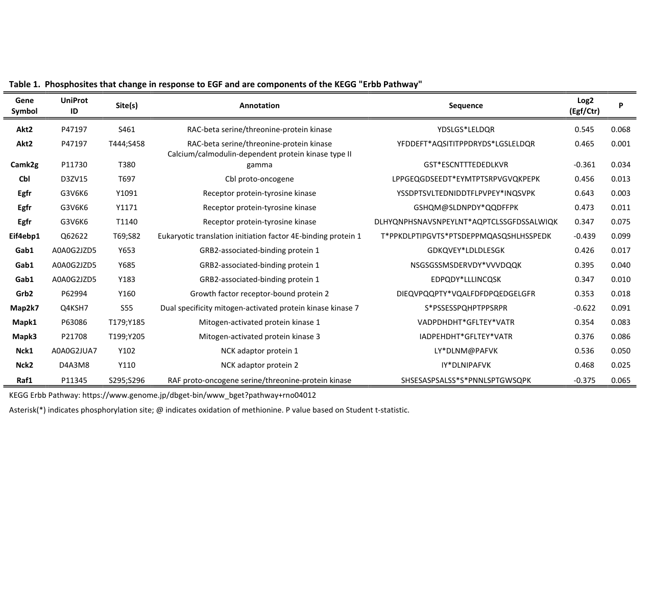
* EGFR phosphorylation increases including Y1091 log2(EGF/control) = +0.643 (p=0.003) and Y1171 = +0.473 (p=0.011) in the ErbB pathway table. (chou2025phosphoproteomicresponseto pages 34-40)
* An immunoblot increase in EGFR pY1091 from ~100 ± 15 to ~183 ± 13 after EGF stimulation. (chou2025phosphoproteomicresponseto pages 30-34, chou2025phosphoproteomicresponseto media 8fe8c363)

Live-cell quantitative pharmacology (2024)

Using BRET biosensors, gefitinib reversed an EGFR SHIP1 recruitment readout with IC50 ≈ 20 nM, and some recruitment/reversal events occurred within minutes (e.g., Grb2 recruitment within ~4 min; reversal within ~3 min after gefitinib addition in the described assay). (gross2024egfrsignalingand pages 4-5)

Rat in vivo EGFR inhibition in kidney disease (2018; still directly rat-relevant)

In the 5/6 nephrectomy rat model, erlotinib reduced macrophage infiltration scores (ED-1 score ~2.06 ± 0.28 to 1.39 ± 0.16; P < 0.05) and reduced multiple pathology measures including fibrosis/inflammation-associated markers and phospho-Akt in renal cortex. (yamamoto2018erlotinibattenuatesthe pages 5-7, yamamoto2018erlotinibattenuatesthe pages 4-5)

10) Evidence summary table

The following table compiles the key functional annotation points for rat Egfr/EGFR, with evidence-linked statements.

Aspect Summary
Identity/domains Rat Egfr (UniProt G3V6K6) matches the canonical epidermal growth factor receptor/ErbB1 receptor tyrosine kinase. EGFR is a single-pass transmembrane receptor with an extracellular ligand-binding region containing cysteine-rich subdomains, a transmembrane helix, and an intracellular tyrosine kinase domain with a C-terminal phosphotyrosine tail for adaptor docking. (kozlova2024celladhesionmolecules pages 4-6, shaban2023targetedinhibitorsof pages 1-4)
Enzymatic activity EGFR is a receptor protein-tyrosine kinase that increases intrinsic kinase activity upon ligand-induced dimerization and catalyzes autophosphorylation on cytoplasmic tyrosines, generating SH2/PTB docking sites for signaling proteins such as GRB2, GAB1, and PLCγ. Key tail phosphosites discussed in recent literature include Y1045, Y1068, Y1086, Y1148, and Y1173; rat phosphoproteomics detected regulated EGFR Y1091 and Y1171. (chou2025phosphoproteomicresponseto pages 1-5, laudadio2024chemicalscaffoldsfor pages 4-7, chou2025phosphoproteomicresponseto pages 34-40)
Activation mechanism Ligand binding to the extracellular domain induces a conformational change that exposes the dimerization arm, promotes receptor dimerization, and enables asymmetric kinase-domain activation followed by tail autophosphorylation. Recent live-cell studies also show ligand bias: EGF and epiregulin trigger distinct potency/efficacy patterns, with epiregulin at least ~100-fold less potent than EGF for SH2-effector recruitment in one biosensor system. (kozlova2024celladhesionmolecules pages 4-6, gross2024egfrsignalingand pages 1-2)
Key downstream pathways Core downstream outputs are RAS-RAF-MEK-ERK, PI3K-AKT-mTOR, and PLCγ/IP3-Ca2+ signaling. In native rat IMCD, EGF-responsive phosphoproteomics strongly enriched ErbB, PI3K-Akt, mTOR, endocytosis, and MAPK pathways, supporting conserved pathway usage in rat tissue. (chou2025phosphoproteomicresponseto pages 1-5, chou2025phosphoproteomicresponseto pages 40-43, chou2025phosphoproteomicresponseto pages 18-21)
Trafficking regulation EGFR signaling is shaped by endocytosis and ubiquitin-dependent sorting. Cbl and Cbl-b are principal E3 ligases for EGFR, but are not fully redundant: Cbl-b preferentially engages pY1045, while Cbl relies more on a GRB2-dependent route; altered ubiquitination affects degradation and endosomal sorting more strongly than initial clathrin-mediated internalization. Ligands also bias fate, with HB-EGF/BTC favoring lysosomal degradation and AREG/TGFα/EPGN favoring recycling. (chastel2024recentadvancesin pages 1-4, pinillamacua2023cblandcblb pages 1-2, pinillamacua2023cblandcblb pages 8-9, kozlova2024celladhesionmolecules pages 6-7)
Subcellular localization EGFR functions primarily at the plasma membrane and remains signaling-competent in early endosomes; receptors can then recycle or progress to lysosomal degradation. In rat collecting duct, EGFR is reported at the basolateral plasma membrane; additional noncanonical trafficking to ER/nuclear membranes and the nucleus has been described in broader EGFR literature. (chou2025phosphoproteomicresponseto pages 1-5, kozlova2024celladhesionmolecules pages 4-6, escoto2024investigatingtherole pages 23-28)
Rat-specific evidence In ex vivo native rat IMCD, EGF treatment activated canonical EGFR signaling and altered 254 phosphosites, consistent with a physiological signaling role in epithelial transport regulation. In an in vivo rat 5/6 nephrectomy CKD model, EGFR inhibition with erlotinib reduced proteinuria, serum creatinine, glomerulosclerosis, tubulointerstitial injury, inflammation, and phospho-Akt, supporting functional importance of rat Egfr in renal pathophysiology. (chou2025phosphoproteomicresponseto pages 1-5, yamamoto2018erlotinibattenuatesthe pages 4-5, yamamoto2018erlotinibattenuatesthe pages 1-2)
Quantitative data points 2024-2025 Rat IMCD phosphoproteomics quantified 29,881 phosphosites across 5,457 proteins, with 135 increased and 119 decreased after EGF; ErbB signaling showed p = 8×10^-7 and 5.48-fold enrichment. EGFR pY1091 increased from about 100 ± 15 to 183 ± 13 by immunoblot, and phosphoproteomics measured EGFR Y1091 log2 +0.643 (p=0.003) and Y1171 log2 +0.473 (p=0.011). In 2024 biosensor assays, gefitinib reversed one EGFR SHIP1 readout with IC50 ~20 nM, and SH2(Grb2) recruitment occurred within ~4 min and reversed within ~3 min after inhibitor addition. (chou2025phosphoproteomicresponseto pages 30-34, chou2025phosphoproteomicresponseto pages 40-43, chou2025phosphoproteomicresponseto pages 34-40, chou2025phosphoproteomicresponseto pages 18-21, gross2024egfrsignalingand pages 4-5, chou2025phosphoproteomicresponseto media e18dc9b0)
Applications Rat Egfr biology is already used in preclinical pharmacology and functional phosphoproteomics. Erlotinib treatment in rats improved CKD-associated phenotypes, while recent phosphoproteomic/network resources in native rat IMCD provide actionable datasets for pathway modeling, biomarker discovery, and interpreting EGFR-targeted interventions. More broadly, current EGFR research is informing next-generation inhibitors and degraders for resistance biology. (yamamoto2018erlotinibattenuatesthe pages 2-4, chou2025phosphoproteomicresponseto pages 1-5, laudadio2024chemicalscaffoldsfor pages 4-7)

Table: This table summarizes core functional annotation for rat Egfr/EGFR, integrating identity, mechanism, signaling, trafficking, localization, and rat-specific evidence. It is useful as a compact evidence-backed reference for narrative gene-function reporting.

11) Visual evidence (figure/table)

The rat IMCD phosphoproteomics paper includes (i) a table listing ErbB-pathway phosphosite changes (including EGFR Y1091/Y1171) and (ii) an immunoblot showing EGF-induced EGFR pY1091 increases, supporting direct activation of rat EGFR signaling in native tissue. (chou2025phosphoproteomicresponseto media e18dc9b0, chou2025phosphoproteomicresponseto media 8fe8c363)

12) Limitations of this evidence set (scope control)

  • The most detailed mechanistic trafficking/ubiquitination evidence retrieved here is primarily from human-cell studies (e.g., Cbl/Cbl-b regulation), but these mechanisms are widely treated as conserved and are consistent with rat tissue signaling outputs (ErbB/MAPK/PI3K enrichment in rat IMCD). (pinillamacua2023cblandcblb pages 1-2, chou2025phosphoproteomicresponseto pages 1-5)
  • For strict enzyme commission (EC) labeling (e.g., EC 2.7.10.1) and full domain names as listed by UniProt/InterPro, the evidence set here supports the receptor tyrosine kinase identity and cysteine-rich extracellular architecture, but EC labeling per se is not explicitly stated in the retrieved articles’ excerpts. (kozlova2024celladhesionmolecules pages 4-6, chou2025phosphoproteomicresponseto pages 1-5)

Key source URLs and publication dates (selection)

  • Kozlova I, Sytnyk V. Cells (Nov 2024). https://doi.org/10.3390/cells13221919 (kozlova2024celladhesionmolecules pages 4-6, kozlova2024celladhesionmolecules pages 6-7)
  • Chastel J, Angers A. Physiology/IntechOpen (Nov 2024). https://doi.org/10.5772/intechopen.114990 (chastel2024recentadvancesin pages 1-4, chastel2024recentadvancesin pages 6-9)
  • Pinilla-Macua I, Sorkin A. Molecular Biology of the Cell (Dec 2023). https://doi.org/10.1091/mbc.e23-02-0058 (pinillamacua2023cblandcblb pages 1-2, pinillamacua2023cblandcblb pages 8-9)
  • Gross F et al. Communications Biology (Mar 2024). https://doi.org/10.1038/s42003-024-05965-5 (gross2024egfrsignalingand pages 4-5, gross2024egfrsignalingand pages 1-2)
  • Chou C-L et al. AJP Renal Physiology (Jan 2025; doi indicates 2024 acceptance). https://doi.org/10.1152/ajprenal.00182.2024 (chou2025phosphoproteomicresponseto pages 1-5, chou2025phosphoproteomicresponseto pages 34-40)
  • Yamamoto Y et al. Nephrology Dialysis Transplantation (Apr 2018). https://doi.org/10.1093/ndt/gfx264 (yamamoto2018erlotinibattenuatesthe pages 2-4, yamamoto2018erlotinibattenuatesthe pages 4-5)

References

  1. (kozlova2024celladhesionmolecules pages 4-6): Irina Kozlova and Vladimir Sytnyk. Cell adhesion molecules as modulators of the epidermal growth factor receptor. Cells, 13:1919, Nov 2024. URL: https://doi.org/10.3390/cells13221919, doi:10.3390/cells13221919. This article has 17 citations.

  2. (shaban2023targetedinhibitorsof pages 1-4): Nina Shaban, Dmitri Kamashev, Aleksandra Emelianova, and Anton Buzdin. Targeted inhibitors of egfr: structure, biology, biomarkers, and clinical applications. Cells, 13:47, Dec 2023. URL: https://doi.org/10.3390/cells13010047, doi:10.3390/cells13010047. This article has 68 citations.

  3. (chou2025phosphoproteomicresponseto pages 1-5): Chung-Lin Chou, Nipun U. Jayatissa, Elena T. Kichula, Shuo-Ming Ou, Kavee Limbutara, and Mark A. Knepper. Phosphoproteomic response to epidermal growth factor in native rat inner medullary collecting duct. Jan 2025. URL: https://doi.org/10.1152/ajprenal.00182.2024, doi:10.1152/ajprenal.00182.2024. This article has 0 citations and is from a peer-reviewed journal.

  4. (chou2025phosphoproteomicresponseto pages 43-45): Chung-Lin Chou, Nipun U. Jayatissa, Elena T. Kichula, Shuo-Ming Ou, Kavee Limbutara, and Mark A. Knepper. Phosphoproteomic response to epidermal growth factor in native rat inner medullary collecting duct. Jan 2025. URL: https://doi.org/10.1152/ajprenal.00182.2024, doi:10.1152/ajprenal.00182.2024. This article has 0 citations and is from a peer-reviewed journal.

  5. (chastel2024recentadvancesin pages 6-9): Julia Chastel and Annie Angers. Recent advances in the importance of ubiquitylation for receptor internalization and signaling. Physiology, Nov 2024. URL: https://doi.org/10.5772/intechopen.114990, doi:10.5772/intechopen.114990. This article has 0 citations and is from a peer-reviewed journal.

  6. (chou2025phosphoproteomicresponseto pages 34-40): Chung-Lin Chou, Nipun U. Jayatissa, Elena T. Kichula, Shuo-Ming Ou, Kavee Limbutara, and Mark A. Knepper. Phosphoproteomic response to epidermal growth factor in native rat inner medullary collecting duct. Jan 2025. URL: https://doi.org/10.1152/ajprenal.00182.2024, doi:10.1152/ajprenal.00182.2024. This article has 0 citations and is from a peer-reviewed journal.

  7. (chou2025phosphoproteomicresponseto pages 40-43): Chung-Lin Chou, Nipun U. Jayatissa, Elena T. Kichula, Shuo-Ming Ou, Kavee Limbutara, and Mark A. Knepper. Phosphoproteomic response to epidermal growth factor in native rat inner medullary collecting duct. Jan 2025. URL: https://doi.org/10.1152/ajprenal.00182.2024, doi:10.1152/ajprenal.00182.2024. This article has 0 citations and is from a peer-reviewed journal.

  8. (chou2025phosphoproteomicresponseto pages 18-21): Chung-Lin Chou, Nipun U. Jayatissa, Elena T. Kichula, Shuo-Ming Ou, Kavee Limbutara, and Mark A. Knepper. Phosphoproteomic response to epidermal growth factor in native rat inner medullary collecting duct. Jan 2025. URL: https://doi.org/10.1152/ajprenal.00182.2024, doi:10.1152/ajprenal.00182.2024. This article has 0 citations and is from a peer-reviewed journal.

  9. (escoto2024investigatingtherole pages 23-28): A Escoto. Investigating the role of nuclear egfr in regulating the tumor immune microenvironment in breast cancer. Unknown journal, 2024.

  10. (yamamoto2018erlotinibattenuatesthe pages 2-4): Yasutaka Yamamoto, Masayuki Iyoda, Shohei Tachibana, Kei Matsumoto, Yukihiro Wada, Taihei Suzuki, Ken Iseri, Tomohiro Saito, Kei Fukuda-Hihara, and Takanori Shibata. Erlotinib attenuates the progression of chronic kidney disease in rats with remnant kidney. Nephrology Dialysis Transplantation, 33:598–606, Apr 2018. URL: https://doi.org/10.1093/ndt/gfx264, doi:10.1093/ndt/gfx264. This article has 20 citations and is from a domain leading peer-reviewed journal.

  11. (yamamoto2018erlotinibattenuatesthe pages 4-5): Yasutaka Yamamoto, Masayuki Iyoda, Shohei Tachibana, Kei Matsumoto, Yukihiro Wada, Taihei Suzuki, Ken Iseri, Tomohiro Saito, Kei Fukuda-Hihara, and Takanori Shibata. Erlotinib attenuates the progression of chronic kidney disease in rats with remnant kidney. Nephrology Dialysis Transplantation, 33:598–606, Apr 2018. URL: https://doi.org/10.1093/ndt/gfx264, doi:10.1093/ndt/gfx264. This article has 20 citations and is from a domain leading peer-reviewed journal.

  12. (pinillamacua2023cblandcblb pages 1-2): Itziar Pinilla-Macua and Alexander Sorkin. Cbl and cbl-b independently regulate egfr through distinct receptor interaction modes. Molecular Biology of the Cell, Dec 2023. URL: https://doi.org/10.1091/mbc.e23-02-0058, doi:10.1091/mbc.e23-02-0058. This article has 28 citations and is from a domain leading peer-reviewed journal.

  13. (pinillamacua2023cblandcblb pages 8-9): Itziar Pinilla-Macua and Alexander Sorkin. Cbl and cbl-b independently regulate egfr through distinct receptor interaction modes. Molecular Biology of the Cell, Dec 2023. URL: https://doi.org/10.1091/mbc.e23-02-0058, doi:10.1091/mbc.e23-02-0058. This article has 28 citations and is from a domain leading peer-reviewed journal.

  14. (chastel2024recentadvancesin pages 1-4): Julia Chastel and Annie Angers. Recent advances in the importance of ubiquitylation for receptor internalization and signaling. Physiology, Nov 2024. URL: https://doi.org/10.5772/intechopen.114990, doi:10.5772/intechopen.114990. This article has 0 citations and is from a peer-reviewed journal.

  15. (chastel2024recentadvancesin pages 9-11): Julia Chastel and Annie Angers. Recent advances in the importance of ubiquitylation for receptor internalization and signaling. Physiology, Nov 2024. URL: https://doi.org/10.5772/intechopen.114990, doi:10.5772/intechopen.114990. This article has 0 citations and is from a peer-reviewed journal.

  16. (gross2024egfrsignalingand pages 1-2): Florence Gross, Arturo Mancini, Billy Breton, Hiroyuki Kobayashi, Pedro Henrique Scarpelli Pereira, Christian Le Gouill, Michel Bouvier, Stephan Schann, Xavier Leroy, and Laurent Sabbagh. Egfr signaling and pharmacology in oncology revealed with innovative bret-based biosensors. Communications Biology, Mar 2024. URL: https://doi.org/10.1038/s42003-024-05965-5, doi:10.1038/s42003-024-05965-5. This article has 11 citations and is from a peer-reviewed journal.

  17. (gross2024egfrsignalingand pages 4-5): Florence Gross, Arturo Mancini, Billy Breton, Hiroyuki Kobayashi, Pedro Henrique Scarpelli Pereira, Christian Le Gouill, Michel Bouvier, Stephan Schann, Xavier Leroy, and Laurent Sabbagh. Egfr signaling and pharmacology in oncology revealed with innovative bret-based biosensors. Communications Biology, Mar 2024. URL: https://doi.org/10.1038/s42003-024-05965-5, doi:10.1038/s42003-024-05965-5. This article has 11 citations and is from a peer-reviewed journal.

  18. (kozlova2024celladhesionmolecules pages 6-7): Irina Kozlova and Vladimir Sytnyk. Cell adhesion molecules as modulators of the epidermal growth factor receptor. Cells, 13:1919, Nov 2024. URL: https://doi.org/10.3390/cells13221919, doi:10.3390/cells13221919. This article has 17 citations.

  19. (chou2025phosphoproteomicresponseto pages 30-34): Chung-Lin Chou, Nipun U. Jayatissa, Elena T. Kichula, Shuo-Ming Ou, Kavee Limbutara, and Mark A. Knepper. Phosphoproteomic response to epidermal growth factor in native rat inner medullary collecting duct. Jan 2025. URL: https://doi.org/10.1152/ajprenal.00182.2024, doi:10.1152/ajprenal.00182.2024. This article has 0 citations and is from a peer-reviewed journal.

  20. (chou2025phosphoproteomicresponseto media 8fe8c363): Chung-Lin Chou, Nipun U. Jayatissa, Elena T. Kichula, Shuo-Ming Ou, Kavee Limbutara, and Mark A. Knepper. Phosphoproteomic response to epidermal growth factor in native rat inner medullary collecting duct. Jan 2025. URL: https://doi.org/10.1152/ajprenal.00182.2024, doi:10.1152/ajprenal.00182.2024. This article has 0 citations and is from a peer-reviewed journal.

  21. (yamamoto2018erlotinibattenuatesthe pages 5-7): Yasutaka Yamamoto, Masayuki Iyoda, Shohei Tachibana, Kei Matsumoto, Yukihiro Wada, Taihei Suzuki, Ken Iseri, Tomohiro Saito, Kei Fukuda-Hihara, and Takanori Shibata. Erlotinib attenuates the progression of chronic kidney disease in rats with remnant kidney. Nephrology Dialysis Transplantation, 33:598–606, Apr 2018. URL: https://doi.org/10.1093/ndt/gfx264, doi:10.1093/ndt/gfx264. This article has 20 citations and is from a domain leading peer-reviewed journal.

  22. (laudadio2024chemicalscaffoldsfor pages 4-7): Emiliano Laudadio, Luca Mangano, and Cristina Minnelli. Chemical scaffolds for the clinical development of mutant-selective and reversible fourth-generation egfr-tkis in nsclc. ACS chemical biology, 19:839-854, Mar 2024. URL: https://doi.org/10.1021/acschembio.4c00028, doi:10.1021/acschembio.4c00028. This article has 28 citations and is from a domain leading peer-reviewed journal.

  23. (yamamoto2018erlotinibattenuatesthe pages 1-2): Yasutaka Yamamoto, Masayuki Iyoda, Shohei Tachibana, Kei Matsumoto, Yukihiro Wada, Taihei Suzuki, Ken Iseri, Tomohiro Saito, Kei Fukuda-Hihara, and Takanori Shibata. Erlotinib attenuates the progression of chronic kidney disease in rats with remnant kidney. Nephrology Dialysis Transplantation, 33:598–606, Apr 2018. URL: https://doi.org/10.1093/ndt/gfx264, doi:10.1093/ndt/gfx264. This article has 20 citations and is from a domain leading peer-reviewed journal.

  24. (chou2025phosphoproteomicresponseto media e18dc9b0): Chung-Lin Chou, Nipun U. Jayatissa, Elena T. Kichula, Shuo-Ming Ou, Kavee Limbutara, and Mark A. Knepper. Phosphoproteomic response to epidermal growth factor in native rat inner medullary collecting duct. Jan 2025. URL: https://doi.org/10.1152/ajprenal.00182.2024, doi:10.1152/ajprenal.00182.2024. This article has 0 citations and is from a peer-reviewed journal.

Artifacts

Citations

  1. kozlova2024celladhesionmolecules pages 4-6
  2. chou2025phosphoproteomicresponseto pages 1-5
  3. escoto2024investigatingtherole pages 23-28
  4. pinillamacua2023cblandcblb pages 8-9
  5. yamamoto2018erlotinibattenuatesthe pages 4-5
  6. chou2025phosphoproteomicresponseto pages 40-43
  7. chou2025phosphoproteomicresponseto pages 34-40
  8. gross2024egfrsignalingand pages 4-5
  9. shaban2023targetedinhibitorsof pages 1-4
  10. chou2025phosphoproteomicresponseto pages 43-45
  11. chastel2024recentadvancesin pages 6-9
  12. chou2025phosphoproteomicresponseto pages 18-21
  13. yamamoto2018erlotinibattenuatesthe pages 2-4
  14. pinillamacua2023cblandcblb pages 1-2
  15. chastel2024recentadvancesin pages 1-4
  16. chastel2024recentadvancesin pages 9-11
  17. gross2024egfrsignalingand pages 1-2
  18. kozlova2024celladhesionmolecules pages 6-7
  19. chou2025phosphoproteomicresponseto pages 30-34
  20. yamamoto2018erlotinibattenuatesthe pages 5-7
  21. laudadio2024chemicalscaffoldsfor pages 4-7
  22. yamamoto2018erlotinibattenuatesthe pages 1-2
  23. https://doi.org/10.3390/cells13221919
  24. https://doi.org/10.5772/intechopen.114990
  25. https://doi.org/10.1091/mbc.e23-02-0058
  26. https://doi.org/10.1038/s42003-024-05965-5
  27. https://doi.org/10.1152/ajprenal.00182.2024
  28. https://doi.org/10.1093/ndt/gfx264
  29. https://doi.org/10.3390/cells13221919,
  30. https://doi.org/10.3390/cells13010047,
  31. https://doi.org/10.1152/ajprenal.00182.2024,
  32. https://doi.org/10.5772/intechopen.114990,
  33. https://doi.org/10.1093/ndt/gfx264,
  34. https://doi.org/10.1091/mbc.e23-02-0058,
  35. https://doi.org/10.1038/s42003-024-05965-5,
  36. https://doi.org/10.1021/acschembio.4c00028,

📚 Additional Documentation

Bioreason Rl Predictions

(Egfr-bioreason-rl-predictions.md)

BioReason Chat Export

Exported on March 22, 2026 at 12:55 AM


Organism: Rattus norvegicus

Sequence:

MRPSGTARTKLLLLLAALCAAGGALEEKKVCQGTSNRLTQLGTFEDHFLSLQRMFNNCEVVLGNLEITYVQRNYDLSFLKTIQEVAGYVLIALNTVERIPLENLQIIRGNALYENTYALAVLSNYGTNKTGLRELPMRNLQEILIGAVRFSNNPILCNMETIQWRDIVQDVFLSNMSMDVQRHLTGCPKCDPSCPNGSCWGRGEENCQKLTKIICAQQCSRRCRGRSPSDCCHNQCAAGCTGPRESDCLVCHRFRDEATCKDTCPPLMLYNPTTYQMDVNPEGKYSFGATCVKKCPRNYVVTDHGSCVRACGPDYYEVEEDGVSKCKKCDGPCRKVCNGIGIGEFKDTLSINATNIKHFKYCTAISGDLHILPVAFKGDSFTRTPPLDPRELEILKTVKEITGFLLIQAWPENWTDLHAFENLEIIRGRTKQHGQFSLAVVGLNITSLGLRSLKEISDGDVIISGNRNLCYANTINWKKLFGTPNQKTKIMNNRAEKDCKATNHVCNPLCSSEGCWGPEPTDCVSCQNVSRGRECVDKCNILEGEPREFVENSECIQCHPECLPQTMNITCTGRGPDNCIKCAHYVDGPHCVKTCPSGIMGENNTLVWKFADANNVCHLCHANCTYGCAGPGLKGCQQPEGPKIPSIATGIVGGLLFIVVVALGIGLFMRRRHIVRKRTLRRLLQERELVEPLTPSGEAPNQAHLRILKETEFKKIKVLGSGAFGTVYKGLWIPEGEKVKIPVAIKELREATSPKANKEILDEAYVMASVDNPHVCRLLGICLTSTVQLITQLMPYGCLLDYVREHKDNIGSQYLLNWCVQIAKGMNYLEDRRLVHRDLAARNVLVKTPQHVKITDFGLAKLLGAEEKEYHAEGGKVPIKWMALESILHRIYTHQSDVWSYGVTVWELMTFGSKPYDGIPASEISSILEKGERLPQPPICTIDVYMIMVKCWMIDADSRPKFRELILEFSKMARDPQRYLVIQGDERMHLPSPTDSNFYRALMEEEDMEDVVDADEYLIPQQGFFNSPSTSRTPLLSSLSANSNSSAVACINRNGSCRVKEDAFLQRYSSDPTSVLTEDNIDDTFLPVPEYINQSVPKRPAGSVQNPVYHNQPLHPAPGRDLHYQNPHSNAVSNPEYLNTAQPTCLSSGFDSSALWIQKGSHQMSLDNPDYQQDFFPKEAKPNGIFKGPTAENAEYLRVAPPSSEFIGA

Thinking Trace

I begin by dissecting the InterPro architecture and its order along the sequence. The N-terminal half is dominated by extracellular receptor modules: IPR036941 (Receptor L-domain superfamily, residues 25–530) encompasses IPR000494 (Receptor L-domain, residues 57–480), establishing tandem L-domains that scaffold ligand-induced dimerization. Embedded within this extracellular scaffold are cysteine-rich segments that typify growth factor receptor ectodomains: IPR009030 (Growth factor receptor cysteine-rich domain superfamily, residues 187–637), IPR006211 (Furin-like cysteine-rich domain, residues 188–338), and IPR006212 (Furin-like repeat, residues 228–653). These repeats stabilize disulfide-bonded folds that promote high-affinity ligand binding and receptor clustering. Distally, IPR032778 (Growth factor receptor domain 4, residues 505–637) completes the ectodomain array that triggers activation.

Immediately after the ectodomain, the membrane-proximal region is specified by IPR044912 (Epidermal growth factor receptor, juxtamembrane domain, residues 644–696) and IPR049328 (Epidermal growth factor receptor-like, transmembrane-juxtamembrane segment, residues 647–682). This architecture enforces single-pass type I topology and couples extracellular conformational changes to intracellular activation via the juxtamembrane switch.

The intracellular portion is a canonical tyrosine kinase engine. IPR011009 (Protein kinase-like domain superfamily, residues 708–1007) houses the catalytic core defined by IPR000719 (Protein kinase domain, residues 713–980), IPR020635 (Tyrosine-protein kinase, catalytic domain, residues 713–969), and IPR001245 (Serine-threonine/tyrosine-protein kinase, catalytic domain, residues 715–967). Catalysis is specified by IPR017441 (Protein kinase, ATP binding site, residues 719–746) and IPR008266 (Tyrosine-protein kinase, active site, residues 834–846), which together enforce ATP-dependent phosphotransfer to tyrosine residues. The overall lineage and breadth of receptor tyrosine kinase features are reinforced by IPR016245 (Tyrosine protein kinase, EGF/ERB/XmrK receptor family, residues 7–1201) and IPR050122 (Receptor Tyrosine Kinase family, residues 58–975).

This ordered architecture causes a precise molecular function: an extracellular ligand-sensing ectodomain drives conformational dimerization that activates an intracellular tyrosine kinase catalytic core. The presence of the ATP-binding pocket and catalytic loop defines ATP-dependent phosphotransfer chemistry characteristic of receptor tyrosine kinases. Therefore, the molecular function is receptor tyrosine kinase activity, which inherently entails ATP binding and catalysis of tyrosine phosphorylation.

From this catalytic activity, the biological process follows. Ligand-induced dimerization and autophosphorylation generate phosphotyrosine docking sites that recruit SH2/PTB-containing effectors, propagating signaling cascades that govern cell proliferation, survival, and migration. This signaling logic is the canonical engine of transmembrane receptor protein tyrosine kinase signaling pathways that orchestrate growth and differentiation programs.

Cellular localization is dictated by the topology and domains. The extracellular L-domains and cysteine-rich repeats require luminal/extracellular exposure, while the juxtamembrane/transmembrane module fixes the protein as a single-pass type I membrane receptor. Such receptors traffic between the plasma membrane and endosomal compartments; the provided single-pass type I topology and the ectodomain suite strongly support residency at the plasma membrane as an integral membrane component.

Mechanistically, I hypothesize that ligand engagement at the L-domain/cysteine-rich ectodomain promotes receptor dimerization and alignment of the intracellular kinase domains, enabling trans-autophosphorylation and creation of high-affinity sites for SH2-domain adaptors. This will couple to MAPK, PI3K–AKT, and STAT pathways. Likely extracellular partners include growth factor-like ligands or accessory matrix components that cluster the ectodomain, while intracellular partners include SH2/PTB adaptors and enzymes that assemble signalosomes at activated receptor clusters.

Functional Summary

A single-pass membrane receptor tyrosine kinase in rat that uses an extracellular ligand-binding scaffold and a juxtamembrane switch to allosterically activate an intracellular kinase domain. Upon ligand-induced clustering, it binds ATP and autophosphorylates tyrosines, creating docking sites that assemble signaling complexes controlling growth and differentiation pathways at the cell surface and along the endomembrane system.

UniProt Summary

Tyrosine-protein kinase that plays a role in the regulation of cell proliferation, differentiation, migration and apoptosis.

InterPro Domains

  • Tyrosine protein kinase, EGF/ERB/XmrK receptor (IPR016245, family) — residues 7-1201
  • Receptor L-domain superfamily (IPR036941, homologous_superfamily) — residues 25-530
  • Receptor L-domain (IPR000494, domain) — residues 57-480
  • Receptor Tyrosine Kinase (IPR050122, family) — residues 58-975
  • Growth factor receptor cysteine-rich domain superfamily (IPR009030, homologous_superfamily) — residues 187-637
  • Furin-like cysteine-rich domain (IPR006211, domain) — residues 188-338
  • Furin-like repeat (IPR006212, repeat) — residues 228-653
  • Growth factor receptor domain 4 (IPR032778, domain) — residues 505-637
  • Epidermal growth factor receptor, juxtamembrane domain (IPR044912, homologous_superfamily) — residues 644-696
  • Epidermal growth factor receptor-like, transmembrane-juxtamembrane segment (IPR049328, domain) — residues 647-682
  • Protein kinase-like domain superfamily (IPR011009, homologous_superfamily) — residues 708-1007
  • Protein kinase domain (IPR000719, domain) — residues 713-980
  • Tyrosine-protein kinase, catalytic domain (IPR020635, domain) — residues 713-969
  • Serine-threonine/tyrosine-protein kinase, catalytic domain (IPR001245, domain) — residues 715-967
  • Protein kinase, ATP binding site (IPR017441, binding_site) — residues 719-746
  • Tyrosine-protein kinase, active site (IPR008266, active_site) — residues 834-846

GO Terms

Molecular Function: molecular_function (GO:0003674), molecular transducer activity (GO:0060089), binding (GO:0005488), catalytic activity (GO:0003824), transferase activity (GO:0016740), protein-containing complex binding (GO:0044877), signaling receptor activity (GO:0038023), catalytic activity, acting on a protein (GO:0140096), hormone binding (GO:0042562), protein binding (GO:0005515), transmembrane signaling receptor activity (GO:0004888), signaling receptor binding (GO:0005102), transferase activity, transferring phosphorus-containing groups (GO:0016772), protein kinase activity (GO:0004672), cell adhesion molecule binding (GO:0050839), enzyme binding (GO:0019899), integrin binding (GO:0005178), calmodulin binding (GO:0005516), growth factor binding (GO:0019838), kinase activity (GO:0016301), transmembrane receptor protein kinase activity (GO:0019199), phosphotransferase activity, alcohol group as acceptor (GO:0016773), kinase binding (GO:0019900), phosphatase binding (GO:0019902), protein tyrosine kinase activity (GO:0004713), receptor tyrosine kinase activity (GO:0004714), protein kinase binding (GO:0019901), protein phosphatase binding (GO:0019903)

Biological Process: biological_process (GO:0008150), signaling (GO:0023052), biological regulation (GO:0065007), response to stimulus (GO:0050896), reproductive process (GO:0022414), negative regulation of biological process (GO:0048519), positive regulation of biological process (GO:0048518), regulation of biological process (GO:0050789), reproduction (GO:0000003), multicellular organismal process (GO:0032501), rhythmic process (GO:0048511), developmental process (GO:0032502), cellular process (GO:0009987), metabolic process (GO:0008152), homeostatic process (GO:0042592), anatomical structure development (GO:0048856), positive regulation of multicellular organismal process (GO:0051240), molting cycle (GO:0042303), cellular component organization or biogenesis (GO:0071840), regulation of multicellular organismal process (GO:0051239), positive regulation of transport (GO:0051050), regulation of biological quality (GO:0065008), regulation of cellular process (GO:0050794), regulation of response to stimulus (GO:0048583), cellular response to stimulus (GO:0051716), negative regulation of cellular process (GO:0048523), cellular developmental process (GO:0048869), response to abiotic stimulus (GO:0009628), biosynthetic process (GO:0009058), positive regulation of response to stimulus (GO:0048584), regulation of metabolic process (GO:0019222), molting cycle process (GO:0022404), cell communication (GO:0007154), positive regulation of cellular process (GO:0048522), response to external stimulus (GO:0009605), anatomical structure morphogenesis (GO:0009653), response to chemical (GO:0042221), nitrogen compound metabolic process (GO:0006807), multicellular organism reproduction (GO:0032504), regulation of developmental process (GO:0050793), response to endogenous stimulus (GO:0009719), regulation of signaling (GO:0023051), signal transduction (GO:0007165), positive regulation of signaling (GO:0023056), multicellular organism development (GO:0007275), cell activation (GO:0001775), regulation of localization (GO:0032879), circadian rhythm (GO:0007623), organic substance metabolic process (GO:0071704), system process (GO:0003008), chemical homeostasis (GO:0048878), cellular metabolic process (GO:0044237), ovulation cycle (GO:0042698), positive regulation of metabolic process (GO:0009893), response to stress (GO:0006950), positive regulation of developmental process (GO:0051094), multicellular organismal reproductive process (GO:0048609), primary metabolic process (GO:0044238), cellular component morphogenesis (GO:0032989), positive regulation of secretion (GO:0051047), hair cycle (GO:0042633), reactive oxygen species metabolic process (GO:0072593), regulation of response to stress (GO:0080134), regulation of system process (GO:0044057), regulation of tissue remodeling (GO:0034103), animal organ development (GO:0048513), regulation of signal transduction (GO:0009966), cellular lipid metabolic process (GO:0044255), hydrogen peroxide metabolic process (GO:0042743), regulation of response to external stimulus (GO:0032101), response to hormone (GO:0009725), response to inorganic substance (GO:0010035), regulation of body fluid levels (GO:0050878), negative regulation of cell cycle (GO:0045786), cellular macromolecule metabolic process (GO:0044260), positive regulation of cell differentiation (GO:0045597), positive regulation of bone resorption (GO:0045780), positive regulation of protein localization (GO:1903829), organic substance biosynthetic process (GO:1901576), regulation of cellular localization (GO:0060341), protein metabolic process (GO:0019538), cellular biosynthetic process (GO:0044249), response to oxygen-containing compound (GO:1901700), cellular nitrogen compound metabolic process (GO:0034641), glial cell activation (GO:0061900), macromolecule metabolic process (GO:0043170), regulation of multicellular organismal development (GO:2000026), cellular response to environmental stimulus (GO:0104004), regulation of cell population proliferation (GO:0042127), lipid metabolic process (GO:0006629), regulation of cell death (GO:0010941), response to nutrient (GO:0007584), response to nitrogen compound (GO:1901698), positive regulation of cellular metabolic process (GO:0031325), regulation of mucus secretion (GO:0070255), positive regulation of cell communication (GO:0010647), regulation of cellular metabolic process (GO:0031323), hair cycle process (GO:0022405), regulation of trans-synaptic signaling (GO:0099177), positive regulation of response to external stimulus (GO:0032103), negative regulation of cell death (GO:0060548), cell development (GO:0048468), response to xenobiotic stimulus (GO:0009410), cell differentiation (GO:0030154), system development (GO:0048731), response to growth factor (GO:0070848), cellular response to endogenous stimulus (GO:0071495), circulatory system process (GO:0003013), phosphorus metabolic process (GO:0006793), tube development (GO:0035295), regulation of cell cycle (GO:0051726), cell morphogenesis (GO:0000902), positive regulation of synaptic transmission (GO:0050806), cellular response to abiotic stimulus (GO:0071214), hair follicle development (GO:0001942), regulation of cell differentiation (GO:0045595), cellular component organization (GO:0016043), cell surface receptor signaling pathway (GO:0007166), positive regulation of defense response (GO:0031349), amide metabolic process (GO:0043603), positive regulation of nervous system development (GO:0051962), cellular response to external stimulus (GO:0071496), regulation of anatomical structure size (GO:0090066), response to salt (GO:1902074), positive regulation of cell population proliferation (GO:0008284), monoatomic ion homeostasis (GO:0050801), positive regulation of signal transduction (GO:0009967), regulation of secretion by cell (GO:1903530), regulation of transport (GO:0051049), organonitrogen compound metabolic process (GO:1901564), intracellular signal transduction (GO:0035556), response to wounding (GO:0009611), positive regulation of hormone secretion (GO:0046887), response to oxidative stress (GO:0006979), defense response (GO:0006952), response to osmotic stress (GO:0006970), inorganic ion homeostasis (GO:0098771), regulation of hormone levels (GO:0010817), response to organic substance (GO:0010033), regulation of hormone secretion (GO:0046883), response to extracellular stimulus (GO:0009991), tissue development (GO:0009888), regulation of cell communication (GO:0010646), cellular response to chemical stimulus (GO:0070887), positive regulation of vasoconstriction (GO:0045907), positive regulation of protein transport (GO:0051222), response to mechanical stimulus (GO:0009612), regeneration (GO:0031099), positive regulation of secretion by cell (GO:1903532), cell projection organization (GO:0030030), isoprenoid metabolic process (GO:0006720), response to ketone (GO:1901654), positive regulation of establishment of protein localization (GO:1904951), magnesium ion homeostasis (GO:0010960), astrocyte activation (GO:0048143), response to steroid hormone (GO:0048545), regulation of glial cell proliferation (GO:0060251), glial cell differentiation (GO:0010001), regulation of smooth muscle cell proliferation (GO:0048660), organonitrogen compound biosynthetic process (GO:1901566), translation (GO:0006412), macromolecule modification (GO:0043412), regulation of protein transport (GO:0051223), negative regulation of mitotic cell cycle (GO:0045930), positive regulation of intracellular signal transduction (GO:1902533), positive regulation of reactive oxygen species metabolic process (GO:2000379), regulation of protein secretion (GO:0050708), cell projection morphogenesis (GO:0048858), cellular response to oxygen-containing compound (GO:1901701), vascular process in circulatory system (GO:0003018), enzyme-linked receptor protein signaling pathway (GO:0007167), regulation of reactive oxygen species metabolic process (GO:2000377), cellular response to organic substance (GO:0071310), regulation of cell development (GO:0060284), sensory organ development (GO:0007423), modulation of chemical synaptic transmission (GO:0050804), positive regulation of smooth muscle cell proliferation (GO:0048661), amide biosynthetic process (GO:0043604), regulation of protein localization (GO:0032880), positive regulation of glial cell proliferation (GO:0060252), hepaticobiliary system development (GO:0061008), protein modification process (GO:0036211), neurogenesis (GO:0022008), cellular response to hormone stimulus (GO:0032870), inflammatory response (GO:0006954), response to lipid (GO:0033993), cellular nitrogen compound biosynthetic process (GO:0044271), cellular macromolecule biosynthetic process (GO:0034645), epithelium development (GO:0060429), cellular response to growth factor stimulus (GO:0071363), respiratory system development (GO:0060541), regulation of defense response (GO:0031347), macromolecule biosynthetic process (GO:0009059), regulation of mitotic cell cycle (GO:0007346), digestive system development (GO:0055123), response to estradiol (GO:0032355), blood circulation (GO:0008015), negative regulation of programmed cell death (GO:0043069), regulation of programmed cell death (GO:0043067), cell part morphogenesis (GO:0032990), regulation of establishment of protein localization (GO:0070201), positive regulation of peptide secretion (GO:0002793), positive regulation of synaptic transmission, glutamatergic (GO:0051968), gland development (GO:0048732), positive regulation of neurogenesis (GO:0050769), skin development (GO:0043588), response to vitamin (GO:0033273), regulation of blood circulation (GO:1903522), neuron development (GO:0048666), animal organ regeneration (GO:0031100), response to calcium ion (GO:0051592), regulation of peptide hormone secretion (GO:0090276), nervous system development (GO:0007399), epidermis development (GO:0008544), response to nutrient levels (GO:0031667), positive regulation of inflammatory response (GO:0050729), central nervous system development (GO:0007417), gene expression (GO:0010467), response to metal ion (GO:0010038), cellular response to xenobiotic stimulus (GO:0071466), peptide metabolic process (GO:0006518), response to cobalamin (GO:0033590), positive regulation of cell development (GO:0010720), glial cell development (GO:0021782), phosphate-containing compound metabolic process (GO:0006796), regulation of nervous system development (GO:0051960), positive regulation of protein secretion (GO:0050714), regulation of tube size (GO:0035150), lung development (GO:0030324), wound healing (GO:0042060), respiratory tube development (GO:0030323), response to organonitrogen compound (GO:0010243), monoatomic cation homeostasis (GO:0055080), regulation of inflammatory response (GO:0050727), regulation of peptide transport (GO:0090087), regulation of bone remodeling (GO:0046850), regulation of secretion (GO:0051046), positive regulation of peptide hormone secretion (GO:0090277), cellular response to mechanical stimulus (GO:0071260), response to organic cyclic compound (GO:0014070), digestive tract development (GO:0048565), neuron differentiation (GO:0030182), regulation of intracellular signal transduction (GO:1902531), regulation of apoptotic process (GO:0042981), response to corticosteroid (GO:0031960), skin epidermis development (GO:0098773), plasma membrane bounded cell projection morphogenesis (GO:0120039), peptidyl-amino acid modification (GO:0018193), phosphorylation (GO:0016310), regulation of bone resorption (GO:0045124), blood vessel diameter maintenance (GO:0097746), liver development (GO:0001889), tongue development (GO:0043586), liver regeneration (GO:0097421), terpenoid metabolic process (GO:0006721), response to phenylpropanoid (GO:0080184), cellular response to ketone (GO:1901655), regulation of MAPK cascade (GO:0043408), peptide biosynthetic process (GO:0043043), midgut development (GO:0007494), plasma membrane bounded cell projection organization (GO:0120036), cellular response to organic cyclic compound (GO:0071407), gliogenesis (GO:0042063), regulation of peptide secretion (GO:0002791), cellular response to steroid hormone stimulus (GO:0071383), neuron projection development (GO:0031175), positive regulation of superoxide anion generation (GO:0032930), neuroinflammatory response (GO:0150076), response to dexamethasone (GO:0071548), astrocyte development (GO:0014002), regulation of tube diameter (GO:0035296), transmembrane receptor protein tyrosine kinase signaling pathway (GO:0007169), regulation of neurogenesis (GO:0050767), positive regulation of MAPK cascade (GO:0043410), generation of neurons (GO:0048699), regulation of superoxide metabolic process (GO:0090322), regulation of vasoconstriction (GO:0019229), negative regulation of apoptotic process (GO:0043066), protein phosphorylation (GO:0006468), cellular response to lipid (GO:0071396), positive regulation of gliogenesis (GO:0014015), regulation of synaptic transmission, glutamatergic (GO:0051966), astrocyte differentiation (GO:0048708), regulation of superoxide anion generation (GO:0032928), neuron projection morphogenesis (GO:0048812), ERBB signaling pathway (GO:0038127), cellular response to dexamethasone stimulus (GO:0071549), peptidyl-tyrosine modification (GO:0018212), peptidyl-tyrosine phosphorylation (GO:0018108), diterpenoid metabolic process (GO:0016101), vasodilation (GO:0042311), response to glucocorticoid (GO:0051384), cellular response to corticosteroid stimulus (GO:0071384), regulation of gliogenesis (GO:0014013), protein autophosphorylation (GO:0046777), cellular response to glucocorticoid stimulus (GO:0071385), epidermal growth factor receptor signaling pathway (GO:0007173)

Cellular Component: cellular_component (GO:0005575), cellular anatomical entity (GO:0110165), intracellular anatomical structure (GO:0005622), basal part of cell (GO:0045178), organelle (GO:0043226), membrane (GO:0016020), cell surface (GO:0009986), cell periphery (GO:0071944), cell junction (GO:0030054), apical part of cell (GO:0045177), cytoplasm (GO:0005737), endomembrane system (GO:0012505), cytoplasmic vesicle (GO:0031410), intracellular organelle (GO:0043229), endosome (GO:0005768), synapse (GO:0045202), apical plasma membrane (GO:0016324), basal plasma membrane (GO:0009925), plasma membrane region (GO:0098590), membrane microdomain (GO:0098857), membrane-bounded organelle (GO:0043227), plasma membrane (GO:0005886), membrane raft (GO:0045121), endocytic vesicle (GO:0030139), intracellular membrane-bounded organelle (GO:0043231), basolateral plasma membrane (GO:0016323), vesicle (GO:0031982), intracellular vesicle (GO:0097708), nucleus (GO:0005634)


Generated by BioReason

Bioreason Rl Review

(Egfr-bioreason-rl-review.md)

BioReason-Pro RL Review: Egfr (rat)

Source: Egfr-bioreason-rl-predictions.md

  • Correctness: 5/5
  • Completeness: 4/5

Functional Summary Review

The functional summary describes:

A single-pass membrane receptor tyrosine kinase in rat that uses an extracellular ligand-binding scaffold and a juxtamembrane switch to allosterically activate an intracellular kinase domain. Upon ligand-induced clustering, it binds ATP and autophosphorylates tyrosines, creating docking sites that assemble signaling complexes controlling growth and differentiation pathways at the cell surface and along the endomembrane system.

This is accurate and well-aligned with the curated review. The ai-review.yaml confirms transmembrane receptor protein tyrosine kinase activity (GO:0004714), protein tyrosine kinase activity (GO:0004713), ATP binding (GO:0005524), plasma membrane (GO:0005886), epidermal growth factor receptor signaling pathway (GO:0007173), positive regulation of MAPK cascade (GO:0043410), and neuron differentiation (GO:0030182).

The mention of "endomembrane system" is a nice touch, consistent with endocytic vesicle (GO:0030139) and endosomal compartments annotated in the curated review.

The summary correctly identifies MAPK, PI3K-AKT, and STAT pathways in the thinking trace (though not in the summary itself), all well-established downstream of EGFR. The description of L-domain ligand binding and cysteine-rich ectodomain architecture is accurate.

Minor gaps: the summary does not mention EGF binding specifically (GO:0048408, annotated via IBA), neuron differentiation (GO:0030182), negative regulation of apoptotic process (GO:0043066), or the ERBB2-EGFR signaling pathway (GO:0038134). These represent important specificity beyond the general receptor tyrosine kinase description.

Comparison with interpro2go:

The interpro2go annotations for Egfr include protein kinase activity (GO:0004672), protein tyrosine kinase activity (GO:0004713), and membrane (GO:0016020). BioReason's summary fully recapitulates and extends these -- correctly identifying receptor tyrosine kinase activity as the specific function, and placing the protein at the plasma membrane. BioReason adds significant value beyond interpro2go by describing the allosteric activation mechanism, juxtamembrane switch, and signaling context.

Notes on thinking trace

The trace provides an excellent domain-by-domain walkthrough of the ectodomain (L-domains, cysteine-rich repeats), juxtamembrane segment, and intracellular kinase core. The mechanistic hypothesis about trans-autophosphorylation and SH2/PTB adaptor recruitment is standard and correct. The reasoning is thorough and well-connected to the domain architecture.

📄 View Raw YAML

id: G3V6K6
gene_symbol: Egfr
product_type: PROTEIN
status: IN_PROGRESS
taxon:
  id: NCBITaxon:10116
  label: Rattus norvegicus
description: Egfr (EGFR/ErbB1) is the rat ortholog of the epidermal growth factor
  receptor, a single-pass type I transmembrane receptor tyrosine kinase (EC 2.7.10.1).
  Its extracellular region contains cysteine-rich (furin-like) subdomains that bind
  EGF-family ligands; ligand binding stabilizes an active conformation, exposes a
  dimerization arm, and drives receptor dimerization. Dimerization activates the
  intracellular tyrosine kinase domain (asymmetric kinase-domain interaction), which
  autophosphorylates C-terminal tail tyrosines, creating SH2/PTB docking sites for
  adaptors (GRB2, SHC1, GAB1, PLCgamma). This couples EGFR to the canonical
  RAS-RAF-MEK-ERK (MAPK), PI3K-AKT-mTOR, and PLCgamma/Ca2+ pathways, governing
  proliferation, survival, motility, and context-dependent transcription. The core
  function is therefore ligand-activated transmembrane receptor tyrosine kinase
  signaling at the plasma membrane. Activated EGFR is internalized by
  clathrin-mediated endocytosis and can continue signaling from early endosomes
  before recycling or Cbl/Cbl-b ubiquitin-dependent lysosomal degradation; this
  trafficking shapes signal duration. In native rat inner medullary collecting duct,
  EGFR is localized to the basolateral plasma membrane and EGF stimulation increases
  EGFR tyrosine phosphorylation (e.g. Y1091/Y1171) with strong ErbB/PI3K-Akt/MAPK
  pathway enrichment. EGFR is highly pleiotropic, contributing to many downstream
  developmental, proliferative, and physiological processes that are not its core
  biochemical function.
existing_annotations:
- term:
    id: GO:0004714
    label: transmembrane receptor protein tyrosine kinase activity
  evidence_type: IBA
  original_reference_id: GO_REF:0000033
  review:
    summary: This is the core molecular function of EGFR - a ligand-activated
      transmembrane receptor tyrosine kinase that transmits a signal across the
      plasma membrane by catalyzing tyrosine autophosphorylation. The IBA inference
      is phylogenetically sound for the EGF receptor subfamily.
    action: ACCEPT
    reason: Core molecular function, well supported by family membership, domain
      architecture, and rat phosphoproteomic evidence of EGF-induced EGFR tyrosine
      phosphorylation.
    supported_by:
    - reference_id: file:rat/Egfr/Egfr-deep-research-falcon.md
      supporting_text: |-
        EGFR is a **receptor protein-tyrosine kinase** that increases intrinsic kinase activity upon ligand-induced dimerization and catalyzes autophosphorylation on cytoplasmic tyrosines, generating SH2/PTB docking sites for signaling proteins such as GRB2, GAB1, and PLCγ.
    - reference_id: file:rat/Egfr/Egfr-deep-research-falcon.md
      supporting_text: |-
        EGFR is a single-pass transmembrane receptor with an extracellular ligand-binding region containing cysteine-rich subdomains, a transmembrane helix, and an intracellular tyrosine kinase domain with a C-terminal phosphotyrosine tail for adaptor docking.
  qualifier: enables
- term:
    id: GO:0005886
    label: plasma membrane
  evidence_type: IBA
  original_reference_id: GO_REF:0000033
  review:
    summary: EGFR is a single-pass type I plasma membrane receptor where ligand
      binding occurs outside the cell and kinase signaling occurs inside. This is
      the core site of its primary function.
    action: ACCEPT
    reason: Core localization for a transmembrane receptor; consistent with UniProt
      cell membrane assignment and rat tissue evidence.
    supported_by:
    - reference_id: file:rat/Egfr/Egfr-deep-research-falcon.md
      supporting_text: |-
        EGFR is primarily a **plasma-membrane** receptor (ligand binding outside the cell; kinase signaling inside), and it is internalized after activation.
  qualifier: is_active_in
- term:
    id: GO:0043235
    label: receptor complex
  evidence_type: IBA
  original_reference_id: GO_REF:0000033
  review:
    summary: Activated EGFR functions as a dimer (homodimer or ErbB heterodimer),
      forming a signaling receptor complex. This is integral to the activation
      mechanism.
    action: ACCEPT
    reason: EGFR signaling requires ligand-induced dimerization into a receptor
      complex; well supported.
    supported_by:
    - reference_id: file:rat/Egfr/Egfr-deep-research-falcon.md
      supporting_text: |-
        EGFR is activated by binding EGF-family ligands, which stabilizes an active extracellular conformation, exposes a dimerization interface, and promotes receptor dimerization.
  qualifier: part_of
- term:
    id: GO:0030182
    label: neuron differentiation
  evidence_type: IBA
  original_reference_id: GO_REF:0000033
  review:
    summary: EGFR signaling contributes to neuronal/glial differentiation in some
      contexts, but this is a downstream pleiotropic developmental outcome rather
      than the core biochemical function of the receptor.
    action: KEEP_AS_NON_CORE
    reason: A downstream developmental process; valid but not part of the core
      receptor tyrosine kinase function.
  qualifier: involved_in
- term:
    id: GO:0043066
    label: negative regulation of apoptotic process
  evidence_type: IBA
  original_reference_id: GO_REF:0000033
  review:
    summary: EGFR-driven PI3K-AKT signaling promotes cell survival, which manifests
      as negative regulation of apoptosis. This is a downstream consequence of
      core signaling rather than the core function itself.
    action: KEEP_AS_NON_CORE
    reason: Downstream survival effect of EGFR signaling; pleiotropic, not core.
  qualifier: involved_in
- term:
    id: GO:0043410
    label: positive regulation of MAPK cascade
  evidence_type: IBA
  original_reference_id: GO_REF:0000033
  review:
    summary: A defining downstream output of EGFR is activation of the RAS-RAF-MEK-ERK
      (MAPK) cascade via GRB2/SHC1 recruitment. This is one of the principal
      signaling consequences of receptor activation, but it is a downstream BP
      consequence of the core RTK activity rather than the core function itself.
    action: KEEP_AS_NON_CORE
    reason: Direct downstream signaling pathway; well-supported in rat tissue
      phosphoproteomics, but represents a BP consequence of core RTK activity
      (which requires RAS/RAF/MEK/ERK, other gene products) rather than the core
      molecular function. Treated consistently with other downstream BP outcomes
      (apoptosis, proliferation) in this review.
    supported_by:
    - reference_id: file:rat/Egfr/Egfr-deep-research-falcon.md
      supporting_text: |-
        Autophosphorylation enables recruitment of adaptor/effector proteins (e.g., GRB2, GAB1, PLCγ), coupling EGFR to major signaling routes including **RAS–RAF–MEK–ERK (MAPK)** and **PI3K–AKT–mTOR**, among others.
    - reference_id: file:rat/Egfr/Egfr-deep-research-falcon.md
      supporting_text: |-
        In rat IMCD, EGF increases phosphorylation on EGFR tyrosines (e.g., Y1091/Y1171 in rat phosphoproteomics), consistent with enhanced kinase activity and generation of SH2-binding motifs that recruit adaptors such as Shc1 and Grb2 to couple EGFR to Ras and downstream MAPK signaling.
  qualifier: involved_in
- term:
    id: GO:0007173
    label: epidermal growth factor receptor signaling pathway
  evidence_type: IBA
  original_reference_id: GO_REF:0000033
  review:
    summary: EGFR is the defining receptor of the epidermal growth factor receptor
      signaling pathway. This is a core biological process for the gene.
    action: ACCEPT
    reason: Core biological process - EGFR is the namesake and central node of this
      pathway.
    supported_by:
    - reference_id: file:rat/Egfr/Egfr-deep-research-falcon.md
      supporting_text: |-
        In native rat IMCD, EGF stimulation produces phosphoproteomic signatures consistent with canonical ErbB signaling (Raf/MEK/ERK; PI3K-Akt; mTOR; endocytosis-associated networks), indicating that rat Egfr is embedded in the conserved EGFR signaling architecture in intact epithelial tissue.
  qualifier: involved_in
- term:
    id: GO:0050679
    label: positive regulation of epithelial cell proliferation
  evidence_type: IBA
  original_reference_id: GO_REF:0000033
  review:
    summary: EGFR mitogenic signaling promotes epithelial cell proliferation. This
      is an important but downstream/pleiotropic consequence of core signaling
      rather than the core biochemical function.
    action: KEEP_AS_NON_CORE
    reason: Downstream proliferative outcome of EGFR signaling; pleiotropic.
  qualifier: involved_in
- term:
    id: GO:0009925
    label: basal plasma membrane
  evidence_type: IBA
  original_reference_id: GO_REF:0000033
  review:
    summary: In polarized epithelia EGFR localizes to the basal/basolateral plasma
      membrane. This is a context-specific refinement of plasma membrane
      localization rather than a distinct core function.
    action: KEEP_AS_NON_CORE
    reason: Context-specific (polarized epithelial) localization refinement;
      supported by rat collecting duct evidence for the basolateral domain.
    supported_by:
    - reference_id: file:rat/Egfr/Egfr-deep-research-falcon.md
      supporting_text: |-
        In native rat IMCD, EGFR is reported at the **basolateral plasma membrane**, consistent with epithelial polarity and paracrine signaling in the collecting duct.
  qualifier: is_active_in
- term:
    id: GO:0048408
    label: epidermal growth factor binding
  evidence_type: IBA
  original_reference_id: GO_REF:0000033
  review:
    summary: EGFR binds EGF-family ligands via its extracellular cysteine-rich
      domains; ligand binding is the obligatory first step of receptor activation.
      This is a core molecular function.
    action: ACCEPT
    reason: Core molecular function - ligand recognition is intrinsic to receptor
      activation.
    supported_by:
    - reference_id: file:rat/Egfr/Egfr-deep-research-falcon.md
      supporting_text: |-
        EGFR is activated by binding EGF-family ligands, which stabilizes an active extracellular conformation, exposes a dimerization interface, and promotes receptor dimerization.
  qualifier: enables
- term:
    id: GO:0000166
    label: nucleotide binding
  evidence_type: IEA
  original_reference_id: GO_REF:0000104
  review:
    summary: This is a generic parent of ATP binding. The informative molecular
      function is ATP binding (GO:0005524) in the kinase active site, which is
      separately annotated.
    action: MARK_AS_OVER_ANNOTATED
    reason: Overly generic - subsumed by the more specific and directly relevant
      ATP binding annotation.
  qualifier: enables
- term:
    id: GO:0004672
    label: protein kinase activity
  evidence_type: IEA
  original_reference_id: GO_REF:0000002
  review:
    summary: Generic parent of the specific transmembrane receptor protein tyrosine
      kinase activity (GO:0004714). EGFR is a tyrosine, not a generic protein,
      kinase.
    action: MARK_AS_OVER_ANNOTATED
    reason: Overly generic; subsumed by the specific GO:0004714 / GO:0004713
      tyrosine kinase annotations.
  qualifier: enables
- term:
    id: GO:0004713
    label: protein tyrosine kinase activity
  evidence_type: IEA
  original_reference_id: GO_REF:0000002
  review:
    summary: EGFR is a protein tyrosine kinase. This is correct but less specific
      than the transmembrane receptor protein tyrosine kinase activity (GO:0004714,
      a direct child of GO:0004713) that captures the receptor nature of EGFR and
      adds no information given GO:0004714.
    action: MARK_AS_OVER_ANNOTATED
    reason: Overly generic; subsumed by and uninformative relative to the more
      specific GO:0004714 (its direct child), consistent with the treatment of the
      analogous generic parent GO:0004672 (protein kinase activity).
  qualifier: enables
- term:
    id: GO:0004714
    label: transmembrane receptor protein tyrosine kinase activity
  evidence_type: IEA
  original_reference_id: GO_REF:0000003
  review:
    summary: Core molecular function (duplicate of the IBA-supported annotation
      above, from EC mapping). EGFR transmits a signal across the membrane by
      tyrosine kinase catalysis.
    action: ACCEPT
    reason: Core molecular function; IEA from EC mapping corroborates the IBA
      annotation.
    supported_by:
    - reference_id: file:rat/Egfr/Egfr-deep-research-falcon.md
      supporting_text: |-
        EGFR catalyzes **protein tyrosine phosphorylation** on its own cytoplasmic tail (autophosphorylation) after ligand-induced activation.
  qualifier: enables
- term:
    id: GO:0005006
    label: epidermal growth factor receptor activity
  evidence_type: IEA
  original_reference_id: GO_REF:0000117
  review:
    summary: This is the most specific molecular function term for EGFR, capturing
      both EGF ligand recognition and the receptor tyrosine kinase activity. A core
      molecular function.
    action: ACCEPT
    reason: Most specific and accurate MF term for this gene product.
    supported_by:
    - reference_id: file:rat/Egfr/Egfr-deep-research-falcon.md
      supporting_text: |-
        EGFR is a **receptor protein-tyrosine kinase** that increases intrinsic kinase activity upon ligand-induced dimerization and catalyzes autophosphorylation on cytoplasmic tyrosines, generating SH2/PTB docking sites for signaling proteins such as GRB2, GAB1, and PLCγ.
  qualifier: enables
- term:
    id: GO:0005524
    label: ATP binding
  evidence_type: IEA
  original_reference_id: GO_REF:0000120
  review:
    summary: ATP binding in the kinase domain active site (residues 719-727, 746 per
      UniProt) is required for the phosphotransfer reaction. A supporting molecular
      function of the kinase activity.
    action: ACCEPT
    reason: Directly supports the catalytic kinase function; ATP is the
      phosphate-donor substrate. Mapped to defined ATP-binding residues in UniProt.
  qualifier: enables
- term:
    id: GO:0005634
    label: nucleus
  evidence_type: IEA
  original_reference_id: GO_REF:0000117
  review:
    summary: A pool of EGFR can undergo noncanonical trafficking to the nucleus where
      it acts as a transcriptional co-activator. This is a real but non-core,
      context-dependent localization; the evidence here is not rat-specific.
    action: KEEP_AS_NON_CORE
    reason: Noncanonical nuclear localization is documented in broader EGFR
      literature but is not the core plasma-membrane signaling function.
    supported_by:
    - reference_id: file:rat/Egfr/Egfr-deep-research-falcon.md
      supporting_text: |-
        additional noncanonical trafficking to ER/nuclear membranes and the nucleus has been described in broader EGFR literature.
  qualifier: located_in
- term:
    id: GO:0005886
    label: plasma membrane
  evidence_type: IEA
  original_reference_id: GO_REF:0000044
  review:
    summary: Core localization (duplicate of the IBA-supported plasma membrane
      annotation above, from UniProt subcellular location mapping).
    action: ACCEPT
    reason: Core localization; IEA corroborates the IBA annotation.
  qualifier: located_in
- term:
    id: GO:0007169
    label: cell surface receptor protein tyrosine kinase signaling pathway
  evidence_type: IEA
  original_reference_id: GO_REF:0000120
  review:
    summary: EGFR signaling is the prototypical cell surface receptor tyrosine kinase
      signaling pathway. This is a correct (more general) parent of the
      EGFR-specific pathway (GO:0007173), which is the preferred term and is
      separately annotated.
    action: KEEP_AS_NON_CORE
    reason: Accurate but redundant with the more specific gene-specific EGFR
      signaling pathway (GO:0007173, ACCEPT); kept as non-core to signal that the
      child term is preferred.
  qualifier: involved_in
- term:
    id: GO:0009986
    label: cell surface
  evidence_type: IEA
  original_reference_id: GO_REF:0000117
  review:
    summary: EGFR is exposed at the cell surface, consistent with its plasma membrane
      receptor role. Supported by rat RGD IDA evidence in UniProt.
    action: KEEP_AS_NON_CORE
    reason: Correct but largely redundant with the more precise plasma membrane
      annotation.
  qualifier: located_in
- term:
    id: GO:0016020
    label: membrane
  evidence_type: IEA
  original_reference_id: GO_REF:0000002
  review:
    summary: Generic membrane localization, subsumed by the specific plasma membrane
      annotation.
    action: MARK_AS_OVER_ANNOTATED
    reason: Overly generic; the informative localization is plasma membrane.
  qualifier: located_in
- term:
    id: GO:0016323
    label: basolateral plasma membrane
  evidence_type: IEA
  original_reference_id: GO_REF:0000117
  review:
    summary: In polarized epithelia (including rat collecting duct) EGFR localizes to
      the basolateral plasma membrane. A context-specific refinement of plasma
      membrane localization.
    action: KEEP_AS_NON_CORE
    reason: Context-specific epithelial localization; supported by rat IMCD evidence
      but not the core localization for the gene generally.
    supported_by:
    - reference_id: file:rat/Egfr/Egfr-deep-research-falcon.md
      supporting_text: |-
        In native rat IMCD, EGFR is reported at the **basolateral plasma membrane**, consistent with epithelial polarity and paracrine signaling in the collecting duct.
  qualifier: located_in
- term:
    id: GO:0021537
    label: telencephalon development
  evidence_type: IEA
  original_reference_id: GO_REF:0000117
  review:
    summary: EGFR signaling contributes to forebrain development in some contexts,
      but this specific developmental annotation from an ARBA ML model is a distal
      pleiotropic outcome, not a core function.
    action: KEEP_AS_NON_CORE
    reason: Distal developmental process; pleiotropic and not core to EGFR
      biochemistry.
  qualifier: involved_in
- term:
    id: GO:0030139
    label: endocytic vesicle
  evidence_type: IEA
  original_reference_id: GO_REF:0000117
  review:
    summary: After ligand activation, EGFR is internalized by clathrin-mediated
      endocytosis into endocytic vesicles/endosomes, where it can continue
      signaling before recycling or degradation. A real, functionally important
      non-core localization.
    action: KEEP_AS_NON_CORE
    reason: Trafficking-associated localization; modulates signal duration but is
      not the core plasma-membrane signaling site.
    supported_by:
    - reference_id: file:rat/Egfr/Egfr-deep-research-falcon.md
      supporting_text: |-
        ligand-activated EGFR undergoes **clathrin-mediated endocytosis (CME)** and can remain signaling-competent in **early endosomes**, with downstream signaling outputs shaped by whether receptors are recycled versus targeted to lysosomal degradation.
  qualifier: located_in
- term:
    id: GO:0038134
    label: ERBB2-EGFR signaling pathway
  evidence_type: IEA
  original_reference_id: GO_REF:0000117
  review:
    summary: EGFR can heterodimerize with ERBB2/HER2 to form a potent signaling unit.
      This is one specific signaling configuration of EGFR rather than its core
      pathway.
    action: KEEP_AS_NON_CORE
    reason: Specific heterodimer signaling configuration; a sub-aspect of EGFR
      signaling, not the core pathway by itself.
  qualifier: involved_in
- term:
    id: GO:0042059
    label: negative regulation of epidermal growth factor receptor signaling pathway
  evidence_type: IEA
  original_reference_id: GO_REF:0000117
  review:
    summary: EGFR signaling is downregulated by Cbl/Cbl-b-mediated ubiquitination and
      receptor degradation, which feed back to limit signaling. EGFR participates in
      its own negative regulation through these trafficking/degradation routes.
    action: KEEP_AS_NON_CORE
    reason: Self-limiting feedback (ubiquitin/degradation) is a regulatory aspect of
      EGFR biology, not its core activating function.
    supported_by:
    - reference_id: file:rat/Egfr/Egfr-deep-research-falcon.md
      supporting_text: |-
        Cbl-b preferentially engages EGFR via **pY1045**, whereas Cbl relies more strongly on a **GRB2-dependent** mechanism.
  qualifier: involved_in
- term:
    id: GO:0048471
    label: perinuclear region of cytoplasm
  evidence_type: IEA
  original_reference_id: GO_REF:0000117
  review:
    summary: Internalized EGFR accumulates in perinuclear endosomal compartments during
      trafficking. A trafficking-associated non-core localization.
    action: KEEP_AS_NON_CORE
    reason: Trafficking-associated localization; not the core signaling site.
  qualifier: located_in
- term:
    id: GO:0050679
    label: positive regulation of epithelial cell proliferation
  evidence_type: IEA
  original_reference_id: GO_REF:0000117
  review:
    summary: Duplicate (IEA) of the IBA epithelial proliferation annotation above; a
      downstream pleiotropic mitogenic outcome.
    action: KEEP_AS_NON_CORE
    reason: Downstream proliferative outcome; pleiotropic, not core.
  qualifier: involved_in
- term:
    id: GO:0005515
    label: protein binding
  evidence_type: IPI
  original_reference_id: PMID:30574069
  review:
    summary: The generic protein binding term is uninformative about the actual
      function. The cited study concerns HIP1R-EGFR interaction in dendritic
      trafficking; the informative functions (receptor tyrosine kinase activity,
      EGF binding, receptor complex formation) are captured by more specific terms.
    action: MARK_AS_OVER_ANNOTATED
    reason: Uninformative generic term; per curation guidelines avoid protein binding
      in favor of specific molecular functions, which are separately annotated.
  qualifier: enables
- term:
    id: GO:0007173
    label: epidermal growth factor receptor signaling pathway
  evidence_type: IDA
  original_reference_id: PMID:20639532
  review:
    summary: Direct experimental annotation of EGFR signaling pathway involvement
      (CPEB3/Stat5b regulation of EGFR in neurons). Core biological process,
      experimentally supported in rat neurons.
    action: ACCEPT
    reason: Core biological process with direct experimental evidence in rat.
  qualifier: involved_in
- term:
    id: GO:0007611
    label: learning or memory
  evidence_type: IDA
  original_reference_id: PMID:20639532
  review:
    summary: EGFR contributes to learning/memory in rat neurons via CPEB3-regulated
      expression. This is a distal pleiotropic, neuron-specific physiological
      outcome rather than the core receptor function.
    action: KEEP_AS_NON_CORE
    reason: Distal physiological/behavioral outcome; pleiotropic, not core
      biochemistry, although experimentally supported.
  qualifier: involved_in
- term:
    id: GO:0071364
    label: cellular response to epidermal growth factor stimulus
  evidence_type: IDA
  original_reference_id: PMID:20639532
  review:
    summary: EGFR is the receptor that mediates the cellular response to EGF, making
      this a core process directly tied to receptor function and supported by direct
      evidence.
    action: ACCEPT
    reason: Core process - EGFR is the proximate transducer of the cellular EGF
      response.
    supported_by:
    - reference_id: file:rat/Egfr/Egfr-deep-research-falcon.md
      supporting_text: |-
        EGFR is a **receptor protein-tyrosine kinase** that increases intrinsic kinase activity upon ligand-induced dimerization and catalyzes autophosphorylation on cytoplasmic tyrosines, generating SH2/PTB docking sites for signaling proteins such as GRB2, GAB1, and PLCγ.
  qualifier: involved_in
core_functions:
- description: Ligand-activated transmembrane receptor tyrosine kinase - EGFR binds
    EGF-family ligands at the cell surface, dimerizes, and autophosphorylates
    C-terminal tyrosines via its intracellular kinase domain, transmitting the
    signal across the plasma membrane.
  molecular_function:
    id: GO:0004714
    label: transmembrane receptor protein tyrosine kinase activity
  supported_by:
  - reference_id: file:rat/Egfr/Egfr-deep-research-falcon.md
    supporting_text: |-
      EGFR is a **receptor protein-tyrosine kinase** that increases intrinsic kinase activity upon ligand-induced dimerization and catalyzes autophosphorylation on cytoplasmic tyrosines, generating SH2/PTB docking sites for signaling proteins such as GRB2, GAB1, and PLCγ.
- description: EGF-family ligand recognition - the extracellular cysteine-rich
    domains bind EGF, the obligatory first step that initiates receptor activation.
  molecular_function:
    id: GO:0048408
    label: epidermal growth factor binding
  supported_by:
  - reference_id: file:rat/Egfr/Egfr-deep-research-falcon.md
    supporting_text: |-
      EGFR is activated by binding EGF-family ligands, which stabilizes an active extracellular conformation, exposes a dimerization interface, and promotes receptor dimerization.
- description: Initiation of EGFR signaling and downstream MAPK/PI3K activation -
    autophosphorylated EGFR recruits GRB2/SHC1/GAB1/PLCgamma to drive the
    RAS-RAF-MEK-ERK and PI3K-AKT-mTOR pathways.
  molecular_function:
    id: GO:0005006
    label: epidermal growth factor receptor activity
  supported_by:
  - reference_id: file:rat/Egfr/Egfr-deep-research-falcon.md
    supporting_text: |-
      Autophosphorylation enables recruitment of adaptor/effector proteins (e.g., GRB2, GAB1, PLCγ), coupling EGFR to major signaling routes including **RAS–RAF–MEK–ERK (MAPK)** and **PI3K–AKT–mTOR**, among others.
references:
- id: GO_REF:0000002
  title: Gene Ontology annotation through association of InterPro records with GO
    terms
  findings: []
- id: GO_REF:0000003
  title: Gene Ontology annotation based on Enzyme Commission mapping
  findings: []
- id: GO_REF:0000033
  title: Annotation inferences using phylogenetic trees
  findings: []
- id: GO_REF:0000044
  title: Gene Ontology annotation based on UniProtKB/Swiss-Prot Subcellular Location
    vocabulary mapping, accompanied by conservative changes to GO terms applied by
    UniProt
  findings: []
- id: GO_REF:0000104
  title: Electronic Gene Ontology annotations created by transferring manual GO annotations
    between related proteins based on shared sequence features
  findings: []
- id: GO_REF:0000117
  title: Electronic Gene Ontology annotations created by ARBA machine learning models
  findings: []
- id: GO_REF:0000120
  title: Combined Automated Annotation using Multiple IEA Methods
  findings: []
- id: PMID:20639532
  title: A novel role of CPEB3 in regulating EGFR gene transcription via association
    with Stat5b in neurons.
  findings: []
- id: PMID:30574069
  title: Endocytic Adaptor Protein HIP1R Controls Intracellular Trafficking of Epidermal
    Growth Factor Receptor in Neuronal Dendritic Development.
  findings: []
- id: file:rat/Egfr/Egfr-deep-research-falcon.md
  title: Falcon (Edison Scientific) deep research report on rat Egfr (G3V6K6)
  findings:
  - statement: |
      Rat Egfr (UniProt G3V6K6) is the canonical EGFR/ErbB1: a single-pass
      transmembrane receptor tyrosine kinase with an extracellular ligand-binding
      region of cysteine-rich subdomains, a transmembrane helix, and an intracellular
      tyrosine kinase domain with a C-terminal phosphotyrosine tail for adaptor docking.
    reference_section_type: OTHER
    supporting_text: |-
      EGFR is a single-pass transmembrane receptor with an extracellular ligand-binding region containing cysteine-rich subdomains, a transmembrane helix, and an intracellular tyrosine kinase domain with a C-terminal phosphotyrosine tail for adaptor docking.
  - statement: |
      EGFR is a receptor protein-tyrosine kinase whose intrinsic kinase activity
      increases upon ligand-induced dimerization, catalyzing autophosphorylation of
      cytoplasmic tyrosines that generate SH2/PTB docking sites for GRB2, GAB1 and PLCgamma.
    reference_section_type: OTHER
    supporting_text: |-
      EGFR is a **receptor protein-tyrosine kinase** that increases intrinsic kinase activity upon ligand-induced dimerization and catalyzes autophosphorylation on cytoplasmic tyrosines, generating SH2/PTB docking sites for signaling proteins such as GRB2, GAB1, and PLCγ.
  - statement: |
      Ligand binding to the extracellular domain stabilizes an active conformation,
      exposes a dimerization interface, and promotes receptor dimerization - the
      obligatory activation mechanism.
    reference_section_type: OTHER
    supporting_text: |-
      EGFR is activated by binding EGF-family ligands, which stabilizes an active extracellular conformation, exposes a dimerization interface, and promotes receptor dimerization.
  - statement: |
      Core downstream outputs of EGFR are the RAS-RAF-MEK-ERK (MAPK), PI3K-AKT-mTOR,
      and PLCgamma/IP3-Ca2+ signaling pathways.
    reference_section_type: OTHER
    supporting_text: |-
      Core downstream outputs are **RAS-RAF-MEK-ERK**, **PI3K-AKT-mTOR**, and **PLCγ/IP3-Ca2+** signaling.
  - statement: |
      EGFR functions primarily at the plasma membrane (extracellular ligand binding,
      intracellular kinase signaling) and is internalized after activation.
    reference_section_type: OTHER
    supporting_text: |-
      EGFR is primarily a **plasma-membrane** receptor (ligand binding outside the cell; kinase signaling inside), and it is internalized after activation.
  - statement: |
      In native rat inner medullary collecting duct, EGFR is localized to the
      basolateral plasma membrane, consistent with epithelial polarity and paracrine
      signaling.
    reference_section_type: OTHER
    supporting_text: |-
      In native rat IMCD, EGFR is reported at the **basolateral plasma membrane**, consistent with epithelial polarity and paracrine signaling in the collecting duct.
  - statement: |
      Activated EGFR undergoes clathrin-mediated endocytosis and can keep signaling
      from early endosomes before recycling or lysosomal degradation, which shapes
      signal duration.
    reference_section_type: OTHER
    supporting_text: |-
      ligand-activated EGFR undergoes **clathrin-mediated endocytosis (CME)** and can remain signaling-competent in **early endosomes**, with downstream signaling outputs shaped by whether receptors are recycled versus targeted to lysosomal degradation.
  - statement: |
      In native rat IMCD, EGF stimulation produces phosphoproteomic signatures
      consistent with canonical ErbB signaling (Raf/MEK/ERK; PI3K-Akt; mTOR;
      endocytosis networks), embedding rat Egfr in the conserved EGFR architecture.
    reference_section_type: OTHER
    supporting_text: |-
      In native rat IMCD, EGF stimulation produces phosphoproteomic signatures consistent with canonical ErbB signaling (Raf/MEK/ERK; PI3K-Akt; mTOR; endocytosis-associated networks), indicating that rat Egfr is embedded in the conserved EGFR signaling architecture in intact epithelial tissue.
  - statement: |
      EGFR signaling is downregulated by Cbl-family E3 ubiquitin ligases acting
      non-redundantly: Cbl-b preferentially engages EGFR via pY1045 while Cbl relies
      on a GRB2-dependent mechanism.
    reference_section_type: OTHER
    supporting_text: |-
      Cbl-b preferentially engages EGFR via **pY1045**, whereas Cbl relies more strongly on a **GRB2-dependent** mechanism.