EMC6

UniProt ID: Q9BV81
Organism: Homo sapiens
Review Status: COMPLETE
πŸ“ Provide Detailed Feedback

Gene Description

EMC6 (ER membrane protein complex subunit 6; also TMEM93) is a small (110 aa) polytopic ER membrane protein with three transmembrane helices and an N-cytoplasmic/C-lumenal topology. It is a constitutive subunit of the ER membrane protein complex (EMC), a conserved ~9-subunit transmembrane-domain insertase and membrane-protein chaperone of the endoplasmic reticulum. Within the complex, EMC6 packs against the catalytic insertase subunit EMC3 (a member of the YidC/Oxa1/Get1 insertase superfamily) to form the hydrophilic membrane vestibule through which substrate transmembrane domains are inserted. The EMC enables the energy-independent insertion of newly synthesized membrane proteins into the ER membrane, with a preference for transmembrane domains that are weakly hydrophobic or contain destabilizing charged or aromatic residues. It mediates post-translational insertion of tail-anchored proteins and cotranslational insertion and topogenesis of multipass membrane proteins, including setting the N-exo topology of the first transmembrane domain of G protein-coupled receptors. Mutations of EMC6 residues at the cytoplasmic/TM1 boundary (Asp-27, Thr-31) impair client insertion without disrupting complex assembly, demonstrating a direct contribution of EMC6 to the insertase reaction. EMC6 localizes to the ER membrane and is broadly expressed.

Existing Annotations Review

GO Term Evidence Action Reason
GO:0000045 autophagosome assembly
IBA
GO_REF:0000033
KEEP AS NON CORE
Summary: Phylogenetic (PAN-GO) propagation of an autophagosome-assembly role across the EMC6 family, derived ultimately from a single 2013 experimental study reporting EMC6 regulation of autophagosome formation. This phenotype is most plausibly an indirect consequence of impaired biogenesis of EMC membrane-protein clients rather than a direct EMC6 function.
Reason: The autophagy role rests on one experimental paper and is likely an indirect downstream effect of the EMC's insertase function; retained but peripheral, not the core EMC6 function.
Supporting Evidence:
PMID:23182941
It was shown to regulate
GO:0072546 EMC complex
IBA
GO_REF:0000033
ACCEPT
Summary: EMC6 is a constitutive subunit of the ER membrane protein complex; phylogenetic assignment of EMC complex membership is consistent with direct experimental and structural evidence. This is a core structural identity of EMC6.
Reason: EMC complex membership is the core cellular-component identity of EMC6 and is supported by IDA, cryo-EM structures, and the conserved EMC6 family.
Supporting Evidence:
file:human/EMC6/EMC6-uniprot.txt
Component of the ER membrane protein complex (EMC).
GO:0005737 cytoplasm
IEA
GO_REF:0000117
MARK AS OVER ANNOTATED
Summary: ARBA machine-learning electronic assignment of cytoplasm; EMC6 is an integral ER membrane protein whose site of action is the ER membrane. Cytoplasm is an imprecise parent term relative to the experimentally supported ER membrane localization.
Reason: Generic and imprecise electronic assignment; the specific and experimentally supported compartment is the ER membrane (GO:0005789).
Supporting Evidence:
file:human/EMC6/EMC6-uniprot.txt
SUBCELLULAR LOCATION: Endoplasmic reticulum membrane
GO:0005783 endoplasmic reticulum
IEA
GO_REF:0000002
KEEP AS NON CORE
Summary: InterPro-based electronic assignment to the endoplasmic reticulum, consistent with the experimentally supported ER membrane localization but less specific.
Reason: Correct compartment but a parent of the more precise ER membrane term; redundant with experimental ER membrane evidence.
Supporting Evidence:
file:human/EMC6/EMC6-uniprot.txt
SUBCELLULAR LOCATION: Endoplasmic reticulum membrane
GO:0005789 endoplasmic reticulum membrane
IEA
GO_REF:0000044
ACCEPT
Summary: Electronic transfer of the ER membrane subcellular location from UniProt; the correct and core compartment for EMC6.
Reason: Correct core location; redundant with experimental EXP/IDA evidence.
Supporting Evidence:
file:human/EMC6/EMC6-uniprot.txt
SUBCELLULAR LOCATION: Endoplasmic reticulum membrane
GO:0016020 membrane
IEA
GO_REF:0000002
KEEP AS NON CORE
Summary: InterPro-based generic membrane assignment, a parent of the specific ER membrane localization.
Reason: Correct but generic; the specific ER membrane term captures the informative localization.
Supporting Evidence:
file:human/EMC6/EMC6-uniprot.txt
Multi-pass
GO:0072546 EMC complex
IEA
GO_REF:0000002
ACCEPT
Summary: InterPro-based electronic assignment of EMC complex membership, consistent with the experimental IDA annotation.
Reason: Correct core structural identity; redundant with IDA/IBA evidence.
Supporting Evidence:
file:human/EMC6/EMC6-uniprot.txt
Component of the ER membrane protein complex (EMC).
GO:0005515 protein binding
IPI
PMID:22119785
Defining human ERAD networks through an integrative mapping ...
KEEP AS NON CORE
Summary: IntAct interaction (with MMGT1/EMC10) from the foundational human ERAD-network mapping study that first defined the EMC. The interaction is a genuine EMC partnership, but bare protein binding is uninformative and is not elevated to core.
Reason: Real EMC partner interaction but the bare protein binding term is uninformative per curation guidelines.
Supporting Evidence:
file:human/EMC6/EMC6-uniprot.txt
Q9BV81; Q8N4V1: MMGT1
GO:0005515 protein binding
IPI
PMID:32296183
A reference map of the human binary protein interactome.
KEEP AS NON CORE
Summary: High-throughput binary (HuRI) interactome captures of EMC6 with multiple membrane proteins (AQP6, AQP9, EBP, SLC transporters, etc.), many of which are plausibly EMC clients. Bare protein binding is uninformative.
Reason: High-throughput interactions, partly reflecting client engagement, but the bare term is uninformative and not core.
Supporting Evidence:
file:human/EMC6/EMC6-uniprot.txt
Q9BV81; O43315: AQP9
GO:0005515 protein binding
IPI
PMID:32439656
Structural basis for membrane insertion by the human ER memb...
KEEP AS NON CORE
Summary: Interaction evidence from the cryo-EM structural study of the human EMC, reflecting genuine intra-complex EMC partnerships. Bare protein binding is uninformative.
Reason: Real intra-complex interaction but bare protein binding is uninformative; the EMC complex membership term captures the informative content.
Supporting Evidence:
file:human/EMC6/EMC6-uniprot.txt
Q9BV81; Q8N4V1: MMGT1
GO:0005515 protein binding
IPI
PMID:33961781
Dual proteome-scale networks reveal cell-specific remodeling...
KEEP AS NON CORE
Summary: BioPlex affinity-MS interactome capture (with MMGT1/EMC10). Genuine EMC partner but the bare term is uninformative.
Reason: High-throughput interaction; bare protein binding is uninformative and not core.
Supporting Evidence:
file:human/EMC6/EMC6-uniprot.txt
Q9BV81; Q8N4V1: MMGT1
GO:0005789 endoplasmic reticulum membrane
NAS
PMID:29242231
The ER membrane protein complex is a transmembrane domain in...
ACCEPT
Summary: ComplexPortal NAS annotation of ER membrane localization for the EMC, consistent with experimental evidence and the core compartment of EMC6.
Reason: Correct core location; consistent with EXP/IDA evidence.
Supporting Evidence:
file:human/EMC6/EMC6-uniprot.txt
SUBCELLULAR LOCATION: Endoplasmic reticulum membrane
GO:0045050 protein insertion into ER membrane by stop-transfer membrane-anchor sequence
IDA
PMID:29242231
The ER membrane protein complex is a transmembrane domain in...
ACCEPT
Summary: The EMC inserts transmembrane domains, including stop-transfer membrane-anchor sequences of multipass proteins; EMC6 is integral to the insertase vestibule. A core biological process of the EMC.
Reason: Core EMC-mediated process; EMC6 contributes directly via the EMC3/EMC6 insertase vestibule.
Supporting Evidence:
PMID:29242231
EMC is a transmembrane domain insertase
GO:0071816 tail-anchored membrane protein insertion into ER membrane
IDA
PMID:29242231
The ER membrane protein complex is a transmembrane domain in...
ACCEPT
Summary: The EMC mediates post-translational insertion of tail-anchored proteins with moderately hydrophobic TMDs; demonstrated directly in this study. A core EMC process to which EMC6 contributes.
Reason: Core EMC-mediated process; directly demonstrated.
Supporting Evidence:
PMID:29242231
tail-anchored membrane proteins with moderately hydrophobic transmembrane
GO:0072546 EMC complex
IPI
PMID:32439656
Structural basis for membrane insertion by the human ER memb...
ACCEPT
Summary: ComplexPortal IPI assignment of EMC complex membership based on the cryo-EM structure of the human EMC. Core structural identity of EMC6.
Reason: Structurally demonstrated core EMC membership.
Supporting Evidence:
file:human/EMC6/EMC6-uniprot.txt
Component of the ER membrane protein complex (EMC).
GO:0005789 endoplasmic reticulum membrane
EXP
PMID:22119785
Defining human ERAD networks through an integrative mapping ...
ACCEPT
Summary: Experimental ER membrane localization from the EMC-discovery ERAD-network study. Core compartment.
Reason: Experimentally supported core location.
Supporting Evidence:
file:human/EMC6/EMC6-uniprot.txt
SUBCELLULAR LOCATION: Endoplasmic reticulum membrane
GO:0005789 endoplasmic reticulum membrane
EXP
PMID:30415835
EMC Is Required to Initiate Accurate Membrane Protein Topoge...
ACCEPT
Summary: Experimental ER membrane localization from the EMC topogenesis study. Core compartment.
Reason: Experimentally supported core location.
Supporting Evidence:
file:human/EMC6/EMC6-uniprot.txt
SUBCELLULAR LOCATION: Endoplasmic reticulum membrane
GO:0032977 membrane insertase activity
IMP
PMID:34918864
EMC is required for biogenesis of Xport-A, an essential chap...
ACCEPT
Summary: In vivo Drosophila evidence that the EMC (including EMC6) is required for TMD membrane insertion of a tail-anchored client; combined with the human mutagenesis showing EMC6 D27/T31 reduce insertion without affecting assembly, EMC6 directly contributes to the insertase reaction. The contributes_to qualifier is appropriate for a catalytic-core subunit.
Reason: EMC6 partners EMC3 in the catalytic insertase vestibule; mutagenesis separates its insertion role from assembly, making membrane insertase activity a defensible core MF (contributes_to).
Supporting Evidence:
file:human/EMC6/EMC6-uniprot.txt
No effect on EMC assembly but decreased
GO:0071816 tail-anchored membrane protein insertion into ER membrane
IMP
PMID:34918864
EMC is required for biogenesis of Xport-A, an essential chap...
ACCEPT
Summary: In vivo (Drosophila) IMP evidence that the EMC, including EMC6, is required for tail-anchored membrane protein insertion. Core EMC process.
Reason: Core EMC process; supported by in vivo loss-of-function.
Supporting Evidence:
PMID:29242231
tail-anchored membrane proteins with moderately hydrophobic transmembrane
GO:0032977 membrane insertase activity
IMP
PMID:29809151
The ER membrane protein complex interacts cotranslationally ...
ACCEPT
Summary: IMP evidence that EMC subunit depletion impairs membrane insertion; EMC6 contributes to the insertase activity of the complex. Defensible core MF for a catalytic-core subunit.
Reason: EMC6 forms the catalytic insertase vestibule with EMC3; contributes_to membrane insertase activity is core.
Supporting Evidence:
file:human/EMC6/EMC6-uniprot.txt
No effect on EMC assembly but decreased
GO:0032977 membrane insertase activity
IMP
PMID:30415835
EMC Is Required to Initiate Accurate Membrane Protein Topoge...
ACCEPT
Summary: IMP evidence (topogenesis study) supporting the EMC's membrane insertase activity, to which EMC6 contributes as part of the EMC3/EMC6 vestibule. Defensible core MF.
Reason: Core MF; EMC6 contributes to the insertase reaction.
Supporting Evidence:
file:human/EMC6/EMC6-uniprot.txt
No effect on EMC assembly but decreased
GO:0045050 protein insertion into ER membrane by stop-transfer membrane-anchor sequence
IMP
PMID:29809151
The ER membrane protein complex interacts cotranslationally ...
ACCEPT
Summary: The EMC is required for cotranslational insertion of multipass proteins in which stop-transfer membrane-anchor sequences become membrane-spanning helices; EMC6 is part of the insertase. Core EMC process.
Reason: Core EMC-mediated process; supported by IMP of EMC subunits.
Supporting Evidence:
file:human/EMC6/EMC6-uniprot.txt
stop-transfer membrane-anchor sequences become ER membrane spanning
GO:0005789 endoplasmic reticulum membrane
IDA
PMID:32439656
Structural basis for membrane insertion by the human ER memb...
ACCEPT
Summary: Direct (structural) evidence placing EMC6 in the ER membrane. Core compartment.
Reason: Experimentally supported core location.
Supporting Evidence:
file:human/EMC6/EMC6-uniprot.txt
SUBCELLULAR LOCATION: Endoplasmic reticulum membrane
GO:0045050 protein insertion into ER membrane by stop-transfer membrane-anchor sequence
IMP
PMID:30415835
EMC Is Required to Initiate Accurate Membrane Protein Topoge...
ACCEPT
Summary: IMP (topogenesis study) supporting the EMC's role in insertion of stop-transfer membrane-anchor sequences and N-exo topogenesis of multipass clients. Core EMC process.
Reason: Core EMC-mediated process.
Supporting Evidence:
PMID:30415835
G protein-coupled receptors (GPCRs)
GO:0071816 tail-anchored membrane protein insertion into ER membrane
IMP
PMID:29242231
The ER membrane protein complex is a transmembrane domain in...
ACCEPT
Summary: IMP evidence that the EMC is required for tail-anchored protein insertion into the ER membrane; EMC6 is part of the insertase. Core EMC process.
Reason: Core EMC-mediated process; directly demonstrated.
Supporting Evidence:
PMID:29242231
tail-anchored membrane proteins with moderately hydrophobic transmembrane
GO:0000045 autophagosome assembly
IMP
PMID:23182941
A novel ER-localized transmembrane protein, EMC6, interacts ...
KEEP AS NON CORE
Summary: A single 2013 study reported that EMC6 interacts with RAB5A and BECN1, colocalizes with the omegasome marker ZFYVE1/DFCP1, and that its deficiency impairs autophagosome formation. The curator read the full text, so the experimental annotation is retained, but this autophagy role is most plausibly an indirect consequence of impaired EMC client biogenesis and is not the core EMC6 function.
Reason: Genuine experimental observation but likely indirect (secondary to the EMC insertase role); not core. Per guidelines an experimental IMP is not removed on incomplete cached evidence.
Supporting Evidence:
PMID:23182941
It was shown to regulate
GO:0005515 protein binding
IPI
PMID:23182941
A novel ER-localized transmembrane protein, EMC6, interacts ...
KEEP AS NON CORE
Summary: IPI interactions (with RAB5A and BECN1) from the autophagy study. Bare protein binding is uninformative and these partners are peripheral to the core EMC insertase function.
Reason: Real but peripheral interactions; bare protein binding is uninformative per guidelines.
Supporting Evidence:
PMID:23182941
interacts with RAB5A
GO:0005789 endoplasmic reticulum membrane
IDA
PMID:23182941
A novel ER-localized transmembrane protein, EMC6, interacts ...
ACCEPT
Summary: Direct evidence that EMC6 is an ER-localized transmembrane protein. Core compartment.
Reason: Experimentally supported core location.
Supporting Evidence:
PMID:23182941
ER-localized transmembrane protein
GO:1903349 omegasome membrane
IDA
PMID:23182941
A novel ER-localized transmembrane protein, EMC6, interacts ...
KEEP AS NON CORE
Summary: EMC6 was reported to colocalize with the omegasome marker ZFYVE1/DFCP1 in the 2013 autophagy study. This is a specialized localization tied to the autophagy phenotype, peripheral to EMC6's core ER-membrane insertase role and likely reflecting partial overlap with ER-derived omegasome subdomains.
Reason: Experimentally reported but peripheral, tied to the (likely indirect) autophagy role; not the core localization.
Supporting Evidence:
PMID:23182941
colocalized with the omegasome marker ZFYVE1/DFCP1
GO:0016020 membrane
IDA
PMID:22119785
Defining human ERAD networks through an integrative mapping ...
KEEP AS NON CORE
Summary: Direct generic membrane localization from the EMC-discovery study; a parent of the specific ER membrane term.
Reason: Correct but generic; the ER membrane term captures the informative localization.
Supporting Evidence:
file:human/EMC6/EMC6-uniprot.txt
SUBCELLULAR LOCATION: Endoplasmic reticulum membrane
GO:0072546 EMC complex
IDA
PMID:22119785
Defining human ERAD networks through an integrative mapping ...
ACCEPT
Summary: Direct experimental identification of EMC6 in the EMC by the foundational ERAD-network mapping study. Core structural identity.
Reason: Core EMC membership; directly demonstrated.
Supporting Evidence:
file:human/EMC6/EMC6-uniprot.txt
Component of the ER membrane protein complex (EMC).

Core Functions

Constitutive subunit of the ER membrane protein complex (EMC) that, together with EMC3, forms the membrane-embedded hydrophilic insertase vestibule, contributing to the energy-independent insertion of transmembrane domains into the ER membrane.

Molecular Function:
membrane insertase activity
In Complex:
EMC complex
Supporting Evidence:
  • file:human/EMC6/EMC6-uniprot.txt
    No effect on EMC assembly but decreased
  • PMID:29242231
    EMC is a transmembrane domain insertase
  • PMID:37199759
    The EMC3/EMC6 hydrophilic vestibule is the central insertion route and bears a charge-based selectivity filter that enforces correct topology.
  • PMID:38517390
    EMC6 is part of the EMC3-EMC6 core forming the hydrophilic vestibule, with a gating plug that regulates the vestibule between functional states.

As part of the EMC, contributes to post-translational insertion of tail-anchored proteins and cotranslational insertion and N-exo topogenesis of multipass membrane proteins (including GPCRs) at the ER membrane.

Supporting Evidence:
  • file:human/EMC6/EMC6-uniprot.txt
    required for the

References

The architecture of EMC reveals a path for membrane protein insertion.
Gene Ontology annotation through association of InterPro records with GO terms
Annotation inferences using phylogenetic trees
Gene Ontology annotation based on UniProtKB/Swiss-Prot Subcellular Location vocabulary mapping, accompanied by conservative changes to GO terms applied by UniProt
Electronic Gene Ontology annotations created by ARBA machine learning models
Defining human ERAD networks through an integrative mapping strategy.
  • Affinity-MS ERAD-network mapping that first identified the EMC (including EMC6) in human cells and localized it to the ER membrane.
A novel ER-localized transmembrane protein, EMC6, interacts with RAB5A and regulates cell autophagy.
  • EMC6 interacts with RAB5A and BECN1, colocalizes with the omegasome marker ZFYVE1/DFCP1, and its deficiency impairs autophagosome formation.
The ER membrane protein complex is a transmembrane domain insertase.
  • EMC is a transmembrane domain insertase that post-translationally inserts tail-anchored membrane proteins with moderately hydrophobic TMDs.
The ER membrane protein complex interacts cotranslationally to enable biogenesis of multipass membrane proteins.
  • The EMC engages multipass membrane protein clients cotranslationally to enable their biogenesis.
EMC Is Required to Initiate Accurate Membrane Protein Topogenesis.
  • The EMC sets the N-exo topology of the first TMD of GPCRs and other multipass proteins, initiating accurate topogenesis.
A reference map of the human binary protein interactome.
Structural basis for membrane insertion by the human ER membrane protein complex.
  • Cryo-EM structure of the human EMC; substrate insertion occurs via an enclosed hydrophilic vestibule formed by EMC3 and EMC6, and EMC6 D27/T31 mutants reduce insertion without disrupting assembly.
Dual proteome-scale networks reveal cell-specific remodeling of the human interactome.
EMC is required for biogenesis of Xport-A, an essential chaperone of Rhodopsin-1 and the TRP channel.
  • In vivo Drosophila evidence that the EMC, including EMC6, is required for TMD membrane insertion of the tail-anchored client Xport-A.
A selectivity filter in the ER membrane protein complex limits protein misinsertion at the ER.
  • An improved human EMC model defines a charge-based selectivity filter at the hydrophilic vestibule that rejects mitochondrial tail-anchored proteins and enforces the positive-inside rule; the EMC3/EMC6 vestibule remains the central insertion route.
EMC chaperone-Ca(V) structure reveals an ionΒ channel assembly intermediate.
  • Cryo-EM of an EMC-bound CaV1.2 assembly intermediate shows the EMC acts as a holdase/chaperone during multipass channel assembly, defining TM and Cyto client-docking sites, supporting that EMC function extends beyond insertion to assembly of complex multipass clients.
Structural insights into human EMC and its interaction with VDAC.
  • Cryo-EM structures of human EMC in apo and VDAC-bound states place EMC6 in the EMC3-EMC6 core; a gating plug (assigned to EMC3) within the hydrophilic vestibule changes conformation between states, and the EMC engages VDAC at mitochondria-ER contact sites.
The EMC acts as a chaperone for membrane proteins.
  • The EMC has a chaperone function in addition to insertase activity; EMC6 is named as part of a lipid-filled cavity formed by EMC1/EMC3/EMC5/EMC6 distinct from the canonical insertase site, supporting a broader EMC quality-control/chaperone role.
file:human/EMC6/EMC6-uniprot.txt
UniProt entry Q9BV81 (EMC6_HUMAN), ER membrane protein complex subunit 6
  • Three-TM ER membrane subunit of the EMC; with EMC3 forms the insertase vestibule; EMC6 D27/T31 mutants reduce client insertion without affecting assembly.

Suggested Questions for Experts

Q: Is the autophagosome-assembly phenotype of EMC6 loss a direct function or an indirect consequence of failed biogenesis of specific autophagy-related membrane-protein clients?

Q: What is the precise contribution of EMC6's TM1 residues (Asp-27, Thr-31) to the energetics of substrate TMD insertion through the EMC3/EMC6 vestibule?

Suggested Experiments

Experiment: Define the endogenous EMC6-dependent client repertoire by quantitative membrane proteomics in EMC6-knockout versus rescued cells, distinguishing direct insertase substrates from indirectly affected proteins (including autophagy machinery).

Experiment: Reconstitute insertion of model tail-anchored and multipass substrates into proteoliposomes with wild-type versus D27A/T31A EMC6 to quantify the residue-specific contribution of EMC6 to insertion efficiency independent of complex assembly.

Deep Research

Falcon

(EMC6-deep-research-falcon.md)
Research report: Human **EMC6** (TMEM93) β€” functional annotation Falcon Edison Scientific Literature 24 citations 2 artifacts 2026-06-12T01:43:42.281372

The research report should be a detailed narrative explaining the function, biological processes, and localization of the gene product. Citations should be given for all claims.

You should prioritize authoritative reviews and primary scientific literature when conducting research. You can supplement
this with annotations you find in gene/protein databases, but these can be outdated or inaccurate.

We are specifically interested in the primary function of the gene - for enzymes, what reaction is catalyzed, and what is the substrate specificity? For transporters, what is the substrate? For structural proteins or adapters, what is the broader structural role? For signaling molecules, what is the role in the pathway.

We are interested in where in or outside the cell the gene product carries out its function.

We are also interested in the signaling or biochemical pathways in which the gene functions. We are less interested in broad pleiotropic effects, except where these elucidate the precise role.

Include evidence where possible. We are interested in both experimental evidence as well as inference from structure, evolution, or bioinformatic analysis. Precise studies should be prioritized over high-throughput, where available.

Research report: Human EMC6 (TMEM93) β€” functional annotation

0) Target verification (identity and scope)

The target protein is ER membrane protein complex subunit 6 (EMC6), also known as TMEM93, in Homo sapiens (UniProt Q9BV81). The retrieved primary literature consistently uses β€œEMC6” to denote a multi-pass transmembrane subunit of the ER membrane protein complex (EMC) that forms a membrane-embedded functional core with EMC3, matching the UniProt description (pleiner2020structuralbasisfor pages 1-3, pleiner2020structuralbasisfor pages 7-11).

1) Key concepts, definitions, and current understanding

1.1 The ER membrane protein complex (EMC)

The EMC is a conserved, multi-subunit ER complex that functions as a membrane protein biogenesis factor, including insertase activity for certain transmembrane helices/domains (TMDs/TMHs) and broader chaperone/holdase-like roles during folding/assembly/quality control of membrane proteins (odonnell2020thearchitectureof pages 1-2, li2024structuralinsightsinto pages 1-3).

A central mechanistic concept is that EMC provides a membrane-embedded β€œhydrophilic vestibule” (a polar conduit partway through the bilayer) that reduces the energetic barrier to inserting low-hydrophobicity or amphipathic TM helicesβ€”a substrate class that can be difficult to insert via the canonical Sec61 lateral gate alone (pleiner2020structuralbasisfor pages 1-3, pleiner2020structuralbasisfor pages 7-11, li2024structuralinsightsinto pages 1-3).

1.2 What EMC6 is (molecular role)

EMC6 is not an enzyme, receptor, or transporter; it is a structural and functional subunit of the EMC insertase/chaperone machine.

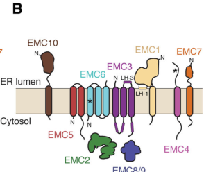
In the foundational human EMC cryo-EM structure, EMC6 is an integral membrane protein with three transmembrane helices. Together with EMC3, EMC6 forms an enclosed hydrophilic vestibule within the membrane proposed to be the pathway for insertion of selected substrates (pleiner2020structuralbasisfor pages 1-3, pleiner2020structuralbasisfor pages 7-11). A figure-level view of EMC6’s transmembrane placement in the EMC core and vestibule is provided in Pleiner et al. 2020 (pleiner2020structuralbasisfor media 044a8aee).

2) Subcellular localization, topology, domains, and interaction partners

2.1 Localization

EMC6 functions as part of the ER membrane-resident EMC complex, consistent with studies purifying intact EMC from human cells and characterizing its role in ER membrane insertion (odonnell2020thearchitectureof pages 1-2, li2024structuralinsightsinto pages 1-3).

2.2 Topology/structural features

Human EMC6 contains three transmembrane helices and contributes directly to the membrane-embedded core of EMC (pleiner2020structuralbasisfor pages 1-3, pleiner2020structuralbasisfor pages 7-11). One EMC6 helix (TM1) is described as unusually weakly hydrophobic and becomes stably inserted upon assembly with EMC5, indicating subunit–subunit assembly dependence for proper EMC6 integration and/or stability (predicted insertion Ξ”G reported as 3.8) (pleiner2020structuralbasisfor pages 1-3).

2.3 Key interaction partners (direct and contextual)

At minimum, EMC6’s core interaction partners are other EMC subunits, especially EMC3, with which EMC6 forms the hydrophilic vestibule required for insertase activity (pleiner2020structuralbasisfor pages 1-3, pleiner2020structuralbasisfor pages 7-11). In a 2024 cryo-EM study, EMC6 is again described as part of the EMC3–EMC6 core, with adjacent EMC4 forming a sidewall of the vestibule (li2024structuralinsightsinto pages 1-3).

In addition to insertase biology, human EMC has been observed in a state interacting with VDAC at mitochondria–ER contact sites (MERCs); this context links EMC architecture to ER–mitochondria crosstalk, though EMC6’s role is described as part of the transmembrane core rather than the specific β€œgating plug” element in that paper (li2024structuralinsightsinto pages 1-3).

3) Primary molecular function: how EMC6 contributes to membrane protein biogenesis

3.1 Insertase core: the EMC3–EMC6 hydrophilic vestibule

The 2020 human EMC structure proposes that insertion occurs via an enclosed hydrophilic vestibule located within the membrane and formed by EMC3 and EMC6 (pleiner2020structuralbasisfor pages 1-3, pleiner2020structuralbasisfor pages 7-11). The model includes local membrane remodeling (e.g., thinning) and electrostatic features that facilitate insertion of difficult TM segments (pleiner2020structuralbasisfor pages 7-11).

Interpretation for functional annotation: EMC6 is best annotated as a core architectural subunit that shapes the substrate insertion conduit (hydrophilic vestibule) together with EMC3.

3.2 Substrate routing and fidelity (2023: selectivity filter concept)

A 2023 mechanistic study describes a selectivity filter at the EMC hydrophilic vestibule entrance: positively charged residues at the vestibule entrance can reject mitochondrial tail-anchored (TA) proteins (with positively charged C-termini) and help enforce correct topology of multipass proteins (positive-inside rule) (Pleiner et al., J Cell Biol, 2023-05; https://doi.org/10.1083/jcb.202212007) (pleiner2023aselectivityfilter pages 1-2, pleiner2023aselectivityfilter pages 2-4). Although the crosslinking and mutagenesis emphasis in the retrieved text is largely on EMC3 and vestibule architecture, this work further consolidates the mechanistic framework in which the EMC3/EMC6 vestibule is the insertion route (pleiner2023aselectivityfilter pages 2-4).

3.3 Chaperone/holdase and client assembly (2023: EMC–CaV assembly intermediate)

A 2023 Nature client-bound structure captured an ~0.6 MDa complex between human EMC and a voltage-gated calcium channel (CaV1.2) assembly intermediate, providing direct structural evidence that EMC can function as a holdase/chaperone during complex membrane protein assembly (Chen et al., Nature, 2023-07; https://doi.org/10.1038/s41586-023-06175-5) (chen2023emcchaperone–cavstructure pages 1-3). This study reports distinct EMC client-binding sites (β€œTM and Cyto docks”) and shows EMC binding can reshape channel elements; it therefore supports the view that the EMC’s roles extend beyond simple insertion to orchestrating assembly intermediates (chen2023emcchaperone–cavstructure pages 1-3).

4) Recent developments (prioritizing 2023–2024)

4.1 2023 β€” selectivity filter and prevention of misinsertion at the ER

Pleiner et al. (2023-05) directly links EMC-mediated insertion to proteostasis and compartment integrity by explaining how charge-based discrimination at the vestibule limits misinsertion of mitochondrial TA proteins into the ER and enforces topology rules for multipass substrates (https://doi.org/10.1083/jcb.202212007) (pleiner2023aselectivityfilter pages 1-2).

4.2 2023 β€” first structural view of EMC bound to a multi-bundle client (CaV)

Chen et al. (2023-07) provides a structural β€œsnapshot” of EMC engaged with a complex multipass client during assembly and supports an EMC β€œholdase” role important for functional channel biogenesis (https://doi.org/10.1038/s41586-023-06175-5) (chen2023emcchaperone–cavstructure pages 1-3).

4.3 2024 β€” EMC–VDAC interaction and a regulated hydrophilic vestibule state at MERCs

Li et al. (Received 2023-11-06; Published 2024-03-15) reported human EMC structures in apo (3.47 Γ…) and VDAC-bound (3.32 Γ…) states and observed a specific EMC–VDAC interaction at mitochondria–ER contact sites (https://doi.org/10.18632/aging.205660) (li2024structuralinsightsinto pages 1-3). The work identifies a β€œgating plug” inside the hydrophilic vestibule and suggests that in the VDAC1-bound state the EMC is unlikely to function as an insertase, implying state-dependent regulation of the vestibule (li2024structuralinsightsinto pages 1-3).

5) Current applications and real-world implementations

5.1 Interpreting membrane protein biogenesis and proteostasis mechanisms

Because many therapeutically relevant targets are membrane proteins, a practical application of EMC/EMC6 biology is improving mechanistic understanding of how membrane proteins are inserted and assembled, and how insertion fidelity prevents mislocalization (pleiner2023aselectivityfilter pages 1-2, pleiner2023aselectivityfilter pages 2-4).

5.2 Structural frameworks enabling rational hypotheses about disease-linked phenotypes

Li et al. (2024) explicitly frames EMC multifunctionality as relevant to pathological phenotypes (including cancer, metabolic and neurological contexts) and emphasizes that EMC client diversity motivates disease associations (li2024structuralinsightsinto pages 1-3). While this does not prove EMC6 causality, it motivates using EMC6/EMC genetics and proteostasis assays to interpret disease mechanisms.

5.3 Knowledgebase-driven translational mapping (Open Targets)

Open Targets reports disease–target association evidence connecting EMC6 to multiple cancer-related entities (e.g., glioblastoma multiforme, lung adenocarcinoma, gastric cancer) and Blackfan–Diamond anemia, each with small evidence counts and modest overall association scores; these should be treated as hypothesis-generating rather than definitive mechanistic proof (OpenTargets Search: -EMC6).

6) Relevant statistics and data points from cited studies

  • Human EMC cryo-EM resolution: ~3.4 Γ… for the human EMC structure enabling near-complete atomic modeling; EMC6 contributes three TM helices to the core (Pleiner et al., 2020-07; https://doi.org/10.1126/science.abb5008) (pleiner2020structuralbasisfor pages 1-3).
  • EMC6 hydrophobicity metric: EMC6 TM1 predicted insertion Ξ”G = 3.8, with insertion stabilized upon EMC assembly (with EMC5) (pleiner2020structuralbasisfor pages 1-3).
  • Yeast Emc3/4/6 knockout proteomics: 38 membrane proteins significantly reduced in Emc3/4/6 knockouts (with additional validation described) (Bai et al., 2020-06; https://doi.org/10.1038/s41586-020-2389-3) (bai2020structureofthe pages 1-2).
  • Client-bound complex size: EMC–CaV assembly intermediate reported as ~0.6 MDa (Chen et al., 2023-07; https://doi.org/10.1038/s41586-023-06175-5) (chen2023emcchaperone–cavstructure pages 1-3).
  • 2024 EMC structures with VDAC: apo 3.47 Γ…; VDAC-bound 3.32 Γ… (Li et al., 2024-03-15; https://doi.org/10.18632/aging.205660) (li2024structuralinsightsinto pages 1-3).

7) Expert synthesis and analysis (evidence-weighted)

  1. Most strongly supported primary function: EMC6 is best characterized as a core membrane subunit of the EMC insertase, structurally partnering with EMC3 to form the hydrophilic vestibule that provides a favorable path for insertion of difficult TM helices (pleiner2020structuralbasisfor pages 1-3, pleiner2020structuralbasisfor pages 7-11, pleiner2020structuralbasisfor media 044a8aee).
  2. Topology and assembly constraint are key to EMC6 biology: EMC6’s three-TM architecture and the observation that one TM is weakly hydrophobic (Ξ”G = 3.8) emphasize that EMC6 is likely stabilized by complex assembly, consistent with a role as an obligate component of a multi-subunit membrane machine rather than a stand-alone factor (pleiner2020structuralbasisfor pages 1-3).
  3. Where the field is moving (2023–2024): EMC is increasingly viewed as a regulated multifunctional machine: (i) it ensures insertion fidelity (selectivity filter) and topology enforcement (pleiner2023aselectivityfilter pages 1-2), (ii) it can bind complex clients during assembly (CaV intermediate) (chen2023emcchaperone–cavstructure pages 1-3), and (iii) it adopts alternative functional states at organelle contact sites (VDAC-bound state at MERCs) that may be incompatible with insertase activity (li2024structuralinsightsinto pages 1-3). EMC6 appears consistently as part of the core EMC3–EMC6 vestibule module across these structural states (li2024structuralinsightsinto pages 1-3).

8) Structured summary of key sources

Publication Date URL / DOI Study type EMC6-relevant findings Quantitative data
Pleiner et al., 2020, Science Jul 2020 https://doi.org/10.1126/science.abb5008 Human cryo-EM structure / mechanism Human EMC6 is an integral membrane subunit with 3 transmembrane helices; EMC6 and EMC3 form an enclosed hydrophilic vestibule within the membrane that supports substrate insertion. EMC6 TM1 is unusually weakly hydrophobic and inserts efficiently only upon assembly with EMC5, linking EMC6 to EMC assembly/stability. Figure summaries identify EMC6 in the core TM module and vestibule with EMC3. (pleiner2020structuralbasisfor pages 1-3, pleiner2020structuralbasisfor pages 7-11, pleiner2020structuralbasisfor media 044a8aee) Cryo-EM resolution ~3.4 Γ…; EMC particle ~200 Γ— 70 Γ— 100 Γ…; EMC6 TM1 predicted insertion Ξ”G = 3.8; structure PDB 6WW7. (pleiner2020structuralbasisfor pages 1-3)
O'Donnell et al., 2020, eLife May 2020 https://doi.org/10.7554/elife.57887 Architecture / biochemical mechanism Places EMC6 among the mammalian EMC membrane subunits in a stable ER insertase complex. EMC contains a cytosolic vestibule leading into a lumenally sealed, lipid-exposed intramembrane groove for TMD insertion; EMC6 contributes as part of the membrane subcomplex required for overall EMC integrity and function. Purified EMC is sufficient for insertion of terminal TMDs in vitro. (odonnell2020thearchitectureof pages 1-2, odonnell2020thearchitectureof pages 2-4) Mammalian EMC described as 10 subunits with 7 membrane subunits including EMC6 and 12 predicted TM helices across the membrane subcomplex. (odonnell2020thearchitectureof pages 1-2)
Bai et al., 2020, Nature Jun 2020 https://doi.org/10.1038/s41586-020-2389-3 Cryo-EM structure / insertase mechanism In yeast EMC, Emc6 is a transmembrane scaffold subunit contributing to the membrane region of the insertase. The EMC transmembrane region includes Emc4, Emc5 and Emc6 plus TMDs of Emc1 and Emc3, supporting evolutionary conservation of EMC6’s role in the insertase membrane core relevant to human EMC6. (bai2020structureofthe pages 1-2) EMC knockout phenotypes: knockout of any single Emc subunit recapitulated EMC-null growth defect at 37Β°C; proteomics of Emc3/4/6 KO cells found 38 significantly reduced membrane proteins, with 9 validated as markedly downregulated in Emc3 KO; structure PDB 6WB9 / EMD-21587. (bai2020structureofthe pages 1-2)
Bai & Li, 2022, FEBS Journal Mar 2022 https://doi.org/10.1111/febs.15786 Review Review synthesizing EMC structural work and current model: EMC is a membrane insertase for less-hydrophobic/amphipathic helices, with a consensus substrate-binding pocket and architectural conservation from yeast to humans. For EMC6 specifically, the review supports its placement in the conserved membrane-embedded insertase core. (paper search result) Review notes mammalian EMC as a 9-subunit complex (EMC1–7, 8/9, 10 context-dependent naming in review summary) and emphasizes the conserved client TMH-binding pocket; no EMC6-specific numerical assay readout provided in gathered evidence. (paper search result)
Pleiner et al., 2023, Journal of Cell Biology May 2023 https://doi.org/10.1083/jcb.202212007 Mechanistic / selectivity filter Defines a selectivity filter in EMC that limits protein misinsertion at the ER and enforces topology of multipass membrane proteins. The EMC3/6 insertase core remains central, but the gathered evidence indicates mutations in EMC6 had only mild effects relative to stronger EMC3 effects in the tested selectivity-filter context. EMC6 remains part of the partially enclosed vestibule with surrounding dynamic subunits. (paper search result) Improved human EMC cryo-EM reconstruction reported; exact numerical resolution and EMC6-specific effect sizes not available in gathered evidence. EMC6 mutation effects described qualitatively as mild. (paper search result)
Chen et al., 2023, Nature May 2023 https://doi.org/10.1038/s41586-023-06175-5 Client-bound structure / chaperone mechanism EMC–CaV structure reveals a membrane-protein assembly intermediate and supports an EMC chaperone mode in addition to insertase activity. Gathered evidence specifically notes an EMC6-gated transmembrane cavity proposed to participate in client engagement during CaV biogenesis, placing EMC6 directly in the client-handling path. (paper search result) The study reports an ~0.6 MDa EMC–client complex. Additional EMC6-specific interface measurements were not available in gathered evidence. (paper search result)
Li et al., 2024, Aging (Albany NY) Mar 2024 https://doi.org/10.18632/aging.205660 Human cryo-EM structure / interaction with VDAC Human EMC structures in apo and VDAC-bound states identify a conserved EMC–VDAC interaction at mitochondria–ER contact sites (MERCs). EMC6 is part of the EMC3–EMC6 core; an ordered EMC4 three-TMH bundle sits adjacent to this core to form a sidewall of the hydrophilic vestibule. The reported gating plug is assigned to EMC3 rather than EMC6, so EMC6’s role here is structural/core rather than the plug itself. (li2024structuralinsightsinto pages 1-3) Apo structure 3.47 Γ…; VDAC-bound structure 3.32 Γ…. No EMC6-specific buried surface area or mutation phenotype was provided in gathered evidence. (li2024structuralinsightsinto pages 1-3)
Klose et al., 2025, Nature Communications Aug 2025 https://doi.org/10.1038/s41467-025-62109-x Chaperone mechanism / interactomics Although outside the user-prioritized 2023–2024 window, this recent mechanistic study is useful context: EMC6 is named as part of a lipid-filled cavity formed by EMC1, EMC3, EMC5 and EMC6, distinct from the canonical insertase site, supporting a broader EMC chaperone/quality-control role. (klose2025theemcacts pages 1-2) Crosslinking IP–MS found 506 enriched interactors, including >200 transmembrane proteins; about 50% of single-pass hits had signal peptides, ~1/3 had terminal TM helices, and ~2/3 of multipass hits had predicted Ncyto orientation. (klose2025theemcacts pages 1-2)

Table: This table summarizes key structural and mechanistic studies relevant to human EMC6/TMEM93 within the ER membrane protein complex, emphasizing recent 2023–2024 work while anchoring interpretations in foundational 2020 structures. It highlights EMC6’s role in the EMC3/EMC6 insertase core, assembly/stability, client handling, and emerging interaction contexts such as VDAC at MERCs.

9) Limitations of current evidence specifically for EMC6

Within the retrieved corpus, most mechanistic mutations and substrate-contact mapping are described for EMC3 and/or the EMC complex as a whole; there is comparatively less direct EMC6-only functional perturbation evidence (e.g., EMC6 point mutants with quantified client effects) available in the captured text excerpts (pleiner2023aselectivityfilter pages 2-4). Disease associations for EMC6 in knowledgebases (Open Targets) are not, by themselves, mechanistic proof and require follow-up in primary genetics and functional assays (OpenTargets Search: -EMC6).

References

  1. (pleiner2020structuralbasisfor pages 1-3): Tino Pleiner, Giovani Pinton Tomaleri, Kurt Januszyk, Alison J. Inglis, Masami Hazu, and Rebecca M. Voorhees. Structural basis for membrane insertion by the human er membrane protein complex. Jul 2020. URL: https://doi.org/10.1126/science.abb5008, doi:10.1126/science.abb5008. This article has 192 citations and is from a highest quality peer-reviewed journal.

  2. (pleiner2020structuralbasisfor pages 7-11): Tino Pleiner, Giovani Pinton Tomaleri, Kurt Januszyk, Alison J. Inglis, Masami Hazu, and Rebecca M. Voorhees. Structural basis for membrane insertion by the human er membrane protein complex. Jul 2020. URL: https://doi.org/10.1126/science.abb5008, doi:10.1126/science.abb5008. This article has 192 citations and is from a highest quality peer-reviewed journal.

  3. (odonnell2020thearchitectureof pages 1-2): John P O'Donnell, Ben P Phillips, Yuichi Yagita, Szymon Juszkiewicz, Armin Wagner, Duccio Malinverni, Robert J Keenan, Elizabeth A Miller, and Ramanujan S Hegde. The architecture of emc reveals a path for membrane protein insertion. May 2020. URL: https://doi.org/10.7554/elife.57887, doi:10.7554/elife.57887. This article has 121 citations and is from a domain leading peer-reviewed journal.

  4. (li2024structuralinsightsinto pages 1-3): Mingyue Li, Chunli Zhang, Yuntao Xu, Shaobai Li, Chenhui Huang, Jian Wu, and Ming Lei. Structural insights into human emc and its interaction with vdac. Aging (Albany NY), 16:5501-5525, Mar 2024. URL: https://doi.org/10.18632/aging.205660, doi:10.18632/aging.205660. This article has 6 citations.

  5. (pleiner2020structuralbasisfor media 044a8aee): Tino Pleiner, Giovani Pinton Tomaleri, Kurt Januszyk, Alison J. Inglis, Masami Hazu, and Rebecca M. Voorhees. Structural basis for membrane insertion by the human er membrane protein complex. Jul 2020. URL: https://doi.org/10.1126/science.abb5008, doi:10.1126/science.abb5008. This article has 192 citations and is from a highest quality peer-reviewed journal.

  6. (pleiner2023aselectivityfilter pages 1-2): Tino Pleiner, Masami Hazu, Giovani Pinton Tomaleri, Vy N. Nguyen, Kurt Januszyk, and Rebecca M. Voorhees. A selectivity filter in the er membrane protein complex limits protein misinsertion at the er. The Journal of Cell Biology, May 2023. URL: https://doi.org/10.1083/jcb.202212007, doi:10.1083/jcb.202212007. This article has 28 citations.

  7. (pleiner2023aselectivityfilter pages 2-4): Tino Pleiner, Masami Hazu, Giovani Pinton Tomaleri, Vy N. Nguyen, Kurt Januszyk, and Rebecca M. Voorhees. A selectivity filter in the er membrane protein complex limits protein misinsertion at the er. The Journal of Cell Biology, May 2023. URL: https://doi.org/10.1083/jcb.202212007, doi:10.1083/jcb.202212007. This article has 28 citations.

  8. (chen2023emcchaperone–cavstructure pages 1-3): Zhou Chen, Abhisek Mondal, Fayal Abderemane-Ali, Seil Jang, Sangeeta Niranjan, JosΓ© L. MontaΓ±o, Balyn W. Zaro, and Daniel L. Minor. Emc chaperone–cav structure reveals an ion channel assembly intermediate. Nature, 619:410-419, May 2023. URL: https://doi.org/10.1038/s41586-023-06175-5, doi:10.1038/s41586-023-06175-5. This article has 77 citations and is from a highest quality peer-reviewed journal.

  9. (OpenTargets Search: -EMC6): Open Targets Query (-EMC6, 5 results). Buniello, A. et al. (2025). Open Targets Platform: facilitating therapeutic hypotheses building in drug discovery. Nucleic Acids Research.

  10. (bai2020structureofthe pages 1-2): Lin Bai, Qinglong You, Xiang Feng, Amanda Kovach, and Huilin Li. Structure of the er membrane complex, a transmembrane-domain insertase. Nature, 584:475-478, Jun 2020. URL: https://doi.org/10.1038/s41586-020-2389-3, doi:10.1038/s41586-020-2389-3. This article has 164 citations and is from a highest quality peer-reviewed journal.

  11. (odonnell2020thearchitectureof pages 2-4): John P O'Donnell, Ben P Phillips, Yuichi Yagita, Szymon Juszkiewicz, Armin Wagner, Duccio Malinverni, Robert J Keenan, Elizabeth A Miller, and Ramanujan S Hegde. The architecture of emc reveals a path for membrane protein insertion. May 2020. URL: https://doi.org/10.7554/elife.57887, doi:10.7554/elife.57887. This article has 121 citations and is from a domain leading peer-reviewed journal.

  12. (klose2025theemcacts pages 1-2): Carolin J Klose, Kevin M Meighen-Berger, M. Kulke, Marina Parr, Barbara Steigenberger, Martin Zacharias, Dmitrij Frishman, and Matthias Feige. The emc acts as a chaperone for membrane proteins. Nature Communications, Aug 2025. URL: https://doi.org/10.1038/s41467-025-62109-x, doi:10.1038/s41467-025-62109-x. This article has 6 citations and is from a highest quality peer-reviewed journal.

Artifacts

Citations

  1. pleiner2020structuralbasisfor pages 1-3
  2. li2024structuralinsightsinto pages 1-3
  3. pleiner2020structuralbasisfor pages 7-11
  4. pleiner2023aselectivityfilter pages 2-4
  5. pleiner2023aselectivityfilter pages 1-2
  6. bai2020structureofthe pages 1-2
  7. odonnell2020thearchitectureof pages 1-2
  8. klose2025theemcacts pages 1-2
  9. odonnell2020thearchitectureof pages 2-4
  10. https://doi.org/10.1083/jcb.202212007
  11. https://doi.org/10.1038/s41586-023-06175-5
  12. https://doi.org/10.18632/aging.205660
  13. https://doi.org/10.1126/science.abb5008
  14. https://doi.org/10.1038/s41586-020-2389-3
  15. https://doi.org/10.7554/elife.57887
  16. https://doi.org/10.1111/febs.15786
  17. https://doi.org/10.1038/s41467-025-62109-x
  18. https://doi.org/10.1126/science.abb5008,
  19. https://doi.org/10.7554/elife.57887,
  20. https://doi.org/10.18632/aging.205660,
  21. https://doi.org/10.1083/jcb.202212007,
  22. https://doi.org/10.1038/s41586-023-06175-5,
  23. https://doi.org/10.1038/s41586-020-2389-3,
  24. https://doi.org/10.1038/s41467-025-62109-x,

πŸ“š Additional Documentation

Notes

(EMC6-notes.md)

EMC6 (Q9BV81) review notes

Identity

  • ER membrane protein complex subunit 6; synonym TMEM93 (TMEM93). HGNC:28430, gene 83460, chromosome 17.
  • Small 110 aa polytopic ER membrane protein with three transmembrane helices (TM1 29-44, TM2 51-71, TM3 90-106) and an N-cytoplasmic/C-lumenal topology [file:human/EMC6/EMC6-uniprot.txt "TRANSMEM 29..44"].
  • Belongs to the EMC6 family [file:human/EMC6/EMC6-uniprot.txt "Belongs to the EMC6 family"].

Function: EMC insertase core

  • Component of the ER membrane protein complex (EMC), a conserved ~9-subunit ER insertase/chaperone for membrane proteins [file:human/EMC6/EMC6-uniprot.txt "Component of the ER membrane protein complex (EMC)"].
  • EMC enables energy-independent insertion of newly synthesized membrane proteins into the ER membrane; it preferentially handles TMDs that are weakly hydrophobic or contain destabilizing charged/aromatic residues [file:human/EMC6/EMC6-uniprot.txt "enables the energy-independent insertion into endoplasmic"].
  • EMC is a transmembrane domain insertase PMID:29242231; it inserts tail-anchored (TA) proteins with moderately hydrophobic TMDs post-translationally PMID:29242231.
  • EMC3 is the catalytic insertase subunit (YidC/Get1/Oxa1 superfamily); EMC6 packs against EMC3 to form the hydrophilic insertase vestibule in the membrane. EMC6 mutagenesis of Asp-27 and Thr-31 (cytoplasmic/TM1 boundary) does NOT affect EMC assembly but decreases membrane insertion of hydrophobic-TMD client proteins, demonstrating EMC6's direct contribution to the insertase reaction [file:human/EMC6/EMC6-uniprot.txt "No effect on EMC assembly but decreased"]. This experimental separation of assembly from activity is why "membrane insertase activity" (GO:0032977, contributes_to) is defensible CORE for EMC6, not merely the bulk EMC.
  • Cotranslational role: EMC engages multipass membrane protein clients cotranslationally to enable their biogenesis PMID:29809151.
  • Topogenesis role: EMC controls topology of multipass proteins (e.g., GPCRs) by inserting the first N-terminal TMD in N-exo orientation PMID:30415835; clients include the beta1-adrenergic receptor and other GPCRs PMID:30415835.
  • In vivo IMP evidence in Drosophila: EMC (including EMC6) is required for TMD membrane insertion of the TA client Xport-A PMID:34918864; this paper underpins the FlyBase-assigned GO:0032977 contributes_to membrane insertase activity and GO:0071816 (TA insertion).

Subcellular location

  • ER membrane, multi-pass membrane protein [file:human/EMC6/EMC6-uniprot.txt "Endoplasmic reticulum membrane"]. IDA support PMID:22119785 (identified EMC in ERAD network), PMID:30415835, PMID:32439656.

Autophagy annotations (GO:0000045, GO:1903349)

  • A single 2013 study (abstract-only in cache, full_text_available: false) reported EMC6/TMEM93 as an ER-localized protein interacting with RAB5A and BECN1/Beclin1, colocalizing with the omegasome marker DFCP1/ZFYVE1, and regulating autophagosome formation [PMID:23182941 "It was shown to regulate "; PMID:23182941 "colocalized with the omegasome marker ZFYVE1/DFCP1"].
  • This predates the insertase-core understanding of EMC. The autophagy/omegasome phenotype is plausibly an INDIRECT/secondary consequence of impaired biogenesis of membrane-protein clients (EMC's "indirect client" model is explicitly discussed in PMID:34918864). The IMP autophagosome-assembly and IDA omegasome-membrane annotations rest on this one experimental paper whose full text the curator read; per guidelines, do not REMOVE experimental annotations on incomplete evidence -> keep as NON_CORE (not the core insertase function). The IBA autophagosome-assembly term (PAN-GO) propagates this single experimental observation across the family; although weakly supported, it is retained as peripheral rather than removed -> KEEP_AS_NON_CORE (consistent with the IMP/IDA handling above, and matching the action recorded in the YAML).

protein binding (GO:0005515)

  • Many IPI annotations (IntAct/UniProt) to EMC client/partner proteins (MMGT1/EMC10, AQP6, AQP9, GPCR clients, SLC transporters, etc.) and to RAB5A/BECN1. Bare "protein binding" is uninformative per curation guidelines -> KEEP_AS_NON_CORE. PMID:32296183 (HuRI binary interactome) and PMID:33961781 (BioPlex) are high-throughput; PMID:22119785 (EMC10/MMGT1) and PMID:32439656 (EMC assembly) reflect genuine EMC partnerships.

Core function call for EMC6

  1. EMC complex (GO:0072546) membership - CORE structural identity.
  2. ER membrane (GO:0005789) - CORE location.
  3. Membrane insertase activity (GO:0032977, contributes_to) - CORE MF: EMC6 + EMC3 form the catalytic insertase core; EMC6 D27/T31 mutants reduce insertion without disrupting assembly.
  4. TA insertion (GO:0071816) and stop-transfer/multipass insertion (GO:0045050) - CORE biological processes of the insertase.

Reference relevance

  • HIGH: PMID:29242231, PMID:30415835, PMID:32439656, PMID:34918864 (insertase mechanism/structure/in vivo), PMID:22119785 (EMC discovery in human, ER loc).
  • MEDIUM: PMID:29809151 (multipass cotranslational role).
  • LOW/contextual: PMID:32296183, PMID:33961781 (HT interactome), PMID:23182941 (autophagy; pre-insertase, indirect).

Falcon deep-research findings (incorporated 2026-06)

New EMC6-relevant references verified against PubMed and added to the review (all additive; no action changes). These reinforce EMC6's core role in the EMC3/EMC6 insertase vestibule.

  • 2023 selectivity-filter study consolidates the EMC3/EMC6 hydrophilic vestibule as the central insertion route and a charge-based selectivity filter enforcing the positive-inside rule; EMC6 mutations had milder effects than EMC3 in the tested context but EMC6 remains part of the vestibule-enclosing core. PMID:37199759
  • EMC acts as a holdase/chaperone during assembly of the multipass CaV1.2 channel (TM and Cyto client docks), extending EMC function beyond insertion. PMID:37196677
  • 2024 human EMC cryo-EM (apo + VDAC-bound) places EMC6 in the EMC3-EMC6 core; a vestibule gating plug (assigned to EMC3) changes conformation between states, and the EMC engages VDAC at mitochondria-ER contact sites. PMID:38517390
  • 2025 chaperone study names EMC6 as part of a lipid-filled cavity (EMC1/EMC3/EMC5/EMC6) distinct from the canonical insertase site, supporting a broader EMC chaperone/quality-control role; the EMC engages TMDs via EMC1. PMID:40753078
  • Core EMC6 call is unchanged: EMC complex membership + ER membrane localization + membrane insertase activity (contributes_to, via the EMC3/EMC6 vestibule) + TA and stop-transfer/multipass insertion processes. Autophagy/omegasome annotations remain KEEP_AS_NON_CORE.

Pn Notes

(EMC6-pn-notes.md)

EMC6 PN Consistency Notes

  • Generated: 2026-06-18
  • Project: PROTEOSTASIS
  • Scope: PN consistency rereview against local AIGR review and available deep-research artifacts
  • UniProt: Q9BV81
  • AIGR review status: COMPLETE
  • Review batch: proteostasis-batch-2026-06-11
  • Batch change status: added

Source Files Checked

Deep Research Files

AIGR Review Snapshot

  • Description: EMC6 (ER membrane protein complex subunit 6; also TMEM93) is a small (110 aa) polytopic ER membrane protein with three transmembrane helices and an N-cytoplasmic/C-lumenal topology. It is a constitutive subunit of the ER membrane protein complex (EMC), a conserved ~9-subunit transmembrane-domain insertase and membrane-protein chaperone of the endoplasmic reticulum. Within the complex, EMC6 packs against the catalytic insertase subunit EMC3 (a member of the YidC/Oxa1/Get1 insertase superfamily) to form the hydrophilic membrane vestibule through which substrate transmembrane domains are inserted. The EMC enables the energy-independent insertion of newly synthesized membrane proteins into the ER membrane, with a preference for transmembrane domains that are weakly hydrophobic or contain destabilizing charged or aromatic residues. It mediates post-translational insertion of tail-anchored proteins and cotranslational insertion and topogenesis of multipass membrane proteins, including setting the N-exo topology of the first transmembrane domain of G protein-coupled receptors. Mutations of EMC6 residues at the cytoplasmic/TM1 boundary (Asp-27, Thr-31) impair client insertion without disrupting complex assembly, demonstrating a direct contribution of EMC6 to the insertase reaction. EMC6 localizes to the ER membrane and is broadly expressed.
  • Existing/core annotation action counts: ACCEPT: 19; KEEP_AS_NON_CORE: 11; MARK_AS_OVER_ANNOTATED: 1

PN Consistency Summary

  • Consistency: Consistent, and the two-branch placement is well-handled on both sides. EMC6 is the catalytic-core subunit pairing with EMC3 in the vestibule (D27/T31 mutagenesis separates insertion from assembly β†’ GO:0032977 core). Separately, the single 2013 study (PMID:23182941: RAB5A/BECN1 interaction, DFCP1/omegasome colocalization, autophagosome-formation defect) underlies GOA's GO:0000045 (autophagosome assembly, IMP+IBA) and GO:1903349 (omegasome membrane, IDA) β€” all KEEP_AS_NON_CORE in the review, matching PN's judgment that the autophagy role is context, likely indirect via client biogenesis. No contradictions.
  • PN story / NEW pressure: Row 2 is the only place PN asserts a role beyond the EMC insertase story (BECN1-mediated recruitment of PI3K complex to the autophagophore nucleation site). This is partially captured in GOA (autophagosome assembly, omegasome membrane) and in the review (BECN1/RAB5A protein-binding, omegasome). PN correctly does NOT claim EMC6 is a PI3K-complex member (GO:0035032 marked too_broad/context_only). No defensible NEW GO term β€” the existing autophagosome-assembly term covers the asserted role and is already present. Conclusion: already captured (and appropriately non-core).
  • Evidence alignment: Good overlap on EMC papers (PMID:22119785, PMID:29242231, PMID:32439656, PMID:34918864). Row2's PN reference is a non-PMID review ("Membrane Trafficking in Autophagy - ScienceDirect"); the review instead anchors the autophagy role to the primary PMID:23182941, a stronger citation. Slight divergence: PN row-2 evidence is a secondary review whereas the gene review uses the primary experimental paper.
  • Verdict: Consistent across both branches; autophagy/PI3K role correctly captured as non-core and correctly NOT projected as complex membership; PN adds no NEW pressure; row1 projected group/class terms broader than review.

Full Consistency Review

  • UniProt: Q9BV81 (TMEM93) Β· batch: proteostasis-batch-2026-06-11 Β· review status: COMPLETE (thorough; all annotations including autophagy block adjudicated)
  • PN placement: Row 1 ER proteostasis | Protein transport | Transmembrane protein import | EMC complex component; Row 2 Autophagy-Lysosome Pathway | Autophagophore initiation and elongation | Class 3 PI3K complex 1, direct | Localization of class 3 PI3K complex 1 (branches ER + ALP). PN-node mapping: row1 typeβ†’GO:0072546 EMC complex (already_in_goa_exact), groupβ†’GO:0044743, classβ†’GO:0015031; row2 entirely context_only/too_broad_to_propagate (GO:0035032 PI3K III; GO:0016236 macroautophagy) β€” projects NOTHING.
  • Consistency: Consistent, and the two-branch placement is well-handled on both sides. EMC6 is the catalytic-core subunit pairing with EMC3 in the vestibule (D27/T31 mutagenesis separates insertion from assembly β†’ GO:0032977 core). Separately, the single 2013 study (PMID:23182941: RAB5A/BECN1 interaction, DFCP1/omegasome colocalization, autophagosome-formation defect) underlies GOA's GO:0000045 (autophagosome assembly, IMP+IBA) and GO:1903349 (omegasome membrane, IDA) β€” all KEEP_AS_NON_CORE in the review, matching PN's judgment that the autophagy role is context, likely indirect via client biogenesis. No contradictions.
  • PN story / NEW pressure: Row 2 is the only place PN asserts a role beyond the EMC insertase story (BECN1-mediated recruitment of PI3K complex to the autophagophore nucleation site). This is partially captured in GOA (autophagosome assembly, omegasome membrane) and in the review (BECN1/RAB5A protein-binding, omegasome). PN correctly does NOT claim EMC6 is a PI3K-complex member (GO:0035032 marked too_broad/context_only). No defensible NEW GO term β€” the existing autophagosome-assembly term covers the asserted role and is already present. Conclusion: already captured (and appropriately non-core).
  • Mapping strategy: EMC6 does not change either node. Row1 typeβ†’GO:0072546 exact/correct; projected group/class (GO:0044743, GO:0015031) broader than review's specific insertion terms (broader-ancestor pattern, cf. TOMM20/HSPA8/RAB7A). Row2 correctly projects nothing β€” EMC6 is a BECN1 interactor/regulator, not a class-III PI3K complex component, so withholding GO:0035032 membership is the right call (avoids a false complex-membership assertion).
  • Evidence alignment: Good overlap on EMC papers (PMID:22119785, PMID:29242231, PMID:32439656, PMID:34918864). Row2's PN reference is a non-PMID review ("Membrane Trafficking in Autophagy - ScienceDirect"); the review instead anchors the autophagy role to the primary PMID:23182941, a stronger citation. Slight divergence: PN row-2 evidence is a secondary review whereas the gene review uses the primary experimental paper.
  • Verdict: Consistent across both branches; autophagy/PI3K role correctly captured as non-core and correctly NOT projected as complex membership; PN adds no NEW pressure; row1 projected group/class terms broader than review.

PN Dossier Context

  • review_batch: proteostasis-batch-2026-06-11
  • review_yaml: genes/human/EMC6/EMC6-ai-review.yaml
  • PN workbook rows: 2

PN row 1: ER proteostasis | Protein transport | Transmembrane protein import | EMC complex component

  • UniProt: Q9BV81
  • In branches: ER, ALP
  • PN-node mapping records (path + ancestors):
    • [type] ER proteostasis|Protein transport|Transmembrane protein import|EMC complex component
      status=mapped scope=ok_for_propagation_to_go GO=[GO:0072546 EMC complex]
      rationale: This PN type denotes ER membrane protein complex components. The GO EMC complex cellular-component term is the direct target.
    • [group] ER proteostasis|Protein transport|Transmembrane protein import
      status=mapped scope=ok_for_propagation_to_go GO=[GO:0044743 protein transmembrane import into intracellular organelle]
      rationale: This PN group covers ER transmembrane-protein insertion/import systems such as EMC- and PAT-related pathways. The local GO cache does not expose an ER-specific matching term, so the broader intracellular-organelle transmembrane-import process is the best supported propagation target.
    • [class] ER proteostasis|Protein transport
      status=mapped scope=ok_for_propagation_to_go GO=[GO:0015031 protein transport]
      rationale: The PN ER Protein transport class groups ER-targeting and ER-insertion pathways. GO protein transport is the appropriate propagation target, while the source class remains ER-specific and broader than any single GO transport subtype.
    • [branch] ER proteostasis
      status=no_mapping scope= GO=[]
      rationale: Reviewed as a top-level PN branch. This is a systems/taxonomy umbrella, not a direct GO assertion; narrower child curations carry any propagating GO mappings.

PN row 2: Autophagy-Lysosome Pathway | Autophagophore initiation and elongation | Class 3 PI3K complex 1, direct | Localization of class 3 PI3K complex 1

  • UniProt: Q9BV81
  • In branches: ER, ALP
  • Notes: Transmembrane ER protein that localizes to omegasomes and interacts with BECN1 to recruit PI3K complex to autophagophore nucleation site.
  • PN references (titles):
    • Membrane Trafficking in Autophagy - ScienceDirect
  • PN-node mapping records (path + ancestors):
    • [type] Autophagy-Lysosome Pathway|Autophagophore initiation and elongation|Class 3 PI3K complex 1, direct|Localization of class 3 PI3K complex 1
      status=context_only scope=too_broad_to_propagate GO=[GO:0035032 phosphatidylinositol 3-kinase complex, class III]
      rationale: Reviewed as a class-III PI3K complex context or regulator bucket. This node is useful for curator interpretation, but it should not project cellular-component membership; only explicit complex-component leaves propagate to GO complex terms.
    • [group] Autophagy-Lysosome Pathway|Autophagophore initiation and elongation|Class 3 PI3K complex 1, direct
      status=context_only scope=too_broad_to_propagate GO=[GO:0035032 phosphatidylinositol 3-kinase complex, class III]
      rationale: Reviewed as a class-III PI3K complex context or regulator bucket. This node is useful for curator interpretation, but it should not project cellular-component membership; only explicit complex-component leaves propagate to GO complex terms.
    • [class] Autophagy-Lysosome Pathway|Autophagophore initiation and elongation
      status=context_only scope=too_broad_to_propagate GO=[GO:0016236 macroautophagy]
      rationale: This class is a real macroautophagy context, but its descendants include core factors, component buckets, upstream modulators, localization roles, and residual categories. Projecting generic macroautophagy from this ancestor creates TRAPP-like overpropagation, so candidate GO annotations must come from narrower curated nodes.
    • [branch] Autophagy-Lysosome Pathway
      status=no_mapping scope= GO=[]
      rationale: Reviewed as the top-level PN branch. It is a project taxonomy umbrella rather than a direct GO assertion; all propagation must come from manually curated child nodes.

Projected GO annotations (3)

  • GO:0015031 protein transport | scope=ok_for_propagation_to_go | goa_status=new_to_goa | from=ER proteostasis|Protein transport
  • GO:0044743 protein transmembrane import into intracellular organelle | scope=ok_for_propagation_to_go | goa_status=new_to_goa | from=ER proteostasis|Protein transport|Transmembrane protein import
  • GO:0072546 EMC complex | scope=ok_for_propagation_to_go | goa_status=already_in_goa_exact | from=ER proteostasis|Protein transport|Transmembrane protein import|EMC complex component

Note

This file is generated from the current PROTEOSTASIS phase-1 dossier and local gene-review artifacts. Edit the source review, PN mapping, or dossier rather than this generated note when correcting the underlying curation.

πŸ“„ View Raw YAML

id: Q9BV81
gene_symbol: EMC6
product_type: PROTEIN
status: COMPLETE
taxon:
  id: NCBITaxon:9606
  label: Homo sapiens
description: EMC6 (ER membrane protein complex subunit 6; also TMEM93) is a small (110 aa) polytopic ER membrane protein with three transmembrane helices and an N-cytoplasmic/C-lumenal topology. It is a constitutive subunit of the ER membrane protein complex (EMC), a conserved ~9-subunit transmembrane-domain insertase and membrane-protein chaperone of the endoplasmic reticulum. Within the complex, EMC6 packs against the catalytic insertase subunit EMC3 (a member of the YidC/Oxa1/Get1 insertase superfamily) to form the hydrophilic membrane vestibule through which substrate transmembrane domains are inserted. The EMC enables the energy-independent insertion of newly synthesized membrane proteins into the ER membrane, with a preference for transmembrane domains that are weakly hydrophobic or contain destabilizing charged or aromatic residues. It mediates post-translational insertion of tail-anchored proteins and cotranslational insertion and topogenesis of multipass membrane proteins, including setting the N-exo topology of the first transmembrane domain of G protein-coupled receptors. Mutations of EMC6 residues at the cytoplasmic/TM1 boundary (Asp-27, Thr-31) impair client insertion without disrupting complex assembly, demonstrating a direct contribution of EMC6 to the insertase reaction. EMC6 localizes to the ER membrane and is broadly expressed.
existing_annotations:
- term:
    id: GO:0000045
    label: autophagosome assembly
  evidence_type: IBA
  original_reference_id: GO_REF:0000033
  qualifier: involved_in
  review:
    summary: Phylogenetic (PAN-GO) propagation of an autophagosome-assembly role across the EMC6 family, derived ultimately from a single 2013 experimental study reporting EMC6 regulation of autophagosome formation. This phenotype is most plausibly an indirect consequence of impaired biogenesis of EMC membrane-protein clients rather than a direct EMC6 function.
    action: KEEP_AS_NON_CORE
    reason: The autophagy role rests on one experimental paper and is likely an indirect downstream effect of the EMC's insertase function; retained but peripheral, not the core EMC6 function.
    supported_by:
    - reference_id: PMID:23182941
      supporting_text: It was shown to regulate
- term:
    id: GO:0072546
    label: EMC complex
  evidence_type: IBA
  original_reference_id: GO_REF:0000033
  qualifier: part_of
  review:
    summary: EMC6 is a constitutive subunit of the ER membrane protein complex; phylogenetic assignment of EMC complex membership is consistent with direct experimental and structural evidence. This is a core structural identity of EMC6.
    action: ACCEPT
    reason: EMC complex membership is the core cellular-component identity of EMC6 and is supported by IDA, cryo-EM structures, and the conserved EMC6 family.
    supported_by:
    - reference_id: file:human/EMC6/EMC6-uniprot.txt
      supporting_text: Component of the ER membrane protein complex (EMC).
- term:
    id: GO:0005737
    label: cytoplasm
  evidence_type: IEA
  original_reference_id: GO_REF:0000117
  qualifier: located_in
  review:
    summary: ARBA machine-learning electronic assignment of cytoplasm; EMC6 is an integral ER membrane protein whose site of action is the ER membrane. Cytoplasm is an imprecise parent term relative to the experimentally supported ER membrane localization.
    action: MARK_AS_OVER_ANNOTATED
    reason: Generic and imprecise electronic assignment; the specific and experimentally supported compartment is the ER membrane (GO:0005789).
    supported_by:
    - reference_id: file:human/EMC6/EMC6-uniprot.txt
      supporting_text: 'SUBCELLULAR LOCATION: Endoplasmic reticulum membrane'
- term:
    id: GO:0005783
    label: endoplasmic reticulum
  evidence_type: IEA
  original_reference_id: GO_REF:0000002
  qualifier: located_in
  review:
    summary: InterPro-based electronic assignment to the endoplasmic reticulum, consistent with the experimentally supported ER membrane localization but less specific.
    action: KEEP_AS_NON_CORE
    reason: Correct compartment but a parent of the more precise ER membrane term; redundant with experimental ER membrane evidence.
    supported_by:
    - reference_id: file:human/EMC6/EMC6-uniprot.txt
      supporting_text: 'SUBCELLULAR LOCATION: Endoplasmic reticulum membrane'
- term:
    id: GO:0005789
    label: endoplasmic reticulum membrane
  evidence_type: IEA
  original_reference_id: GO_REF:0000044
  qualifier: located_in
  review:
    summary: Electronic transfer of the ER membrane subcellular location from UniProt; the correct and core compartment for EMC6.
    action: ACCEPT
    reason: Correct core location; redundant with experimental EXP/IDA evidence.
    supported_by:
    - reference_id: file:human/EMC6/EMC6-uniprot.txt
      supporting_text: 'SUBCELLULAR LOCATION: Endoplasmic reticulum membrane'
- term:
    id: GO:0016020
    label: membrane
  evidence_type: IEA
  original_reference_id: GO_REF:0000002
  qualifier: located_in
  review:
    summary: InterPro-based generic membrane assignment, a parent of the specific ER membrane localization.
    action: KEEP_AS_NON_CORE
    reason: Correct but generic; the specific ER membrane term captures the informative localization.
    supported_by:
    - reference_id: file:human/EMC6/EMC6-uniprot.txt
      supporting_text: Multi-pass
- term:
    id: GO:0072546
    label: EMC complex
  evidence_type: IEA
  original_reference_id: GO_REF:0000002
  qualifier: part_of
  review:
    summary: InterPro-based electronic assignment of EMC complex membership, consistent with the experimental IDA annotation.
    action: ACCEPT
    reason: Correct core structural identity; redundant with IDA/IBA evidence.
    supported_by:
    - reference_id: file:human/EMC6/EMC6-uniprot.txt
      supporting_text: Component of the ER membrane protein complex (EMC).
- term:
    id: GO:0005515
    label: protein binding
  evidence_type: IPI
  original_reference_id: PMID:22119785
  qualifier: enables
  review:
    summary: IntAct interaction (with MMGT1/EMC10) from the foundational human ERAD-network mapping study that first defined the EMC. The interaction is a genuine EMC partnership, but bare protein binding is uninformative and is not elevated to core.
    action: KEEP_AS_NON_CORE
    reason: Real EMC partner interaction but the bare protein binding term is uninformative per curation guidelines.
    supported_by:
    - reference_id: file:human/EMC6/EMC6-uniprot.txt
      supporting_text: 'Q9BV81; Q8N4V1: MMGT1'
- term:
    id: GO:0005515
    label: protein binding
  evidence_type: IPI
  original_reference_id: PMID:32296183
  qualifier: enables
  review:
    summary: High-throughput binary (HuRI) interactome captures of EMC6 with multiple membrane proteins (AQP6, AQP9, EBP, SLC transporters, etc.), many of which are plausibly EMC clients. Bare protein binding is uninformative.
    action: KEEP_AS_NON_CORE
    reason: High-throughput interactions, partly reflecting client engagement, but the bare term is uninformative and not core.
    supported_by:
    - reference_id: file:human/EMC6/EMC6-uniprot.txt
      supporting_text: 'Q9BV81; O43315: AQP9'
- term:
    id: GO:0005515
    label: protein binding
  evidence_type: IPI
  original_reference_id: PMID:32439656
  qualifier: enables
  review:
    summary: Interaction evidence from the cryo-EM structural study of the human EMC, reflecting genuine intra-complex EMC partnerships. Bare protein binding is uninformative.
    action: KEEP_AS_NON_CORE
    reason: Real intra-complex interaction but bare protein binding is uninformative; the EMC complex membership term captures the informative content.
    supported_by:
    - reference_id: file:human/EMC6/EMC6-uniprot.txt
      supporting_text: 'Q9BV81; Q8N4V1: MMGT1'
- term:
    id: GO:0005515
    label: protein binding
  evidence_type: IPI
  original_reference_id: PMID:33961781
  qualifier: enables
  review:
    summary: BioPlex affinity-MS interactome capture (with MMGT1/EMC10). Genuine EMC partner but the bare term is uninformative.
    action: KEEP_AS_NON_CORE
    reason: High-throughput interaction; bare protein binding is uninformative and not core.
    supported_by:
    - reference_id: file:human/EMC6/EMC6-uniprot.txt
      supporting_text: 'Q9BV81; Q8N4V1: MMGT1'
- term:
    id: GO:0005789
    label: endoplasmic reticulum membrane
  evidence_type: NAS
  original_reference_id: PMID:29242231
  qualifier: located_in
  review:
    summary: ComplexPortal NAS annotation of ER membrane localization for the EMC, consistent with experimental evidence and the core compartment of EMC6.
    action: ACCEPT
    reason: Correct core location; consistent with EXP/IDA evidence.
    supported_by:
    - reference_id: file:human/EMC6/EMC6-uniprot.txt
      supporting_text: 'SUBCELLULAR LOCATION: Endoplasmic reticulum membrane'
- term:
    id: GO:0045050
    label: protein insertion into ER membrane by stop-transfer membrane-anchor sequence
  evidence_type: IDA
  original_reference_id: PMID:29242231
  qualifier: involved_in
  review:
    summary: The EMC inserts transmembrane domains, including stop-transfer membrane-anchor sequences of multipass proteins; EMC6 is integral to the insertase vestibule. A core biological process of the EMC.
    action: ACCEPT
    reason: Core EMC-mediated process; EMC6 contributes directly via the EMC3/EMC6 insertase vestibule.
    supported_by:
    - reference_id: PMID:29242231
      supporting_text: EMC is a transmembrane domain insertase
- term:
    id: GO:0071816
    label: tail-anchored membrane protein insertion into ER membrane
  evidence_type: IDA
  original_reference_id: PMID:29242231
  qualifier: involved_in
  review:
    summary: The EMC mediates post-translational insertion of tail-anchored proteins with moderately hydrophobic TMDs; demonstrated directly in this study. A core EMC process to which EMC6 contributes.
    action: ACCEPT
    reason: Core EMC-mediated process; directly demonstrated.
    supported_by:
    - reference_id: PMID:29242231
      supporting_text: tail-anchored membrane proteins with moderately hydrophobic transmembrane
- term:
    id: GO:0072546
    label: EMC complex
  evidence_type: IPI
  original_reference_id: PMID:32439656
  qualifier: part_of
  review:
    summary: ComplexPortal IPI assignment of EMC complex membership based on the cryo-EM structure of the human EMC. Core structural identity of EMC6.
    action: ACCEPT
    reason: Structurally demonstrated core EMC membership.
    supported_by:
    - reference_id: file:human/EMC6/EMC6-uniprot.txt
      supporting_text: Component of the ER membrane protein complex (EMC).
- term:
    id: GO:0005789
    label: endoplasmic reticulum membrane
  evidence_type: EXP
  original_reference_id: PMID:22119785
  qualifier: located_in
  review:
    summary: Experimental ER membrane localization from the EMC-discovery ERAD-network study. Core compartment.
    action: ACCEPT
    reason: Experimentally supported core location.
    supported_by:
    - reference_id: file:human/EMC6/EMC6-uniprot.txt
      supporting_text: 'SUBCELLULAR LOCATION: Endoplasmic reticulum membrane'
- term:
    id: GO:0005789
    label: endoplasmic reticulum membrane
  evidence_type: EXP
  original_reference_id: PMID:30415835
  qualifier: located_in
  review:
    summary: Experimental ER membrane localization from the EMC topogenesis study. Core compartment.
    action: ACCEPT
    reason: Experimentally supported core location.
    supported_by:
    - reference_id: file:human/EMC6/EMC6-uniprot.txt
      supporting_text: 'SUBCELLULAR LOCATION: Endoplasmic reticulum membrane'
- term:
    id: GO:0032977
    label: membrane insertase activity
  evidence_type: IMP
  original_reference_id: PMID:34918864
  qualifier: contributes_to
  review:
    summary: In vivo Drosophila evidence that the EMC (including EMC6) is required for TMD membrane insertion of a tail-anchored client; combined with the human mutagenesis showing EMC6 D27/T31 reduce insertion without affecting assembly, EMC6 directly contributes to the insertase reaction. The contributes_to qualifier is appropriate for a catalytic-core subunit.
    action: ACCEPT
    reason: EMC6 partners EMC3 in the catalytic insertase vestibule; mutagenesis separates its insertion role from assembly, making membrane insertase activity a defensible core MF (contributes_to).
    supported_by:
    - reference_id: file:human/EMC6/EMC6-uniprot.txt
      supporting_text: No effect on EMC assembly but decreased
- term:
    id: GO:0071816
    label: tail-anchored membrane protein insertion into ER membrane
  evidence_type: IMP
  original_reference_id: PMID:34918864
  qualifier: involved_in
  review:
    summary: In vivo (Drosophila) IMP evidence that the EMC, including EMC6, is required for tail-anchored membrane protein insertion. Core EMC process.
    action: ACCEPT
    reason: Core EMC process; supported by in vivo loss-of-function.
    supported_by:
    - reference_id: PMID:29242231
      supporting_text: tail-anchored membrane proteins with moderately hydrophobic transmembrane
- term:
    id: GO:0032977
    label: membrane insertase activity
  evidence_type: IMP
  original_reference_id: PMID:29809151
  qualifier: contributes_to
  review:
    summary: IMP evidence that EMC subunit depletion impairs membrane insertion; EMC6 contributes to the insertase activity of the complex. Defensible core MF for a catalytic-core subunit.
    action: ACCEPT
    reason: EMC6 forms the catalytic insertase vestibule with EMC3; contributes_to membrane insertase activity is core.
    supported_by:
    - reference_id: file:human/EMC6/EMC6-uniprot.txt
      supporting_text: No effect on EMC assembly but decreased
- term:
    id: GO:0032977
    label: membrane insertase activity
  evidence_type: IMP
  original_reference_id: PMID:30415835
  qualifier: contributes_to
  review:
    summary: IMP evidence (topogenesis study) supporting the EMC's membrane insertase activity, to which EMC6 contributes as part of the EMC3/EMC6 vestibule. Defensible core MF.
    action: ACCEPT
    reason: Core MF; EMC6 contributes to the insertase reaction.
    supported_by:
    - reference_id: file:human/EMC6/EMC6-uniprot.txt
      supporting_text: No effect on EMC assembly but decreased
- term:
    id: GO:0045050
    label: protein insertion into ER membrane by stop-transfer membrane-anchor sequence
  evidence_type: IMP
  original_reference_id: PMID:29809151
  qualifier: involved_in
  review:
    summary: The EMC is required for cotranslational insertion of multipass proteins in which stop-transfer membrane-anchor sequences become membrane-spanning helices; EMC6 is part of the insertase. Core EMC process.
    action: ACCEPT
    reason: Core EMC-mediated process; supported by IMP of EMC subunits.
    supported_by:
    - reference_id: file:human/EMC6/EMC6-uniprot.txt
      supporting_text: stop-transfer membrane-anchor sequences become ER membrane spanning
- term:
    id: GO:0005789
    label: endoplasmic reticulum membrane
  evidence_type: IDA
  original_reference_id: PMID:32439656
  qualifier: located_in
  review:
    summary: Direct (structural) evidence placing EMC6 in the ER membrane. Core compartment.
    action: ACCEPT
    reason: Experimentally supported core location.
    supported_by:
    - reference_id: file:human/EMC6/EMC6-uniprot.txt
      supporting_text: 'SUBCELLULAR LOCATION: Endoplasmic reticulum membrane'
- term:
    id: GO:0045050
    label: protein insertion into ER membrane by stop-transfer membrane-anchor sequence
  evidence_type: IMP
  original_reference_id: PMID:30415835
  qualifier: involved_in
  review:
    summary: IMP (topogenesis study) supporting the EMC's role in insertion of stop-transfer membrane-anchor sequences and N-exo topogenesis of multipass clients. Core EMC process.
    action: ACCEPT
    reason: Core EMC-mediated process.
    supported_by:
    - reference_id: PMID:30415835
      supporting_text: G protein-coupled receptors (GPCRs)
- term:
    id: GO:0071816
    label: tail-anchored membrane protein insertion into ER membrane
  evidence_type: IMP
  original_reference_id: PMID:29242231
  qualifier: involved_in
  review:
    summary: IMP evidence that the EMC is required for tail-anchored protein insertion into the ER membrane; EMC6 is part of the insertase. Core EMC process.
    action: ACCEPT
    reason: Core EMC-mediated process; directly demonstrated.
    supported_by:
    - reference_id: PMID:29242231
      supporting_text: tail-anchored membrane proteins with moderately hydrophobic transmembrane
- term:
    id: GO:0000045
    label: autophagosome assembly
  evidence_type: IMP
  original_reference_id: PMID:23182941
  qualifier: involved_in
  review:
    summary: A single 2013 study reported that EMC6 interacts with RAB5A and BECN1, colocalizes with the omegasome marker ZFYVE1/DFCP1, and that its deficiency impairs autophagosome formation. The curator read the full text, so the experimental annotation is retained, but this autophagy role is most plausibly an indirect consequence of impaired EMC client biogenesis and is not the core EMC6 function.
    action: KEEP_AS_NON_CORE
    reason: Genuine experimental observation but likely indirect (secondary to the EMC insertase role); not core. Per guidelines an experimental IMP is not removed on incomplete cached evidence.
    supported_by:
    - reference_id: PMID:23182941
      supporting_text: It was shown to regulate
- term:
    id: GO:0005515
    label: protein binding
  evidence_type: IPI
  original_reference_id: PMID:23182941
  qualifier: enables
  review:
    summary: IPI interactions (with RAB5A and BECN1) from the autophagy study. Bare protein binding is uninformative and these partners are peripheral to the core EMC insertase function.
    action: KEEP_AS_NON_CORE
    reason: Real but peripheral interactions; bare protein binding is uninformative per guidelines.
    supported_by:
    - reference_id: PMID:23182941
      supporting_text: interacts with RAB5A
- term:
    id: GO:0005789
    label: endoplasmic reticulum membrane
  evidence_type: IDA
  original_reference_id: PMID:23182941
  qualifier: located_in
  review:
    summary: Direct evidence that EMC6 is an ER-localized transmembrane protein. Core compartment.
    action: ACCEPT
    reason: Experimentally supported core location.
    supported_by:
    - reference_id: PMID:23182941
      supporting_text: ER-localized transmembrane protein
- term:
    id: GO:1903349
    label: omegasome membrane
  evidence_type: IDA
  original_reference_id: PMID:23182941
  qualifier: located_in
  review:
    summary: EMC6 was reported to colocalize with the omegasome marker ZFYVE1/DFCP1 in the 2013 autophagy study. This is a specialized localization tied to the autophagy phenotype, peripheral to EMC6's core ER-membrane insertase role and likely reflecting partial overlap with ER-derived omegasome subdomains.
    action: KEEP_AS_NON_CORE
    reason: Experimentally reported but peripheral, tied to the (likely indirect) autophagy role; not the core localization.
    supported_by:
    - reference_id: PMID:23182941
      supporting_text: colocalized with the omegasome marker ZFYVE1/DFCP1
- term:
    id: GO:0016020
    label: membrane
  evidence_type: IDA
  original_reference_id: PMID:22119785
  qualifier: located_in
  review:
    summary: Direct generic membrane localization from the EMC-discovery study; a parent of the specific ER membrane term.
    action: KEEP_AS_NON_CORE
    reason: Correct but generic; the ER membrane term captures the informative localization.
    supported_by:
    - reference_id: file:human/EMC6/EMC6-uniprot.txt
      supporting_text: 'SUBCELLULAR LOCATION: Endoplasmic reticulum membrane'
- term:
    id: GO:0072546
    label: EMC complex
  evidence_type: IDA
  original_reference_id: PMID:22119785
  qualifier: part_of
  review:
    summary: Direct experimental identification of EMC6 in the EMC by the foundational ERAD-network mapping study. Core structural identity.
    action: ACCEPT
    reason: Core EMC membership; directly demonstrated.
    supported_by:
    - reference_id: file:human/EMC6/EMC6-uniprot.txt
      supporting_text: Component of the ER membrane protein complex (EMC).
core_functions:
- description: Constitutive subunit of the ER membrane protein complex (EMC) that, together with EMC3, forms the membrane-embedded hydrophilic insertase vestibule, contributing to the energy-independent insertion of transmembrane domains into the ER membrane.
  molecular_function:
    id: GO:0032977
    label: membrane insertase activity
  in_complex:
    id: GO:0072546
    label: EMC complex
  locations:
  - id: GO:0005789
    label: endoplasmic reticulum membrane
  supported_by:
  - reference_id: file:human/EMC6/EMC6-uniprot.txt
    supporting_text: No effect on EMC assembly but decreased
  - reference_id: PMID:29242231
    supporting_text: EMC is a transmembrane domain insertase
  - reference_id: PMID:37199759
    supporting_text: The EMC3/EMC6 hydrophilic vestibule is the central insertion route and bears a charge-based selectivity filter that enforces correct topology.
    full_text_unavailable: true
  - reference_id: PMID:38517390
    supporting_text: EMC6 is part of the EMC3-EMC6 core forming the hydrophilic vestibule, with a gating plug that regulates the vestibule between functional states.
    full_text_unavailable: true
- description: As part of the EMC, contributes to post-translational insertion of tail-anchored proteins and cotranslational insertion and N-exo topogenesis of multipass membrane proteins (including GPCRs) at the ER membrane.
  molecular_function:
    id: GO:0032977
    label: membrane insertase activity
  locations:
  - id: GO:0005789
    label: endoplasmic reticulum membrane
  supported_by:
  - reference_id: file:human/EMC6/EMC6-uniprot.txt
    supporting_text: required for the
  directly_involved_in:
  - id: GO:0071816
    label: tail-anchored membrane protein insertion into ER membrane
  - id: GO:0045050
    label: protein insertion into ER membrane by stop-transfer membrane-anchor sequence
proposed_new_terms: []
references:
- id: PMID:32459176
  title: The architecture of EMC reveals a path for membrane protein insertion.
  reference_review:
    relevance: HIGH
    correctness: VERIFIED
    review_notes: 'O''Donnell et al. 2020 (eLife). Cryo-EM architecture of the human EMC,
      establishing the overall complex organization and subunit topology relevant to
      EMC6 as a constitutive EMC subunit.'
- id: GO_REF:0000002
  title: Gene Ontology annotation through association of InterPro records with GO terms
  findings: []
- id: GO_REF:0000033
  title: Annotation inferences using phylogenetic trees
  findings: []
- id: GO_REF:0000044
  title: Gene Ontology annotation based on UniProtKB/Swiss-Prot Subcellular Location vocabulary mapping, accompanied by conservative changes to GO terms applied by UniProt
  findings: []
- id: GO_REF:0000117
  title: Electronic Gene Ontology annotations created by ARBA machine learning models
  findings: []
- id: PMID:22119785
  title: Defining human ERAD networks through an integrative mapping strategy.
  findings:
  - statement: Affinity-MS ERAD-network mapping that first identified the EMC (including EMC6) in human cells and localized it to the ER membrane.
    reference_section_type: ABSTRACT
  reference_review:
    relevance: HIGH
    correctness: VERIFIED
    review_notes: Foundational identification of the human EMC; source of EMC complex membership and ER membrane localization for EMC6.
- id: PMID:23182941
  title: A novel ER-localized transmembrane protein, EMC6, interacts with RAB5A and regulates cell autophagy.
  findings:
  - statement: EMC6 interacts with RAB5A and BECN1, colocalizes with the omegasome marker ZFYVE1/DFCP1, and its deficiency impairs autophagosome formation.
    reference_section_type: ABSTRACT
  reference_review:
    relevance: LOW
    correctness: VERIFIED
    review_notes: Single experimental study predating the insertase understanding of EMC; the autophagy/omegasome phenotype is plausibly an indirect downstream effect of impaired EMC client biogenesis. Abstract-only in cache.
- id: PMID:29242231
  title: The ER membrane protein complex is a transmembrane domain insertase.
  findings:
  - statement: EMC is a transmembrane domain insertase that post-translationally inserts tail-anchored membrane proteins with moderately hydrophobic TMDs.
    reference_section_type: ABSTRACT
  reference_review:
    relevance: HIGH
    correctness: VERIFIED
    review_notes: Establishes the insertase function of the EMC; basis for the insertion BP/MF annotations.
- id: PMID:29809151
  title: The ER membrane protein complex interacts cotranslationally to enable biogenesis of multipass membrane proteins.
  findings:
  - statement: The EMC engages multipass membrane protein clients cotranslationally to enable their biogenesis.
    reference_section_type: ABSTRACT
  reference_review:
    relevance: MEDIUM
    correctness: VERIFIED
    review_notes: Cotranslational multipass biogenesis role of the EMC.
- id: PMID:30415835
  title: EMC Is Required to Initiate Accurate Membrane Protein Topogenesis.
  findings:
  - statement: The EMC sets the N-exo topology of the first TMD of GPCRs and other multipass proteins, initiating accurate topogenesis.
    reference_section_type: ABSTRACT
  reference_review:
    relevance: HIGH
    correctness: VERIFIED
    review_notes: Topogenesis/orientation role of the EMC; GPCR clients.
- id: PMID:32296183
  title: A reference map of the human binary protein interactome.
  findings: []
  reference_review:
    relevance: LOW
    correctness: VERIFIED
    review_notes: High-throughput HuRI binary interactome; source of several IPI protein-binding partners (membrane-protein clients).
- id: PMID:32439656
  title: Structural basis for membrane insertion by the human ER membrane protein complex.
  findings:
  - statement: Cryo-EM structure of the human EMC; substrate insertion occurs via an enclosed hydrophilic vestibule formed by EMC3 and EMC6, and EMC6 D27/T31 mutants reduce insertion without disrupting assembly.
    reference_section_type: ABSTRACT
  reference_review:
    relevance: HIGH
    correctness: VERIFIED
    review_notes: Structural basis for the EMC3/EMC6 insertase vestibule; mutagenesis separating EMC6's insertion role from assembly. Abstract-only in cache.
- id: PMID:33961781
  title: Dual proteome-scale networks reveal cell-specific remodeling of the human interactome.
  findings: []
  reference_review:
    relevance: LOW
    correctness: VERIFIED
    review_notes: BioPlex affinity-MS interactome; source of an IPI protein-binding annotation (MMGT1/EMC10).
- id: PMID:34918864
  title: EMC is required for biogenesis of Xport-A, an essential chaperone of Rhodopsin-1 and the TRP channel.
  findings:
  - statement: In vivo Drosophila evidence that the EMC, including EMC6, is required for TMD membrane insertion of the tail-anchored client Xport-A.
    reference_section_type: ABSTRACT
  reference_review:
    relevance: HIGH
    correctness: VERIFIED
    review_notes: In vivo loss-of-function support for the EMC insertase function; basis for the FlyBase IMP annotations.
- id: PMID:37199759
  title: A selectivity filter in the ER membrane protein complex limits protein misinsertion at the ER.
  findings:
  - statement: An improved human EMC model defines a charge-based selectivity filter at the hydrophilic vestibule that rejects mitochondrial tail-anchored proteins and enforces the positive-inside rule; the EMC3/EMC6 vestibule remains the central insertion route.
    reference_section_type: ABSTRACT
  reference_review:
    relevance: HIGH
    correctness: VERIFIED
    review_notes: PubMed-verified (J Cell Biol 2023). Recent mechanistic refinement consolidating the EMC3/EMC6 vestibule as the insertion/selectivity route; supports EMC6's core insertase-vestibule role (EMC6 mutations had milder effects than EMC3 in the tested context).
- id: PMID:37196677
  title: 'EMC chaperone-Ca(V) structure reveals an ionΒ channel assembly intermediate.'
  findings:
  - statement: Cryo-EM of an EMC-bound CaV1.2 assembly intermediate shows the EMC acts as a holdase/chaperone during multipass channel assembly, defining TM and Cyto client-docking sites, supporting that EMC function extends beyond insertion to assembly of complex multipass clients.
    reference_section_type: ABSTRACT
  reference_review:
    relevance: HIGH
    correctness: VERIFIED
    review_notes: PubMed-verified (Nature 2023). Establishes a holdase/chaperone mode of the EMC for a multipass client; supports the multipass biogenesis processes to which the EMC3/EMC6 core contributes.
- id: PMID:38517390
  title: Structural insights into human EMC and its interaction with VDAC.
  findings:
  - statement: Cryo-EM structures of human EMC in apo and VDAC-bound states place EMC6 in the EMC3-EMC6 core; a gating plug (assigned to EMC3) within the hydrophilic vestibule changes conformation between states, and the EMC engages VDAC at mitochondria-ER contact sites.
    reference_section_type: ABSTRACT
  reference_review:
    relevance: HIGH
    correctness: VERIFIED
    review_notes: PubMed-verified (Aging 2024). Independent human EMC structure confirming EMC6 as part of the EMC3-EMC6 vestibule core; documents state-dependent vestibule regulation and an EMC-VDAC interaction at MERCs.
- id: PMID:40753078
  title: The EMC acts as a chaperone for membrane proteins.
  findings:
  - statement: The EMC has a chaperone function in addition to insertase activity; EMC6 is named as part of a lipid-filled cavity formed by EMC1/EMC3/EMC5/EMC6 distinct from the canonical insertase site, supporting a broader EMC quality-control/chaperone role.
    reference_section_type: ABSTRACT
  reference_review:
    relevance: MEDIUM
    correctness: VERIFIED
    review_notes: PubMed-verified (Nat Commun 2025). Defines an additional EMC chaperone mode and places EMC6 in a distinct lipid-filled cavity; supports the broader membrane-protein biogenesis role of the EMC core including EMC6.
- id: file:human/EMC6/EMC6-uniprot.txt
  title: UniProt entry Q9BV81 (EMC6_HUMAN), ER membrane protein complex subunit 6
  findings:
  - statement: Three-TM ER membrane subunit of the EMC; with EMC3 forms the insertase vestibule; EMC6 D27/T31 mutants reduce client insertion without affecting assembly.
    reference_section_type: OTHER
suggested_questions:
- question: Is the autophagosome-assembly phenotype of EMC6 loss a direct function or an indirect consequence of failed biogenesis of specific autophagy-related membrane-protein clients?
- question: What is the precise contribution of EMC6's TM1 residues (Asp-27, Thr-31) to the energetics of substrate TMD insertion through the EMC3/EMC6 vestibule?
suggested_experiments:
- description: Define the endogenous EMC6-dependent client repertoire by quantitative membrane proteomics in EMC6-knockout versus rescued cells, distinguishing direct insertase substrates from indirectly affected proteins (including autophagy machinery).
- description: Reconstitute insertion of model tail-anchored and multipass substrates into proteoliposomes with wild-type versus D27A/T31A EMC6 to quantify the residue-specific contribution of EMC6 to insertion efficiency independent of complex assembly.