EMC8 (ER membrane protein complex subunit 8; also known as C16orf2/C16orf4, COX4NB/NOC4 "Neighbor of COX4", FAM158B) is a small (210 aa) cytosolic, peripherally membrane-associated subunit of the endoplasmic reticulum membrane protein complex (EMC). The EMC is a conserved, nine-subunit ER transmembrane-domain insertase and membrane-protein chaperone that mediates energy-independent insertion of newly synthesized membrane proteins into the ER membrane, with a preference for transmembrane domains that are weakly hydrophobic or carry destabilizing charged/aromatic residues. The complex acts both co-translationally on multipass membrane proteins (where stop-transfer/membrane-anchor sequences become spanning helices, controlling N-exo topology of substrates such as G protein-coupled receptors) and post-translationally on tail-anchored proteins. Within EMC, EMC8 lies on the cytoplasmic face of the complex and is a non-catalytic accessory subunit; it contains an MPN domain (a JAMM/MPN-related fold) but lacks the catalytic metalloprotease residues, and the substrate-insertion vestibule is provided by the transmembrane subunits EMC3/EMC6 rather than by EMC8. EMC8 and its paralog EMC9 are mutually exclusive subunits that occupy the same position, defining alternative EMC variants. EMC8 docks onto the EMC2 scaffold subunit, its principal and well-documented protein interaction. EMC8 is broadly expressed across human tissues.
| GO Term | Evidence | Action | Reason |
|---|---|---|---|
|
GO:0032977
membrane insertase activity
|
IBA
GO_REF:0000033 |
KEEP AS NON CORE |
Summary: Phylogenetic (IBA) assignment of membrane insertase activity with the contributes_to qualifier, reflecting EMC8 participation in the EMC insertase complex rather than an intrinsic enzymatic activity of EMC8 itself.
Reason: Correct use of contributes_to for a non-catalytic subunit of the EMC insertase. EMC8 is a cytosolic accessory subunit and is not the catalytic insertase (the insertion vestibule is formed by EMC3/EMC6), so this complex-level MF is retained but not as the standalone core function. The core descriptor for EMC8 is EMC complex membership plus ER membrane localization.
Supporting Evidence:
file:human/EMC8/EMC8-uniprot.txt
enables the energy-independent insertion into endoplasmic
|
|
GO:0045050
protein insertion into ER membrane by stop-transfer membrane-anchor sequence
|
IBA
GO_REF:0000033 |
ACCEPT |
Summary: Phylogenetic assignment of the EMC role in cotranslational insertion of multipass membrane proteins via stop-transfer/membrane-anchor sequences; a process EMC8 participates in as part of the complex.
Reason: Consistent with the experimentally supported EMC function; the complex mediates cotranslational insertion of multipass proteins in which stop-transfer anchors become membrane-spanning helices. This is a complex-level process EMC8 is involved in.
Supporting Evidence:
file:human/EMC8/EMC8-uniprot.txt
cotranslational insertion of multi-pass membrane proteins in which
|
|
GO:0071816
tail-anchored membrane protein insertion into ER membrane
|
IBA
GO_REF:0000033 |
ACCEPT |
Summary: Phylogenetic assignment of the EMC role in post-translational insertion of tail-anchored proteins; EMC8 participates as part of the complex.
Reason: Consistent with experimental evidence that EMC is required for post-translational insertion of tail-anchored proteins into the ER membrane.
Supporting Evidence:
file:human/EMC8/EMC8-uniprot.txt
post-translational insertion of tail-anchored/TA proteins in
|
|
GO:0072546
EMC complex
|
IBA
GO_REF:0000033 |
ACCEPT |
Summary: Phylogenetic assignment of EMC complex membership, the defining and core cellular-component association for EMC8.
Reason: Core localization/membership for EMC8; directly demonstrated experimentally (IDA PMID:22119785; IPI PMID:32439656) and conserved across the EMC8/EMC9 family.
Supporting Evidence:
file:human/EMC8/EMC8-uniprot.txt
Component of the ER membrane protein complex (EMC)
|
|
GO:0005789
endoplasmic reticulum membrane
|
IEA
GO_REF:0000044 |
ACCEPT |
Summary: Electronic transfer of the ER membrane localization from the UniProt subcellular location, consistent with EMC8 being a peripheral, cytoplasmic-side subunit of the ER-resident EMC.
Reason: Correct core compartment; EMC8 is a peripheral membrane protein at the ER membrane. Redundant with EXP/NAS ER membrane annotations.
Supporting Evidence:
file:human/EMC8/EMC8-uniprot.txt
SUBCELLULAR LOCATION: Endoplasmic reticulum membrane
|
|
GO:0072546
EMC complex
|
IEA
GO_REF:0000002 |
ACCEPT |
Summary: InterPro-based (IPR005366, EMC8/9 family) electronic assignment to the EMC complex, consistent with the experimental membership annotation.
Reason: Correct core membership; redundant with IDA/IPI/IBA evidence for EMC complex membership.
Supporting Evidence:
file:human/EMC8/EMC8-uniprot.txt
Component of the ER membrane protein complex (EMC)
|
|
GO:0005515
protein binding
|
IPI
PMID:16189514 Towards a proteome-scale map of the human protein-protein in... |
KEEP AS NON CORE |
Summary: High-throughput yeast two-hybrid human interactome capturing the EMC8-EMC2 interaction (WITH/FROM UniProtKB:Q15006). Bare protein binding is uninformative, though the EMC2 partner is the scaffold EMC8 docks onto within the complex.
Reason: Records a real interaction with EMC2 (the EMC scaffold) but bare protein binding does not convey function; per guidelines not elevated to core. The functional content is captured by EMC complex membership.
Supporting Evidence:
file:human/EMC8/EMC8-uniprot.txt
O43402; Q15006: EMC2; NbExp=17
|
|
GO:0005515
protein binding
|
IPI
PMID:22119785 Defining human ERAD networks through an integrative mapping ... |
KEEP AS NON CORE |
Summary: EMC8-EMC2 interaction (WITH/FROM UniProtKB:Q15006) from the integrative ERAD/EMC mapping study that originally placed EMC8 (as COX4NB) in the mammalian EMC. Bare protein binding is uninformative.
Reason: Real, biologically meaningful interaction with the EMC2 scaffold, but the bare MF term is uninformative; the membership is better captured by GO:0072546.
Supporting Evidence:
file:human/EMC8/EMC8-uniprot.txt
O43402; Q15006: EMC2; NbExp=17
|
|
GO:0005515
protein binding
|
IPI
PMID:26496610 A human interactome in three quantitative dimensions organiz... |
KEEP AS NON CORE |
Summary: EMC8-EMC2 interaction (WITH/FROM UniProtKB:Q15006) from a quantitative stoichiometry-resolved interactome. Bare protein binding is uninformative.
Reason: Genuine EMC2 interaction from a high-throughput study, but bare protein binding is uninformative; intra-EMC contact captured by complex membership.
Supporting Evidence:
file:human/EMC8/EMC8-uniprot.txt
O43402; Q15006: EMC2; NbExp=17
|
|
GO:0005515
protein binding
|
IPI
PMID:28514442 Architecture of the human interactome defines protein commun... |
KEEP AS NON CORE |
Summary: EMC8-EMC2 interaction (WITH/FROM UniProtKB:Q15006) from the BioPlex interactome/community analysis. Bare protein binding is uninformative.
Reason: Real EMC2 interaction from a high-throughput AP-MS network, but bare protein binding is uninformative; not elevated to core.
Supporting Evidence:
file:human/EMC8/EMC8-uniprot.txt
O43402; Q15006: EMC2; NbExp=17
|
|
GO:0005515
protein binding
|
IPI
PMID:30021884 Histone Interaction Landscapes Visualized by Crosslinking Ma... |
KEEP AS NON CORE |
Summary: EMC8-EMC2 interaction (WITH/FROM UniProtKB:Q15006) entered into IntAct from a crosslinking-MS study. The paper title concerns histone interaction landscapes, an unexpected source, but the curated datapoint records an EMC8-EMC2 contact.
Reason: Curator-entered experimental IPI to the known EMC2 partner; per guidelines an experimental IPI is not removed merely because the abstract foregrounds a different topic. Bare protein binding remains uninformative, so kept as non-core.
Supporting Evidence:
file:human/EMC8/EMC8-uniprot.txt
O43402; Q15006: EMC2; NbExp=17
|
|
GO:0005515
protein binding
|
IPI
PMID:32296183 A reference map of the human binary protein interactome. |
KEEP AS NON CORE |
Summary: EMC8-EMC2 interaction (WITH/FROM UniProtKB:Q15006) from the HuRI reference binary interactome. Bare protein binding is uninformative.
Reason: Genuine binary EMC2 interaction, but bare protein binding is uninformative; complex membership captures the functional content.
Supporting Evidence:
file:human/EMC8/EMC8-uniprot.txt
O43402; Q15006: EMC2; NbExp=17
|
|
GO:0005515
protein binding
|
IPI
PMID:32439656 Structural basis for membrane insertion by the human ER memb... |
KEEP AS NON CORE |
Summary: EMC8-EMC2 interaction (WITH/FROM UniProtKB:Q15006) consistent with the cryo-EM structure of the human EMC, which resolves EMC8 contacting the EMC2 scaffold. Bare protein binding is uninformative.
Reason: Structurally corroborated EMC2 interaction, but bare protein binding is uninformative; the structural membership is captured by GO:0072546 EMC complex.
Supporting Evidence:
file:human/EMC8/EMC8-uniprot.txt
O43402; Q15006: EMC2; NbExp=17
|
|
GO:0005515
protein binding
|
IPI
PMID:33961781 Dual proteome-scale networks reveal cell-specific remodeling... |
KEEP AS NON CORE |
Summary: EMC8-EMC2 interaction (WITH/FROM UniProtKB:Q15006) from BioPlex 3 dual proteome-scale networks. Bare protein binding is uninformative.
Reason: Real EMC2 interaction from a large AP-MS network, but bare protein binding is uninformative; not elevated to core.
Supporting Evidence:
file:human/EMC8/EMC8-uniprot.txt
O43402; Q15006: EMC2; NbExp=17
|
|
GO:0005515
protein binding
|
IPI
PMID:35271311 OpenCell: Endogenous tagging for the cartography of human ce... |
KEEP AS NON CORE |
Summary: EMC8-EMC2 interaction (WITH/FROM UniProtKB:Q15006) from OpenCell endogenous-tagging interactome/localization. Bare protein binding is uninformative.
Reason: Genuine endogenous EMC2 interaction, but bare protein binding is uninformative; the functional content is captured by EMC complex membership.
Supporting Evidence:
file:human/EMC8/EMC8-uniprot.txt
O43402; Q15006: EMC2; NbExp=17
|
|
GO:0005783
endoplasmic reticulum
|
IDA
GO_REF:0000052 |
ACCEPT |
Summary: Direct immunofluorescence (HPA) evidence for ER localization, consistent with EMC8 being a subunit of the ER-resident EMC.
Reason: Correct compartment; the more specific ER membrane (GO:0005789) better captures the peripheral, cytoplasmic-side association of EMC8.
Supporting Evidence:
file:human/EMC8/EMC8-uniprot.txt
SUBCELLULAR LOCATION: Endoplasmic reticulum membrane
|
|
GO:0005789
endoplasmic reticulum membrane
|
NAS
PMID:29242231 The ER membrane protein complex is a transmembrane domain in... |
ACCEPT |
Summary: ComplexPortal (NAS) assertion of EMC8 ER membrane localization, drawn from the EMC insertase characterization. Core compartment for EMC8.
Reason: Correct core compartment; EMC8 is a peripheral membrane protein on the cytoplasmic side of the ER membrane. Consistent with EXP/IEA ER membrane annotations.
Supporting Evidence:
file:human/EMC8/EMC8-uniprot.txt
SUBCELLULAR LOCATION: Endoplasmic reticulum membrane
|
|
GO:0045050
protein insertion into ER membrane by stop-transfer membrane-anchor sequence
|
IDA
PMID:29242231 The ER membrane protein complex is a transmembrane domain in... |
ACCEPT |
Summary: ComplexPortal IDA for the EMC role in cotranslational insertion of multipass proteins via stop-transfer anchors; EMC8 participates as part of the complex.
Reason: Consistent with experimentally demonstrated EMC insertase function; a complex-level process EMC8 is involved in.
Supporting Evidence:
file:human/EMC8/EMC8-uniprot.txt
cotranslational insertion of multi-pass membrane proteins in which
|
|
GO:0071816
tail-anchored membrane protein insertion into ER membrane
|
IDA
PMID:29242231 The ER membrane protein complex is a transmembrane domain in... |
ACCEPT |
Summary: ComplexPortal IDA for the EMC role in post-translational insertion of tail-anchored proteins, the function for which Guna et al. demonstrated EMC is necessary and sufficient. EMC8 participates as part of the complex.
Reason: Directly supported by the reconstitution evidence that EMC inserts tail-anchored substrates; a complex-level process EMC8 is involved in.
Supporting Evidence:
file:human/EMC8/EMC8-uniprot.txt
post-translational insertion of tail-anchored/TA proteins in
|
|
GO:0072546
EMC complex
|
IPI
PMID:32439656 Structural basis for membrane insertion by the human ER memb... |
ACCEPT |
Summary: ComplexPortal IPI placing EMC8 in the EMC, corroborated by the cryo-EM structure of the human nine-subunit complex. Core membership.
Reason: Core, structurally demonstrated EMC complex membership; EMC8 (chain H) is resolved in the human EMC structures.
Supporting Evidence:
file:human/EMC8/EMC8-uniprot.txt
ComplexPortal; CPX-5848; Endoplasmic reticulum membrane complex, EMC8 variant.
|
|
GO:0005789
endoplasmic reticulum membrane
|
EXP
PMID:22119785 Defining human ERAD networks through an integrative mapping ... |
ACCEPT |
Summary: Experimental localization of EMC8 (identified as COX4NB) to the ER membrane in the integrative ERAD/EMC mapping study. Core compartment.
Reason: Core compartment supported by direct experimental evidence; EMC8 associates peripherally with the ER membrane as part of EMC.
Supporting Evidence:
PMID:22119785
we identified 5 additional HCIPs (TTC35, TMEM32/MMGT1, TMEM85, C15orf24 and COX4NB)
|
|
GO:0032977
membrane insertase activity
|
IMP
PMID:29809151 The ER membrane protein complex interacts cotranslationally ... |
KEEP AS NON CORE |
Summary: IMP (loss-of-function) evidence that EMC subunits contribute to the membrane insertase activity of the complex, assigned to EMC8 with the contributes_to qualifier. The full text (subunit perturbation in cotranslational multipass biogenesis) was read by the curator.
Reason: Appropriate contributes_to assignment for a non-catalytic accessory subunit of the EMC insertase. EMC8 is not itself catalytic (the EMC3/EMC6 vestibule performs insertion), so this complex-level MF is retained but not as the standalone core function. Not removed, since contributes_to is the intended qualifier for subunit contributions to a complex activity.
Supporting Evidence:
file:human/EMC8/EMC8-uniprot.txt
enables the energy-independent insertion into endoplasmic
|
|
GO:0032977
membrane insertase activity
|
IMP
PMID:30415835 EMC Is Required to Initiate Accurate Membrane Protein Topoge... |
KEEP AS NON CORE |
Summary: IMP evidence (EMC knockout/depletion in GPCR topogenesis) that the EMC has membrane insertase activity, assigned to EMC8 with contributes_to. Full text read by the curator.
Reason: Correct contributes_to assignment for a non-catalytic accessory subunit; the catalytic insertase activity is a property of the complex, not of EMC8 alone. Kept but not core for the individual subunit.
Supporting Evidence:
file:human/EMC8/EMC8-uniprot.txt
the topology of multi-pass
|
|
GO:0045050
protein insertion into ER membrane by stop-transfer membrane-anchor sequence
|
IMP
PMID:29809151 The ER membrane protein complex interacts cotranslationally ... |
ACCEPT |
Summary: IMP evidence that EMC (including EMC8) is required for cotranslational insertion of multipass membrane proteins via stop-transfer anchors.
Reason: Consistent with the experimentally supported EMC function; loss of EMC subunits impairs cotranslational multipass insertion. A complex-level process EMC8 is involved in.
Supporting Evidence:
file:human/EMC8/EMC8-uniprot.txt
cotranslational insertion of multi-pass membrane proteins in which
|
|
GO:0045050
protein insertion into ER membrane by stop-transfer membrane-anchor sequence
|
IMP
PMID:30415835 EMC Is Required to Initiate Accurate Membrane Protein Topoge... |
ACCEPT |
Summary: IMP evidence (EMC knockout impairs accurate GPCR/multipass topogenesis) supporting the EMC role in cotranslational insertion via stop-transfer anchors; EMC8 participates as part of the complex.
Reason: Consistent with experimental topogenesis evidence; EMC mediates cotranslational insertion that sets multipass topology. A complex-level process EMC8 is involved in.
Supporting Evidence:
file:human/EMC8/EMC8-uniprot.txt
the proper cotranslational insertion of N-terminal
|
|
GO:0016020
membrane
|
HDA
PMID:19946888 Defining the membrane proteome of NK cells. |
KEEP AS NON CORE |
Summary: High-throughput membrane-proteome (NK-cell) MS dataset assigning generic membrane localization. Correct but a generic parent of the ER membrane localization.
Reason: Accurate but low-information; the specific ER membrane (GO:0005789) better captures the localization of EMC8.
Supporting Evidence:
file:human/EMC8/EMC8-uniprot.txt
SUBCELLULAR LOCATION: Endoplasmic reticulum membrane
|
|
GO:0005737
cytoplasm
|
IDA
PMID:22119785 Defining human ERAD networks through an integrative mapping ... |
KEEP AS NON CORE |
Summary: Direct evidence consistent with EMC8 being on the cytoplasmic side of the ER membrane; cytoplasm is a correct but less specific compartment than ER membrane.
Reason: Correct (EMC8 is a peripheral, cytoplasmic-side subunit) but generic; the ER membrane localization and EMC complex membership are the informative core terms.
Supporting Evidence:
file:human/EMC8/EMC8-uniprot.txt
Cytoplasmic side
|
|
GO:0072546
EMC complex
|
IDA
PMID:22119785 Defining human ERAD networks through an integrative mapping ... |
ACCEPT |
Summary: Direct experimental identification of EMC8 (as COX4NB) as a component of the mammalian EMC; the foundational evidence for the core complex membership of EMC8.
Reason: Core, directly demonstrated EMC complex membership; this is the defining cellular-component association for EMC8.
Supporting Evidence:
PMID:22119785
we identified 5 additional HCIPs (TTC35, TMEM32/MMGT1, TMEM85, C15orf24 and COX4NB)
|
Q: What is the specific functional role of EMC8 (versus its mutually exclusive paralog EMC9) within the EMC, and do EMC8- and EMC9-containing complexes differ in substrate preference or regulation?
Q: Does the degenerate MPN domain of EMC8 retain any binding or regulatory function, or is it purely structural for incorporation into the complex?
Experiment: Compare substrate-insertion profiles (e.g., ribosome profiling / proteomics of membrane-protein biogenesis) in EMC8-knockout versus EMC9-knockout versus EMC8/EMC9 double-knockout cells to define paralog-specific contributions to EMC function.
Experiment: Reconstitute EMC insertase activity in liposomes with and without EMC8 (and with EMC9 substituted) to test whether EMC8 is required for complex assembly, stability, or insertion efficiency of defined substrates.
Experiment: Use cryo-EM and crosslinking-MS to map the EMC8-EMC2 interface and determine how EMC8 incorporation affects the architecture of the cytosolic cap of the EMC.
The research report should be a detailed narrative explaining the function, biological processes, and localization of the gene product. Citations should be given for all claims.
You should prioritize authoritative reviews and primary scientific literature when conducting research. You can supplement
this with annotations you find in gene/protein databases, but these can be outdated or inaccurate.
We are specifically interested in the primary function of the gene - for enzymes, what reaction is catalyzed, and what is the substrate specificity? For transporters, what is the substrate? For structural proteins or adapters, what is the broader structural role? For signaling molecules, what is the role in the pathway.
We are interested in where in or outside the cell the gene product carries out its function.
We are also interested in the signaling or biochemical pathways in which the gene functions. We are less interested in broad pleiotropic effects, except where these elucidate the precise role.
Include evidence where possible. We are interested in both experimental evidence as well as inference from structure, evolution, or bioinformatic analysis. Precise studies should be prioritized over high-throughput, where available.
Target verified: The evidence assembled here concerns human EMC8, annotated as ER membrane protein complex subunit 8, a soluble/cytosolic subunit of the ER membrane protein complex (EMC), and a paralog of EMC9 in mammals. Multiple primary studies explicitly place EMC8 among the cytosolic EMC subunits (with EMC2 and EMC9), consistent with the UniProt-provided identity (O43402; alt names include FAM158B/COX4NB). (tian2019proteomicanalysisidentifies pages 1-3, odonnell2020thearchitectureof pages 2-4)
The endoplasmic reticulum (ER) is the major site for membrane protein insertion, folding, and assembly. Many membrane proteins require dedicated insertases and membrane chaperones to reduce the energetic cost of inserting transmembrane domains (TMDs) and to stabilize partially assembled helices/complexes. A core conceptual distinction is between SecY/Sec61-family systems (full aqueous channel suited for longer translocation) and Oxa1/YidC-like insertases that provide a partial, hydrophilic vestibule that accommodates shorter segments/loops adjacent to TMDs and supports lateral release into the membrane. (Hegde 2022; publication date June 2022; URL https://doi.org/10.1146/annurev-biochem-032620-104553) (hegde2022thefunctionstructure pages 2-4)
In this framework, EMC is an abundant, conserved multi-subunit ER factor implicated in TMD insertion and also in later steps of membrane protein folding/assembly. (Hegde 2022; URL above) (hegde2022thefunctionstructure pages 4-6)
Across studies of mammalian EMC, EMC8 is a cytosolic subunit (not membrane-spanning) and is closely related to EMC9. In mammals, EMC8 and EMC9 are described as partially redundant/paralogous, and EMC8 is ~23 kDa with ~44% identity to EMC9. (OβDonnell et al., May 2020; URL https://doi.org/10.7554/eLife.57887) (odonnell2020thearchitectureof pages 2-4, odonnell2020thearchitectureof pages 1-2)
Human EMC architecture reveals a cytosolic vestibule/cap that binds client TMDs and connects to an intramembrane path for insertion. In a detailed biochemical/cryo-EM architecture study, the EMC cytosolic region forms a βlarge, moderately hydrophobic vestibuleβ that can bind substrate TMDs and leads into a lumenally sealed, lipid-exposed intramembrane groove that can accommodate a single TMD; this provides a plausible pathway for energy-independent membrane insertion consistent with EMC lacking nucleotide-binding domains. (OβDonnell et al., May 2020; URL https://doi.org/10.7554/eLife.57887) (odonnell2020thearchitectureof pages 1-2)
Within the human EMC cytosolic basket, EMC8 is positioned at the cytosolic face as part of the structural scaffold: EMC2 forms an Ξ±-solenoid that clamps around EMC8, and EMC8 participates in a composite interface with other cytosolic features of the complex. Structure-guided mutational perturbations at interfaces involving EMC8 can disrupt subunit binding in vitro, supporting that EMC8 contributes to complex assembly/stability needed for EMC activity. (Pleiner et al., July 2020; URL https://doi.org/10.1126/science.abb5008) (pleiner2020structuralbasisfor pages 1-3)
Structure-function analysis across EMC client classes indicates that features near the cytosolic cap (including regions adjacent to EMC8/9) modulate client stability (e.g., increasing SQS levels when altered), supporting that the cytosolic cap is functionally coupled to insertase/chaperone outcomes. (Miller-Vedam et al., Sep 2020; URL https://doi.org/10.1101/2020.09.02.280008) (millervedam2020structuralandmechanistic pages 25-28)
Across mammalian EMC studies, EMC8 is described as cytosolic/soluble, consistent with localization on the cytosolic face of the ER membrane as part of the EMC cytosolic domain. (Tian et al., Sep 2019; URL https://doi.org/10.1016/j.celrep.2019.08.006) (tian2019proteomicanalysisidentifies pages 1-3)
A quantitative proteomics analysis identified 36 EMC-dependent versus 171 EMC-independent membrane proteins. This work supported a mechanistic model in which EMC promotes biogenesis/integration especially for TMDs containing polar/charged residues, and it showed that TMD mutagenesis can convert proteins between EMC-dependent and independent categories, indicating that TMD physicochemical features can drive EMC reliance. (Tian et al., Sep 2019; URL https://doi.org/10.1016/j.celrep.2019.08.006) (tian2019proteomicanalysisidentifies pages 1-3)
Although this proteomics evidence is primarily about EMC as a complex, it is relevant to EMC8 because EMC8 is one of the soluble subunits required for intact EMC assembly and therefore for these client outcomes. (tian2019proteomicanalysisidentifies pages 1-3, pleiner2020structuralbasisfor pages 1-3)
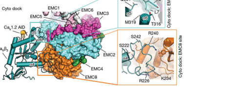
A major advance was the client-bound cryo-EM structure of an EMC chaperone complex with a high-voltage activated calcium channel assembly intermediate. In this structure, EMC8 contributes directly to a cytosolic docking interface (βCyto dockβ) with CaVΞ²3, demonstrating a specific assembly/chaperone role beyond generic insertase concepts. (Chen et al., May 2023; URL https://doi.org/10.1038/s41586-023-06175-5) (chen2023emcchaperoneβcavstructure pages 3-4)
Key quantitative structural findings include:
- The Cyto dock interface is ~1,500 Γ
Β² total. (chen2023emcchaperoneβcavstructure pages 3-4)
- The EMC8-centered subsite (βEMC8 siteβ) is 962 Γ
Β², centered on the last helix of EMC8. (chen2023emcchaperoneβcavstructure pages 3-4)
- Two CaVΞ² loops bind the EMC8 site: Ξ²7βΞ±4 (Thr218βAla243) and Ξ²8βΞ²9 (Pro277βLys282), forming hydrogen-bond and salt-bridge networks involving seven conserved CaVΞ² residues. (chen2023emcchaperoneβcavstructure pages 3-4)
- A CaVΞ² region (Lys225βSer245) that is disordered in isolated CaVΞ² becomes ordered upon EMC8 binding, implying EMC8-mediated stabilization of an assembly intermediate. (chen2023emcchaperoneβcavstructure pages 3-4)
Visual evidence: The retrieved figure panels illustrate the Cyto dock and EMC8-site interaction geometry and contact details. (chen2023emcchaperoneβcavstructure media 6624cdd7, chen2023emcchaperoneβcavstructure media f7eece75, chen2023emcchaperoneβcavstructure media b43f9754, chen2023emcchaperoneβcavstructure media 579bfd7d)
A 2024 cryo-EM study reported apo and VDAC1-bound human EMC structures at 3.47 Γ and 3.32 Γ , respectively, and described a specific interaction with VDAC proteins at mitochondriaβER contact sites. The study proposed that a βgating plugβ inside the EMC hydrophilic vestibule changes conformation upon VDAC binding and may regulate EMC functional modes (including an insertase vs alternative state). The provided evidence does not resolve an EMC8-specific mechanistic assignment within this interaction, but it adds to 2023β2024 views that EMC has multiple regulated functional states. (Li et al., Mar 2024; URL https://doi.org/10.18632/aging.205660) (OpenTargets Search: -EMC8)
Authoritative review synthesis emphasizes that EMC supports insertion/biogenesis of multi-pass membrane proteins and that validated EMC-dependent client lists remain relatively limited compared with the breadth of reported phenotypes; this is consistent with EMC acting at a fundamental and failure-prone step in membrane protein biogenesis, where disruption can yield broad downstream effects. (Hegde 2022; URL https://doi.org/10.1146/annurev-biochem-032620-104553) (hegde2022thefunctionstructure pages 19-20)
Within the retrieved evidence set, direct, mechanistically validated human Mendelian disease links for EMC8 were not available. However, Open Targets reports low-scoring disease associations for EMC8 (ENSG00000131148), including exfoliation syndrome, jaw disease, diverticular disease, retinitis pigmentosa with erythrocytic microcytosis, and familial ocular anterior segment mesenchymal dysgenesis, each supported by a small number of evidence entries in the platform output (without literature identifiers in the retrieved snippet). (Open Targets; retrieved context) (OpenTargets Search: -EMC8)
Interpretation: These Open Targets associations should be treated as hypothesis-generating rather than definitive, because the retrieved evidence does not include study-level details (e.g., variant pathogenicity, functional validation). (OpenTargets Search: -EMC8)
Primary supported role: EMC8 is a cytosolic structural and client-interaction subunit of the ER membrane protein complex. Evidence supports EMC8 as part of the cytosolic cap/vestibule essential for EMC assembly and for the EMCβs ability to engage substrates during membrane protein insertion and assembly. (pleiner2020structuralbasisfor pages 1-3, odonnell2020thearchitectureof pages 1-2)
Most direct EMC8-specific mechanistic evidence (2023β2024): EMC8 provides a defined client-binding surface for CaVΞ²3 in an EMCβCaV assembly intermediate, consistent with a chaperone/assembly function in addition to insertase activity attributed to the overall complex. (chen2023emcchaperoneβcavstructure pages 3-4, chen2023emcchaperoneβcavstructure media 6624cdd7, chen2023emcchaperoneβcavstructure media f7eece75, chen2023emcchaperoneβcavstructure media b43f9754, chen2023emcchaperoneβcavstructure media 579bfd7d)
Evidence gaps (transparent limitations): Direct EMC8-specific human disease mechanisms and a curated list of EMC8-specific clients (distinct from general EMC dependence) remain limited in the retrieved literature set; Open Targets provides low-score associations without study-level detail in the snippet. (OpenTargets Search: -EMC8, hegde2022thefunctionstructure pages 19-20)
| Claim/insight about EMC8 | Evidence type | Key quantitative details | System/clients studied | Citation |
|---|---|---|---|---|
| EMC8 is a cytosolic subunit of the human ER membrane protein complex and is functionally redundant with EMC9 in mammals. | Cryo-EM/biochemical architecture | EMC8 is ~23 kDa and ~44% identical to EMC9; no free EMC subunits detected, indicating stable complex assembly. | Human EMC; general membrane protein biogenesis | O'Donnell et al., 2020, eLife, https://doi.org/10.7554/eLife.57887 (odonnell2020thearchitectureof pages 2-4, odonnell2020thearchitectureof pages 1-2) |
| EMC8 helps form the cytosolic vestibule/cap that engages substrate transmembrane domains during EMC-mediated insertion. | Cryo-EM/biochemical mechanism | EMC has a βlarge, moderately hydrophobic vestibuleβ leading to a lumenally sealed, lipid-exposed intramembrane groove; insertase reaction described as energy independent. | Human EMC; tail-anchored client SQS and terminal TMD substrates | O'Donnell et al., 2020, eLife, https://doi.org/10.7554/eLife.57887 (odonnell2020thearchitectureof pages 2-4, odonnell2020thearchitectureof pages 1-2) |
| EMC8 is a structural assembly factor in the human EMC cytosolic basket, binding EMC2 and contributing to complex stability. | Cryo-EM/structure-guided mutagenesis | Human EMC resolved at 3.4 Γ ; EMC2 clamps around EMC8 via an extensive hydrophobic surface, and mutations at EMC8/EMC2/EMC3/EMC5 interfaces disrupted subunit binding in vitro. | Human EMC reconstituted in lipid nanodiscs | Pleiner et al., 2020, Science, https://doi.org/10.1126/science.abb5008 (pleiner2020structuralbasisfor pages 1-3) |
| In mammals, EMC8 is one of the soluble/cytosolic EMC components rather than a membrane-spanning subunit. | Quantitative proteomics/background functional study | Mammalian EMC described with 10 subunits; EMC2, EMC8, and EMC9 are cytosolic; Tian et al. defined 36 EMC-dependent versus 171 EMC-independent membrane proteins. | Mammalian EMC; broad membrane protein client sets | Tian et al., 2019, Cell Reports, https://doi.org/10.1016/j.celrep.2019.08.006 (tian2019proteomicanalysisidentifies pages 1-3) |
| EMC dependency is linked to difficult transmembrane segments containing polar/charged residues, supporting an EMC8-containing cytosolic recognition/chaperoning role in the intact complex. | Proteomics/mutagenesis | 36 EMC-dependent and 171 EMC-independent membrane proteins identified; TMD mutagenesis could convert EMC dependency. | Diverse membrane proteins; disease-relevant examples include CFTR and connexin-32 at EMC-complex level | Tian et al., 2019, Cell Reports, https://doi.org/10.1016/j.celrep.2019.08.006 (tian2019proteomicanalysisidentifies pages 1-3) |
| Reviews place EMC8 within the cytosolic subcomplex of EMC and interpret EMC as both an insertase and membrane-protein biogenesis factor/chaperone. | Authoritative review | EMC is considered a nine-protein complex in many species with EMC8/EMC9 as mutually exclusive paralogs; validated EMC-dependent proteins remain limited to roughly ~a dozen in the literature summarized. | EMC broadly; multipass membrane proteins and cotranslational biogenesis | Hegde, 2022, Annual Review of Biochemistry, https://doi.org/10.1146/annurev-biochem-032620-104553 (hegde2022thefunctionstructure pages 19-20, hegde2022thefunctionstructure pages 4-6, hegde2022thefunctionstructure pages 2-4) |
| Structure-function mapping places EMC8 near the cytoplasmic cap adjacent to EMC3, a region that influences client-specific biogenesis. | Cryo-EM/functional mutagenesis | Mutating the cytoplasmic cap near EMC8/9 increased SQS levels; distinct EMC regions separately affected tail-anchored, N-terminal, and polytopic clients. | SQS, B1AR, TMEM97 | Miller-Vedam et al., 2020, eLife preprint record, https://doi.org/10.1101/2020.09.02.280008 (millervedam2020structuralandmechanistic pages 25-28) |
| 2023 cryo-EM shows EMC8 directly contacts CaVΞ²3 in a client-bound EMC assembly intermediate, demonstrating a specific EMC8-dependent cytosolic docking role. | Cryo-EM/client-bound structure | Cyto dock area ~1,500 Γ ^2; EMC8-centered subsite 962 Γ ^2; adjacent EMC2 site ~550 Γ ^2; overall maps at 3.4 Γ /3.3 Γ with local 2.4β2.0 Γ . | Human EMCβCaV1.2(ΞC)βCaVΞ²3 complex | Chen et al., 2023, Nature, https://doi.org/10.1038/s41586-023-06175-5 (chen2023emcchaperoneβcavstructure pages 3-4, chen2023emcchaperoneβcavstructure media 6624cdd7, chen2023emcchaperoneβcavstructure media f7eece75, chen2023emcchaperoneβcavstructure media b43f9754, chen2023emcchaperoneβcavstructure media 579bfd7d) |
| EMC8 binding stabilizes otherwise disordered regions of CaVΞ²3, supporting a chaperone/assembly role beyond simple TMD insertion. | Cryo-EM/client interaction analysis | Two CaVΞ² loops (Ξ²7βΞ±4, Thr218βAla243; Ξ²8βΞ²9, Pro277βLys282) bind the EMC8 site; a CaVΞ² region Lys225βSer245 becomes ordered on EMC8 binding; seven conserved CaVΞ² residues participate in H-bond/salt-bridge networks. | Human CaV1.2/CaVΞ²3 assembly intermediate | Chen et al., 2023, Nature, https://doi.org/10.1038/s41586-023-06175-5 (chen2023emcchaperoneβcavstructure pages 3-4, chen2023emcchaperoneβcavstructure media 6624cdd7, chen2023emcchaperoneβcavstructure media f7eece75, chen2023emcchaperoneβcavstructure media b43f9754, chen2023emcchaperoneβcavstructure media 579bfd7d) |
| A 2024 structural study supports multifunctionality of human EMC and identifies a VDAC-bound state at mitochondriaβER contact sites, but the provided snippet does not assign a specific direct mechanistic role to EMC8. | Cryo-EM/structural study | Apo and VDAC1-bound human EMC structures at 3.47 Γ and 3.32 Γ ; VDAC interaction observed at mitochondria-ER contact sites; gating-plug conformational change proposed to regulate function. | Human EMC; VDAC1 | Li et al., 2024, Aging (Albany NY), https://doi.org/10.18632/aging.205660 |
| Disease links for EMC8 specifically are currently weak/indirect in the provided evidence, with Open Targets listing only low-score associations and no literature details in the retrieved snippet. | Disease-target database association | Example association scores: jaw disease 0.07599; diverticular disease 0.03701; retinitis pigmentosa and erythrocytic microcytosis 0.03655; familial ocular anterior segment mesenchymal dysgenesis 0.03620; exfoliation syndrome 0.03484. | Human EMC8 disease associations | Open Targets platform result for EMC8 (context output) (OpenTargets Search: -EMC8) |
Table: This table compiles the main structural, mechanistic, proteomic, and disease-association evidence for human EMC8 from the provided sources. It highlights where EMC8 is best supported as a cytosolic structural/chaperone component of the ER membrane protein complex and where evidence remains indirect or limited.
References
(tian2019proteomicanalysisidentifies pages 1-3): Songhai Tian, Quan Wu, Bo Zhou, Mei Yuk Choi, Bo Ding, Wei Yang, and Min Dong. Proteomic analysis identifies membrane proteins dependent on the er membrane protein complex. Cell reports, 28:2517-2526.e5, Sep 2019. URL: https://doi.org/10.1016/j.celrep.2019.08.006, doi:10.1016/j.celrep.2019.08.006. This article has 79 citations and is from a highest quality peer-reviewed journal.
(odonnell2020thearchitectureof pages 2-4): John P O'Donnell, Ben P Phillips, Yuichi Yagita, Szymon Juszkiewicz, Armin Wagner, Duccio Malinverni, Robert J Keenan, Elizabeth A Miller, and Ramanujan S Hegde. The architecture of emc reveals a path for membrane protein insertion. May 2020. URL: https://doi.org/10.7554/elife.57887, doi:10.7554/elife.57887. This article has 121 citations and is from a domain leading peer-reviewed journal.
(hegde2022thefunctionstructure pages 2-4): Ramanujan S. Hegde. The function, structure, and origins of the er membrane protein complex. Annual Review of Biochemistry, 91:651-678, Jun 2022. URL: https://doi.org/10.1146/annurev-biochem-032620-104553, doi:10.1146/annurev-biochem-032620-104553. This article has 65 citations and is from a domain leading peer-reviewed journal.
(hegde2022thefunctionstructure pages 4-6): Ramanujan S. Hegde. The function, structure, and origins of the er membrane protein complex. Annual Review of Biochemistry, 91:651-678, Jun 2022. URL: https://doi.org/10.1146/annurev-biochem-032620-104553, doi:10.1146/annurev-biochem-032620-104553. This article has 65 citations and is from a domain leading peer-reviewed journal.
(odonnell2020thearchitectureof pages 1-2): John P O'Donnell, Ben P Phillips, Yuichi Yagita, Szymon Juszkiewicz, Armin Wagner, Duccio Malinverni, Robert J Keenan, Elizabeth A Miller, and Ramanujan S Hegde. The architecture of emc reveals a path for membrane protein insertion. May 2020. URL: https://doi.org/10.7554/elife.57887, doi:10.7554/elife.57887. This article has 121 citations and is from a domain leading peer-reviewed journal.
(pleiner2020structuralbasisfor pages 1-3): Tino Pleiner, Giovani Pinton Tomaleri, Kurt Januszyk, Alison J. Inglis, Masami Hazu, and Rebecca M. Voorhees. Structural basis for membrane insertion by the human er membrane protein complex. Jul 2020. URL: https://doi.org/10.1126/science.abb5008, doi:10.1126/science.abb5008. This article has 192 citations and is from a highest quality peer-reviewed journal.
(millervedam2020structuralandmechanistic pages 25-28): Lakshmi E. Miller-Vedam, Bastian BrΓ€uning, Katerina D. Popova, Nicole T. Schirle Oakdale, Jessica L. Bonnar, Jesuraj Rajan Prabu, Elizabeth A. Boydston, Natalia Sevillano, Matthew J. Shurtleff, Robert M. Stroud, Charles S. Craik, Brenda A. Schulman, Adam Frost, and Jonathan S. Weissman. Structural and mechanistic basis of the emc-dependent biogenesis of distinct transmembrane clients. eLife, Sep 2020. URL: https://doi.org/10.1101/2020.09.02.280008, doi:10.1101/2020.09.02.280008. This article has 102 citations and is from a domain leading peer-reviewed journal.
(chen2023emcchaperoneβcavstructure pages 3-4): Zhou Chen, Abhisek Mondal, Fayal Abderemane-Ali, Seil Jang, Sangeeta Niranjan, JosΓ© L. MontaΓ±o, Balyn W. Zaro, and Daniel L. Minor. Emc chaperoneβcav structure reveals an ion channel assembly intermediate. Nature, 619:410-419, May 2023. URL: https://doi.org/10.1038/s41586-023-06175-5, doi:10.1038/s41586-023-06175-5. This article has 77 citations and is from a highest quality peer-reviewed journal.
(chen2023emcchaperoneβcavstructure media 6624cdd7): Zhou Chen, Abhisek Mondal, Fayal Abderemane-Ali, Seil Jang, Sangeeta Niranjan, JosΓ© L. MontaΓ±o, Balyn W. Zaro, and Daniel L. Minor. Emc chaperoneβcav structure reveals an ion channel assembly intermediate. Nature, 619:410-419, May 2023. URL: https://doi.org/10.1038/s41586-023-06175-5, doi:10.1038/s41586-023-06175-5. This article has 77 citations and is from a highest quality peer-reviewed journal.
(chen2023emcchaperoneβcavstructure media f7eece75): Zhou Chen, Abhisek Mondal, Fayal Abderemane-Ali, Seil Jang, Sangeeta Niranjan, JosΓ© L. MontaΓ±o, Balyn W. Zaro, and Daniel L. Minor. Emc chaperoneβcav structure reveals an ion channel assembly intermediate. Nature, 619:410-419, May 2023. URL: https://doi.org/10.1038/s41586-023-06175-5, doi:10.1038/s41586-023-06175-5. This article has 77 citations and is from a highest quality peer-reviewed journal.
(chen2023emcchaperoneβcavstructure media b43f9754): Zhou Chen, Abhisek Mondal, Fayal Abderemane-Ali, Seil Jang, Sangeeta Niranjan, JosΓ© L. MontaΓ±o, Balyn W. Zaro, and Daniel L. Minor. Emc chaperoneβcav structure reveals an ion channel assembly intermediate. Nature, 619:410-419, May 2023. URL: https://doi.org/10.1038/s41586-023-06175-5, doi:10.1038/s41586-023-06175-5. This article has 77 citations and is from a highest quality peer-reviewed journal.
(chen2023emcchaperoneβcavstructure media 579bfd7d): Zhou Chen, Abhisek Mondal, Fayal Abderemane-Ali, Seil Jang, Sangeeta Niranjan, JosΓ© L. MontaΓ±o, Balyn W. Zaro, and Daniel L. Minor. Emc chaperoneβcav structure reveals an ion channel assembly intermediate. Nature, 619:410-419, May 2023. URL: https://doi.org/10.1038/s41586-023-06175-5, doi:10.1038/s41586-023-06175-5. This article has 77 citations and is from a highest quality peer-reviewed journal.
(OpenTargets Search: -EMC8): Open Targets Query (-EMC8, 5 results). Buniello, A. et al. (2025). Open Targets Platform: facilitating therapeutic hypotheses building in drug discovery. Nucleic Acids Research.
(hegde2022thefunctionstructure pages 19-20): Ramanujan S. Hegde. The function, structure, and origins of the er membrane protein complex. Annual Review of Biochemistry, 91:651-678, Jun 2022. URL: https://doi.org/10.1146/annurev-biochem-032620-104553, doi:10.1146/annurev-biochem-032620-104553. This article has 65 citations and is from a domain leading peer-reviewed journal.
contributes_to qualifier is exactly for a subunit contributing to a complex MF; full text of the IMP papers (knockdown of EMC subunits) was read by curators.GO:0005515 protein binding IPI annotations have WITH/FROM = UniProtKB:Q15006 (EMC2) and come from high-throughput interactome / structural mapping papers (PMID:16189514 Rual Y2H map; 22119785 ERAD network; 26496610 BioPlex stoichiometry; 28514442 interactome communities; 30021884 histone XL-MS; 32296183 HuRI binary; 32439656 EMC structure; 33961781 BioPlex 3; 35271311 OpenCell). EMC2 is the EMC scaffold that EMC8 docks onto, so the interaction is real and biologically meaningful (it is how EMC8 joins the complex), but bare "protein binding" is uninformative -> KEEP_AS_NON_CORE per guidelines. The UniProt INTERACTION block confirms the EMC2 partner [file:human/EMC8/EMC8-uniprot.txt "O43402; Q15006: EMC2; NbExp=17"].GO:0016020 membrane (HDA, PMID:19946888 NK-cell membrane proteome) is a generic parent, correct but low-information [file:human/EMC8/EMC8-uniprot.txt "SUBCELLULAR LOCATION: Endoplasmic reticulum membrane"].contributes_to membrane insertase activity (GO:0032977) capturing its participation in the complex MF.protein binding GO:0005515 IPI (bare term, but real EMC2 interaction); membrane HDA (generic parent).ER proteostasis|Protein transport|Transmembrane protein import|EMC complex component ; PN-node mapping: type β GO:0072546 (EMC complex); group β GO:0044743 (protein transmembrane import into intracellular organelle); class β GO:0015031 (protein transport); branch=no_mapping.This file is generated from the current PROTEOSTASIS phase-1 dossier and local gene-review artifacts. Edit the source review, PN mapping, or dossier rather than this generated note when correcting the underlying curation.
id: O43402
gene_symbol: EMC8
product_type: PROTEIN
status: COMPLETE
taxon:
id: NCBITaxon:9606
label: Homo sapiens
description: 'EMC8 (ER membrane protein complex subunit 8; also known as C16orf2/C16orf4, COX4NB/NOC4 "Neighbor of COX4", FAM158B) is a small (210 aa) cytosolic, peripherally membrane-associated subunit of the endoplasmic reticulum membrane protein complex (EMC). The EMC is a conserved, nine-subunit ER transmembrane-domain insertase and membrane-protein chaperone that mediates energy-independent insertion of newly synthesized membrane proteins into the ER membrane, with a preference for transmembrane domains that are weakly hydrophobic or carry destabilizing charged/aromatic residues. The complex acts both co-translationally on multipass membrane proteins (where stop-transfer/membrane-anchor sequences become spanning helices, controlling N-exo topology of substrates such as G protein-coupled receptors) and post-translationally on tail-anchored proteins. Within EMC, EMC8 lies on the cytoplasmic face of the complex and is a non-catalytic accessory subunit; it contains an MPN domain (a JAMM/MPN-related fold) but lacks the catalytic metalloprotease residues, and the substrate-insertion vestibule is provided by the transmembrane subunits EMC3/EMC6 rather than by EMC8. EMC8 and its paralog EMC9 are mutually exclusive subunits that occupy the same position, defining alternative EMC variants. EMC8 docks onto the EMC2 scaffold subunit, its principal and well-documented protein interaction. EMC8 is broadly expressed across human tissues.'
alternative_products:
- name: '1'
id: O43402-1
- name: '2'
id: O43402-2
sequence_note: VSP_045089
existing_annotations:
- term:
id: GO:0032977
label: membrane insertase activity
evidence_type: IBA
original_reference_id: GO_REF:0000033
qualifier: contributes_to
review:
summary: Phylogenetic (IBA) assignment of membrane insertase activity with the contributes_to qualifier, reflecting EMC8 participation in the EMC insertase complex rather than an intrinsic enzymatic activity of EMC8 itself.
action: KEEP_AS_NON_CORE
reason: Correct use of contributes_to for a non-catalytic subunit of the EMC insertase. EMC8 is a cytosolic accessory subunit and is not the catalytic insertase (the insertion vestibule is formed by EMC3/EMC6), so this complex-level MF is retained but not as the standalone core function. The core descriptor for EMC8 is EMC complex membership plus ER membrane localization.
supported_by:
- reference_id: file:human/EMC8/EMC8-uniprot.txt
supporting_text: enables the energy-independent insertion into endoplasmic
- term:
id: GO:0045050
label: protein insertion into ER membrane by stop-transfer membrane-anchor sequence
evidence_type: IBA
original_reference_id: GO_REF:0000033
qualifier: involved_in
review:
summary: Phylogenetic assignment of the EMC role in cotranslational insertion of multipass membrane proteins via stop-transfer/membrane-anchor sequences; a process EMC8 participates in as part of the complex.
action: ACCEPT
reason: Consistent with the experimentally supported EMC function; the complex mediates cotranslational insertion of multipass proteins in which stop-transfer anchors become membrane-spanning helices. This is a complex-level process EMC8 is involved in.
supported_by:
- reference_id: file:human/EMC8/EMC8-uniprot.txt
supporting_text: cotranslational insertion of multi-pass membrane proteins in which
- term:
id: GO:0071816
label: tail-anchored membrane protein insertion into ER membrane
evidence_type: IBA
original_reference_id: GO_REF:0000033
qualifier: involved_in
review:
summary: Phylogenetic assignment of the EMC role in post-translational insertion of tail-anchored proteins; EMC8 participates as part of the complex.
action: ACCEPT
reason: Consistent with experimental evidence that EMC is required for post-translational insertion of tail-anchored proteins into the ER membrane.
supported_by:
- reference_id: file:human/EMC8/EMC8-uniprot.txt
supporting_text: post-translational insertion of tail-anchored/TA proteins in
- term:
id: GO:0072546
label: EMC complex
evidence_type: IBA
original_reference_id: GO_REF:0000033
qualifier: part_of
review:
summary: Phylogenetic assignment of EMC complex membership, the defining and core cellular-component association for EMC8.
action: ACCEPT
reason: Core localization/membership for EMC8; directly demonstrated experimentally (IDA PMID:22119785; IPI PMID:32439656) and conserved across the EMC8/EMC9 family.
supported_by:
- reference_id: file:human/EMC8/EMC8-uniprot.txt
supporting_text: Component of the ER membrane protein complex (EMC)
- term:
id: GO:0005789
label: endoplasmic reticulum membrane
evidence_type: IEA
original_reference_id: GO_REF:0000044
qualifier: located_in
review:
summary: Electronic transfer of the ER membrane localization from the UniProt subcellular location, consistent with EMC8 being a peripheral, cytoplasmic-side subunit of the ER-resident EMC.
action: ACCEPT
reason: Correct core compartment; EMC8 is a peripheral membrane protein at the ER membrane. Redundant with EXP/NAS ER membrane annotations.
supported_by:
- reference_id: file:human/EMC8/EMC8-uniprot.txt
supporting_text: 'SUBCELLULAR LOCATION: Endoplasmic reticulum membrane'
- term:
id: GO:0072546
label: EMC complex
evidence_type: IEA
original_reference_id: GO_REF:0000002
qualifier: part_of
review:
summary: InterPro-based (IPR005366, EMC8/9 family) electronic assignment to the EMC complex, consistent with the experimental membership annotation.
action: ACCEPT
reason: Correct core membership; redundant with IDA/IPI/IBA evidence for EMC complex membership.
supported_by:
- reference_id: file:human/EMC8/EMC8-uniprot.txt
supporting_text: Component of the ER membrane protein complex (EMC)
- term:
id: GO:0005515
label: protein binding
evidence_type: IPI
original_reference_id: PMID:16189514
qualifier: enables
review:
summary: High-throughput yeast two-hybrid human interactome capturing the EMC8-EMC2 interaction (WITH/FROM UniProtKB:Q15006). Bare protein binding is uninformative, though the EMC2 partner is the scaffold EMC8 docks onto within the complex.
action: KEEP_AS_NON_CORE
reason: Records a real interaction with EMC2 (the EMC scaffold) but bare protein binding does not convey function; per guidelines not elevated to core. The functional content is captured by EMC complex membership.
supported_by:
- reference_id: file:human/EMC8/EMC8-uniprot.txt
supporting_text: 'O43402; Q15006: EMC2; NbExp=17'
- term:
id: GO:0005515
label: protein binding
evidence_type: IPI
original_reference_id: PMID:22119785
qualifier: enables
review:
summary: EMC8-EMC2 interaction (WITH/FROM UniProtKB:Q15006) from the integrative ERAD/EMC mapping study that originally placed EMC8 (as COX4NB) in the mammalian EMC. Bare protein binding is uninformative.
action: KEEP_AS_NON_CORE
reason: Real, biologically meaningful interaction with the EMC2 scaffold, but the bare MF term is uninformative; the membership is better captured by GO:0072546.
supported_by:
- reference_id: file:human/EMC8/EMC8-uniprot.txt
supporting_text: 'O43402; Q15006: EMC2; NbExp=17'
- term:
id: GO:0005515
label: protein binding
evidence_type: IPI
original_reference_id: PMID:26496610
qualifier: enables
review:
summary: EMC8-EMC2 interaction (WITH/FROM UniProtKB:Q15006) from a quantitative stoichiometry-resolved interactome. Bare protein binding is uninformative.
action: KEEP_AS_NON_CORE
reason: Genuine EMC2 interaction from a high-throughput study, but bare protein binding is uninformative; intra-EMC contact captured by complex membership.
supported_by:
- reference_id: file:human/EMC8/EMC8-uniprot.txt
supporting_text: 'O43402; Q15006: EMC2; NbExp=17'
- term:
id: GO:0005515
label: protein binding
evidence_type: IPI
original_reference_id: PMID:28514442
qualifier: enables
review:
summary: EMC8-EMC2 interaction (WITH/FROM UniProtKB:Q15006) from the BioPlex interactome/community analysis. Bare protein binding is uninformative.
action: KEEP_AS_NON_CORE
reason: Real EMC2 interaction from a high-throughput AP-MS network, but bare protein binding is uninformative; not elevated to core.
supported_by:
- reference_id: file:human/EMC8/EMC8-uniprot.txt
supporting_text: 'O43402; Q15006: EMC2; NbExp=17'
- term:
id: GO:0005515
label: protein binding
evidence_type: IPI
original_reference_id: PMID:30021884
qualifier: enables
review:
summary: EMC8-EMC2 interaction (WITH/FROM UniProtKB:Q15006) entered into IntAct from a crosslinking-MS study. The paper title concerns histone interaction landscapes, an unexpected source, but the curated datapoint records an EMC8-EMC2 contact.
action: KEEP_AS_NON_CORE
reason: Curator-entered experimental IPI to the known EMC2 partner; per guidelines an experimental IPI is not removed merely because the abstract foregrounds a different topic. Bare protein binding remains uninformative, so kept as non-core.
supported_by:
- reference_id: file:human/EMC8/EMC8-uniprot.txt
supporting_text: 'O43402; Q15006: EMC2; NbExp=17'
- term:
id: GO:0005515
label: protein binding
evidence_type: IPI
original_reference_id: PMID:32296183
qualifier: enables
review:
summary: EMC8-EMC2 interaction (WITH/FROM UniProtKB:Q15006) from the HuRI reference binary interactome. Bare protein binding is uninformative.
action: KEEP_AS_NON_CORE
reason: Genuine binary EMC2 interaction, but bare protein binding is uninformative; complex membership captures the functional content.
supported_by:
- reference_id: file:human/EMC8/EMC8-uniprot.txt
supporting_text: 'O43402; Q15006: EMC2; NbExp=17'
- term:
id: GO:0005515
label: protein binding
evidence_type: IPI
original_reference_id: PMID:32439656
qualifier: enables
review:
summary: EMC8-EMC2 interaction (WITH/FROM UniProtKB:Q15006) consistent with the cryo-EM structure of the human EMC, which resolves EMC8 contacting the EMC2 scaffold. Bare protein binding is uninformative.
action: KEEP_AS_NON_CORE
reason: Structurally corroborated EMC2 interaction, but bare protein binding is uninformative; the structural membership is captured by GO:0072546 EMC complex.
supported_by:
- reference_id: file:human/EMC8/EMC8-uniprot.txt
supporting_text: 'O43402; Q15006: EMC2; NbExp=17'
- term:
id: GO:0005515
label: protein binding
evidence_type: IPI
original_reference_id: PMID:33961781
qualifier: enables
review:
summary: EMC8-EMC2 interaction (WITH/FROM UniProtKB:Q15006) from BioPlex 3 dual proteome-scale networks. Bare protein binding is uninformative.
action: KEEP_AS_NON_CORE
reason: Real EMC2 interaction from a large AP-MS network, but bare protein binding is uninformative; not elevated to core.
supported_by:
- reference_id: file:human/EMC8/EMC8-uniprot.txt
supporting_text: 'O43402; Q15006: EMC2; NbExp=17'
- term:
id: GO:0005515
label: protein binding
evidence_type: IPI
original_reference_id: PMID:35271311
qualifier: enables
review:
summary: EMC8-EMC2 interaction (WITH/FROM UniProtKB:Q15006) from OpenCell endogenous-tagging interactome/localization. Bare protein binding is uninformative.
action: KEEP_AS_NON_CORE
reason: Genuine endogenous EMC2 interaction, but bare protein binding is uninformative; the functional content is captured by EMC complex membership.
supported_by:
- reference_id: file:human/EMC8/EMC8-uniprot.txt
supporting_text: 'O43402; Q15006: EMC2; NbExp=17'
- term:
id: GO:0005783
label: endoplasmic reticulum
evidence_type: IDA
original_reference_id: GO_REF:0000052
qualifier: located_in
review:
summary: Direct immunofluorescence (HPA) evidence for ER localization, consistent with EMC8 being a subunit of the ER-resident EMC.
action: ACCEPT
reason: Correct compartment; the more specific ER membrane (GO:0005789) better captures the peripheral, cytoplasmic-side association of EMC8.
supported_by:
- reference_id: file:human/EMC8/EMC8-uniprot.txt
supporting_text: 'SUBCELLULAR LOCATION: Endoplasmic reticulum membrane'
- term:
id: GO:0005789
label: endoplasmic reticulum membrane
evidence_type: NAS
original_reference_id: PMID:29242231
qualifier: located_in
review:
summary: ComplexPortal (NAS) assertion of EMC8 ER membrane localization, drawn from the EMC insertase characterization. Core compartment for EMC8.
action: ACCEPT
reason: Correct core compartment; EMC8 is a peripheral membrane protein on the cytoplasmic side of the ER membrane. Consistent with EXP/IEA ER membrane annotations.
supported_by:
- reference_id: file:human/EMC8/EMC8-uniprot.txt
supporting_text: 'SUBCELLULAR LOCATION: Endoplasmic reticulum membrane'
- term:
id: GO:0045050
label: protein insertion into ER membrane by stop-transfer membrane-anchor sequence
evidence_type: IDA
original_reference_id: PMID:29242231
qualifier: involved_in
review:
summary: ComplexPortal IDA for the EMC role in cotranslational insertion of multipass proteins via stop-transfer anchors; EMC8 participates as part of the complex.
action: ACCEPT
reason: Consistent with experimentally demonstrated EMC insertase function; a complex-level process EMC8 is involved in.
supported_by:
- reference_id: file:human/EMC8/EMC8-uniprot.txt
supporting_text: cotranslational insertion of multi-pass membrane proteins in which
- term:
id: GO:0071816
label: tail-anchored membrane protein insertion into ER membrane
evidence_type: IDA
original_reference_id: PMID:29242231
qualifier: involved_in
review:
summary: ComplexPortal IDA for the EMC role in post-translational insertion of tail-anchored proteins, the function for which Guna et al. demonstrated EMC is necessary and sufficient. EMC8 participates as part of the complex.
action: ACCEPT
reason: Directly supported by the reconstitution evidence that EMC inserts tail-anchored substrates; a complex-level process EMC8 is involved in.
supported_by:
- reference_id: file:human/EMC8/EMC8-uniprot.txt
supporting_text: post-translational insertion of tail-anchored/TA proteins in
- term:
id: GO:0072546
label: EMC complex
evidence_type: IPI
original_reference_id: PMID:32439656
qualifier: part_of
review:
summary: ComplexPortal IPI placing EMC8 in the EMC, corroborated by the cryo-EM structure of the human nine-subunit complex. Core membership.
action: ACCEPT
reason: Core, structurally demonstrated EMC complex membership; EMC8 (chain H) is resolved in the human EMC structures.
supported_by:
- reference_id: file:human/EMC8/EMC8-uniprot.txt
supporting_text: ComplexPortal; CPX-5848; Endoplasmic reticulum membrane complex, EMC8 variant.
- term:
id: GO:0005789
label: endoplasmic reticulum membrane
evidence_type: EXP
original_reference_id: PMID:22119785
qualifier: located_in
review:
summary: Experimental localization of EMC8 (identified as COX4NB) to the ER membrane in the integrative ERAD/EMC mapping study. Core compartment.
action: ACCEPT
reason: Core compartment supported by direct experimental evidence; EMC8 associates peripherally with the ER membrane as part of EMC.
supported_by:
- reference_id: PMID:22119785
supporting_text: we identified 5 additional HCIPs (TTC35, TMEM32/MMGT1, TMEM85, C15orf24 and COX4NB)
- term:
id: GO:0032977
label: membrane insertase activity
evidence_type: IMP
original_reference_id: PMID:29809151
qualifier: contributes_to
review:
summary: IMP (loss-of-function) evidence that EMC subunits contribute to the membrane insertase activity of the complex, assigned to EMC8 with the contributes_to qualifier. The full text (subunit perturbation in cotranslational multipass biogenesis) was read by the curator.
action: KEEP_AS_NON_CORE
reason: Appropriate contributes_to assignment for a non-catalytic accessory subunit of the EMC insertase. EMC8 is not itself catalytic (the EMC3/EMC6 vestibule performs insertion), so this complex-level MF is retained but not as the standalone core function. Not removed, since contributes_to is the intended qualifier for subunit contributions to a complex activity.
supported_by:
- reference_id: file:human/EMC8/EMC8-uniprot.txt
supporting_text: enables the energy-independent insertion into endoplasmic
- term:
id: GO:0032977
label: membrane insertase activity
evidence_type: IMP
original_reference_id: PMID:30415835
qualifier: contributes_to
review:
summary: IMP evidence (EMC knockout/depletion in GPCR topogenesis) that the EMC has membrane insertase activity, assigned to EMC8 with contributes_to. Full text read by the curator.
action: KEEP_AS_NON_CORE
reason: Correct contributes_to assignment for a non-catalytic accessory subunit; the catalytic insertase activity is a property of the complex, not of EMC8 alone. Kept but not core for the individual subunit.
supported_by:
- reference_id: file:human/EMC8/EMC8-uniprot.txt
supporting_text: the topology of multi-pass
- term:
id: GO:0045050
label: protein insertion into ER membrane by stop-transfer membrane-anchor sequence
evidence_type: IMP
original_reference_id: PMID:29809151
qualifier: involved_in
review:
summary: IMP evidence that EMC (including EMC8) is required for cotranslational insertion of multipass membrane proteins via stop-transfer anchors.
action: ACCEPT
reason: Consistent with the experimentally supported EMC function; loss of EMC subunits impairs cotranslational multipass insertion. A complex-level process EMC8 is involved in.
supported_by:
- reference_id: file:human/EMC8/EMC8-uniprot.txt
supporting_text: cotranslational insertion of multi-pass membrane proteins in which
- term:
id: GO:0045050
label: protein insertion into ER membrane by stop-transfer membrane-anchor sequence
evidence_type: IMP
original_reference_id: PMID:30415835
qualifier: involved_in
review:
summary: IMP evidence (EMC knockout impairs accurate GPCR/multipass topogenesis) supporting the EMC role in cotranslational insertion via stop-transfer anchors; EMC8 participates as part of the complex.
action: ACCEPT
reason: Consistent with experimental topogenesis evidence; EMC mediates cotranslational insertion that sets multipass topology. A complex-level process EMC8 is involved in.
supported_by:
- reference_id: file:human/EMC8/EMC8-uniprot.txt
supporting_text: the proper cotranslational insertion of N-terminal
- term:
id: GO:0016020
label: membrane
evidence_type: HDA
original_reference_id: PMID:19946888
qualifier: located_in
review:
summary: High-throughput membrane-proteome (NK-cell) MS dataset assigning generic membrane localization. Correct but a generic parent of the ER membrane localization.
action: KEEP_AS_NON_CORE
reason: Accurate but low-information; the specific ER membrane (GO:0005789) better captures the localization of EMC8.
supported_by:
- reference_id: file:human/EMC8/EMC8-uniprot.txt
supporting_text: 'SUBCELLULAR LOCATION: Endoplasmic reticulum membrane'
- term:
id: GO:0005737
label: cytoplasm
evidence_type: IDA
original_reference_id: PMID:22119785
qualifier: located_in
review:
summary: Direct evidence consistent with EMC8 being on the cytoplasmic side of the ER membrane; cytoplasm is a correct but less specific compartment than ER membrane.
action: KEEP_AS_NON_CORE
reason: Correct (EMC8 is a peripheral, cytoplasmic-side subunit) but generic; the ER membrane localization and EMC complex membership are the informative core terms.
supported_by:
- reference_id: file:human/EMC8/EMC8-uniprot.txt
supporting_text: Cytoplasmic side
- term:
id: GO:0072546
label: EMC complex
evidence_type: IDA
original_reference_id: PMID:22119785
qualifier: part_of
review:
summary: Direct experimental identification of EMC8 (as COX4NB) as a component of the mammalian EMC; the foundational evidence for the core complex membership of EMC8.
action: ACCEPT
reason: Core, directly demonstrated EMC complex membership; this is the defining cellular-component association for EMC8.
supported_by:
- reference_id: PMID:22119785
supporting_text: we identified 5 additional HCIPs (TTC35, TMEM32/MMGT1, TMEM85, C15orf24 and COX4NB)
references:
- id: GO_REF:0000002
title: Gene Ontology annotation through association of InterPro records with GO terms
findings: []
- id: GO_REF:0000033
title: Annotation inferences using phylogenetic trees
findings: []
- id: GO_REF:0000044
title: Gene Ontology annotation based on UniProtKB/Swiss-Prot Subcellular Location vocabulary mapping, accompanied by conservative changes to GO terms applied by UniProt
findings: []
- id: GO_REF:0000052
title: Gene Ontology annotation based on curation of immunofluorescence data
findings: []
- id: PMID:16189514
title: Towards a proteome-scale map of the human protein-protein interaction network.
findings: []
reference_review:
relevance: LOW
correctness: VERIFIED
review_notes: High-throughput Y2H interactome; source of an EMC8-EMC2 protein binding IPI. Not informative about the molecular function of EMC8 beyond complex membership.
- id: PMID:19946888
title: Defining the membrane proteome of NK cells.
findings: []
reference_review:
relevance: LOW
correctness: VERIFIED
review_notes: NK-cell membrane proteome MS dataset; source of the generic membrane (GO:0016020) HDA annotation. Abstract-only in cache.
- id: PMID:22119785
title: Defining human ERAD networks through an integrative mapping strategy.
findings:
- statement: Identified EMC8 (as COX4NB) as a high-confidence component of the mammalian ER membrane protein complex (mEMC), establishing EMC complex membership and ER membrane/cytoplasm localization.
reference_section_type: RESULTS
reference_review:
relevance: HIGH
correctness: VERIFIED
review_notes: Foundational integrative MS study that placed EMC8 (COX4NB) in the mammalian EMC; source of the IDA EMC-complex membership and cytoplasm/ER membrane annotations.
- id: PMID:26496610
title: A human interactome in three quantitative dimensions organized by stoichiometries and abundances.
findings: []
reference_review:
relevance: LOW
correctness: VERIFIED
review_notes: Quantitative interactome; source of an EMC8-EMC2 protein binding IPI. Background interaction data only.
- id: PMID:28514442
title: Architecture of the human interactome defines protein communities and disease networks.
findings: []
reference_review:
relevance: LOW
correctness: VERIFIED
review_notes: BioPlex interactome/community analysis; source of an EMC8-EMC2 protein binding IPI.
- id: PMID:29242231
title: The ER membrane protein complex is a transmembrane domain insertase.
findings:
- statement: The EMC is a transmembrane-domain insertase that catalyzes insertion of tail-anchored and other substrates into the ER membrane in a reconstituted system; EMC8 is a subunit of this complex.
reference_section_type: ABSTRACT
reference_review:
relevance: HIGH
correctness: VERIFIED
review_notes: Definitive demonstration that EMC is a TMD insertase; basis for the complex-level insertase MF and TA-insertion BP annotations EMC8 contributes to.
- id: PMID:29809151
title: The ER membrane protein complex interacts cotranslationally to enable biogenesis of multipass membrane proteins.
findings:
- statement: EMC engages ribosomes cotranslationally and is required for biogenesis of multipass membrane proteins; depletion of EMC subunits impairs insertion.
reference_section_type: ABSTRACT
reference_review:
relevance: HIGH
correctness: VERIFIED
review_notes: Source of IMP membrane insertase activity (contributes_to) and cotranslational multipass insertion BP annotations.
- id: PMID:30021884
title: Histone Interaction Landscapes Visualized by Crosslinking Mass Spectrometry in Intact Cell Nuclei.
findings: []
reference_review:
relevance: LOW
correctness: VERIFIED
review_notes: Crosslinking-MS study; an unexpected source for an ER-complex subunit, but curated in IntAct as an EMC8-EMC2 protein binding datapoint. Experimental IPI retained as non-core, not removed.
- id: PMID:30415835
title: EMC Is Required to Initiate Accurate Membrane Protein Topogenesis.
findings:
- statement: EMC mediates cotranslational insertion of the first TMD of GPCRs and other multipass proteins and cooperates with Sec61 to ensure accurate topogenesis; EMC knockout impairs correct topology.
reference_section_type: ABSTRACT
reference_review:
relevance: HIGH
correctness: VERIFIED
review_notes: Source of IMP membrane insertase activity (contributes_to) and stop-transfer multipass insertion BP annotations.
- id: PMID:32296183
title: A reference map of the human binary protein interactome.
findings: []
reference_review:
relevance: LOW
correctness: VERIFIED
review_notes: HuRI binary interactome; source of an EMC8-EMC2 protein binding IPI.
- id: PMID:32439656
title: Structural basis for membrane insertion by the human ER membrane protein complex.
findings:
- statement: Cryo-EM structure of the human nine-subunit EMC; substrate insertion occurs via an enclosed hydrophilic vestibule formed by EMC3 and EMC6, with EMC8 resolved as a cytosolic-facing subunit.
reference_section_type: ABSTRACT
reference_review:
relevance: HIGH
correctness: VERIFIED
review_notes: Structure of the human EMC (EMC8 variant); shows the catalytic vestibule is EMC3/EMC6, confirming EMC8 is a non-catalytic subunit. Abstract-only in cache.
- id: PMID:33961781
title: Dual proteome-scale networks reveal cell-specific remodeling of the human interactome.
findings: []
reference_review:
relevance: LOW
correctness: VERIFIED
review_notes: BioPlex 3 interactome; source of an EMC8-EMC2 protein binding IPI.
- id: PMID:35271311
title: 'OpenCell: Endogenous tagging for the cartography of human cellular organization.'
findings: []
reference_review:
relevance: LOW
correctness: VERIFIED
review_notes: OpenCell endogenous-tagging interactome/localization; source of an EMC8-EMC2 protein binding IPI consistent with EMC membership.
- id: PMID:37196677
title: 'EMC chaperone-Ca(V) structure reveals an ionΒ channel assembly intermediate.'
findings:
- statement: Cryo-EM of human EMC bound to a CaV1.2-CaVbeta3 assembly intermediate; EMC8 forms a specific cytosolic client-docking interface ("Cyto dock"/"EMC8 site", ~962 A^2 centered on the last helix of EMC8) with CaVbeta3, binding two CaVbeta loops and ordering an otherwise disordered CaVbeta region, demonstrating an EMC8-specific chaperone/assembly role beyond generic insertion.
reference_section_type: RESULTS
reference_review:
relevance: HIGH
correctness: VERIFIED
review_notes: PubMed-verified (Nature 2023, PMID:37196677). Provides direct EMC8-specific structural evidence for a client-binding (CaVbeta3) surface, establishing a chaperone/assembly function for EMC8 distinct from the EMC3/EMC6 insertase core.
- id: PMID:32459176
title: The architecture of EMC reveals a path for membrane protein insertion.
findings:
- statement: EMC2 forms an alpha-solenoid that clamps around EMC8 in the cytosolic basket; EMC8 is a ~23 kDa monomer ~44% identical to EMC9, and EMC8 and EMC9 are mutually exclusive subunits (no ternary EMC2-EMC8-EMC9 complex forms), supporting EMC8 as a structural assembly/scaffold subunit of the cytosolic module.
reference_section_type: RESULTS
reference_review:
relevance: HIGH
correctness: VERIFIED
review_notes: PubMed-verified (eLife 2020, PMID:32459176). Source of the EMC8-EMC2 clamp interaction and EMC8/EMC9 mutual exclusivity; supports EMC8's cytosolic structural/assembly role.
- id: PMID:35287476
title: The Function, Structure, and Origins of the ER Membrane Protein Complex.
findings:
- statement: Authoritative review of EMC function, structure, and evolution; places EMC2, EMC8, and EMC9 as the cytosolic subunits and EMC8/EMC9 as mutually exclusive paralogs of the cytosolic cradle that engages membrane-protein clients before insertion.
reference_section_type: LITERATURE_REVIEW
reference_review:
relevance: MEDIUM
correctness: VERIFIED
review_notes: PubMed-verified (Annu Rev Biochem 2022, PMID:35287476). Review supporting EMC8's cytosolic placement and EMC8/EMC9 paralog relationship already described in the review.
- id: PMID:38517390
title: Structural insights into human EMC and its interaction with VDAC.
findings:
- statement: Cryo-EM of human EMC apo and VDAC1-bound states at mitochondria-ER contact sites, with a hydrophilic-vestibule "gating plug" conformational change proposed to regulate EMC function; EMC8 is a cytosolic subunit of the complexes studied (no EMC8-specific mechanistic role assigned in this study).
reference_section_type: RESULTS
reference_review:
relevance: LOW
correctness: VERIFIED
review_notes: PubMed-verified (Aging (Albany NY) 2024, PMID:38517390). Structural study of EMC-VDAC1; does not assign an EMC8-specific role but provides context for alternative EMC functional states. Relevance to EMC8 specifically is low.
- id: file:human/EMC8/EMC8-uniprot.txt
title: UniProt entry O43402 (EMC8_HUMAN), ER membrane protein complex subunit 8
findings:
- statement: EMC8 is a cytosolic, peripheral subunit (cytoplasmic side) of the ER membrane protein complex (EMC), an energy-independent TMD insertase for newly synthesized membrane proteins; mediates cotranslational multipass and post-translational tail-anchored insertion. EMC8 and EMC9 are mutually exclusive subunits; EMC8 belongs to the EMC8/EMC9 family and contains an MPN domain.
reference_section_type: OTHER
core_functions:
- description: Cytosolic, non-catalytic accessory subunit of the ER membrane protein complex (EMC); its core role is to be a constitutive component of the EMC, an ER transmembrane-domain insertase/chaperone, docking onto the EMC2 scaffold on the cytoplasmic side of the ER membrane and providing a cytosolic client-docking surface (e.g., the EMC8 "Cyto dock" for CaVbeta3) during membrane-protein assembly.
supported_by:
- reference_id: file:human/EMC8/EMC8-uniprot.txt
supporting_text: Component of the ER membrane protein complex (EMC)
- reference_id: PMID:22119785
supporting_text: we identified 5 additional HCIPs (TTC35, TMEM32/MMGT1, TMEM85, C15orf24 and COX4NB)
in_complex:
id: GO:0072546
label: EMC complex
locations:
- id: GO:0005789
label: endoplasmic reticulum membrane
- description: As part of the EMC, contributes to membrane insertase activity, participating in energy-independent insertion of newly synthesized membrane proteins into the ER membrane (cotranslational multipass insertion via stop-transfer anchors and post-translational tail-anchored protein insertion). EMC8 is not itself catalytic; the insertion vestibule is provided by EMC3/EMC6.
contributes_to_molecular_function:
id: GO:0032977
label: membrane insertase activity
locations:
- id: GO:0005789
label: endoplasmic reticulum membrane
supported_by:
- reference_id: file:human/EMC8/EMC8-uniprot.txt
supporting_text: enables the energy-independent insertion into endoplasmic
directly_involved_in:
- id: GO:0071816
label: tail-anchored membrane protein insertion into ER membrane
- id: GO:0045050
label: protein insertion into ER membrane by stop-transfer membrane-anchor sequence
proposed_new_terms: []
suggested_questions:
- question: What is the specific functional role of EMC8 (versus its mutually exclusive paralog EMC9) within the EMC, and do EMC8- and EMC9-containing complexes differ in substrate preference or regulation?
- question: Does the degenerate MPN domain of EMC8 retain any binding or regulatory function, or is it purely structural for incorporation into the complex?
suggested_experiments:
- description: Compare substrate-insertion profiles (e.g., ribosome profiling / proteomics of membrane-protein biogenesis) in EMC8-knockout versus EMC9-knockout versus EMC8/EMC9 double-knockout cells to define paralog-specific contributions to EMC function.
- description: Reconstitute EMC insertase activity in liposomes with and without EMC8 (and with EMC9 substituted) to test whether EMC8 is required for complex assembly, stability, or insertion efficiency of defined substrates.
- description: Use cryo-EM and crosslinking-MS to map the EMC8-EMC2 interface and determine how EMC8 incorporation affects the architecture of the cytosolic cap of the EMC.