P3H1

UniProt ID: Q32P28
Organism: Homo sapiens
Review Status: COMPLETE
πŸ“ Provide Detailed Feedback

Gene Description

P3H1 (prolyl 3-hydroxylase 1; gene LEPRE1, also known as leprecan-1 and growth suppressor 1) is an endoplasmic-reticulum-lumenal 2-oxoglutarate/Fe(II)-dependent dioxygenase (EC 1.14.11.7) of the leprecan family. It catalyzes the post-translational formation of 3-hydroxyproline at specific -Xaa-Pro-Gly- prolines in procollagen chains, most notably the alpha1(I)Pro986 residue of type I and type II collagen, a modification required for proper collagen triple-helix folding, assembly and stability. P3H1 is the catalytic core of the ER collagen prolyl 3-hydroxylation complex (the "PCP complex"), a 1:1:1 ternary assembly with cartilage-associated protein (CRTAP) and peptidyl-prolyl cis-trans isomerase B (PPIB/cyclophilin B). Within this complex P3H1 contributes prolyl 3-hydroxylase activity while PPIB provides cis-trans isomerase activity and CRTAP stabilizes the assembly and helps recruit collagen substrate; the complex also functions as a collagen chaperone. P3H1 carries a C-terminal KDEL ER-retrieval signal that retains it (and, with it, CRTAP) in the ER lumen. Catalysis requires a non-heme Fe(II) center, the co-substrate 2-oxoglutarate, molecular oxygen, and L-ascorbate (vitamin C) as cofactor. Loss-of-function mutations in LEPRE1 abolish alpha1(I)Pro986 3-hydroxylation, delay collagen folding and cause overmodification of the collagen helix, resulting in autosomal recessive osteogenesis imperfecta type 8 (OI8). A secreted chondroitin sulfate proteoglycan form of leprecan can also be deposited in the extracellular matrix.

Existing Annotations Review

GO Term Evidence Action Reason
GO:0005783 endoplasmic reticulum
IBA
GO_REF:0000033
ACCEPT
Summary: P3H1 acts in the ER lumen, where it 3-hydroxylates procollagen as part of the CRTAP/PPIB complex. The phylogenetic ER localization agrees with direct experimental evidence and the KDEL ER-retention signal.
Reason: Correct site of action; corroborated by EXP/IDA ER annotations and the KDEL retention motif retaining isoform 1 in the ER.
Supporting Evidence:
file:human/P3H1/P3H1-uniprot.txt
SUBCELLULAR LOCATION: [Isoform 1]: Endoplasmic reticulum
GO:0019797 procollagen-proline 3-dioxygenase activity
IBA
GO_REF:0000033
ACCEPT
Summary: Core molecular function of P3H1, conserved across the leprecan/P3H family; it hydroxylates the 3-position of specific procollagen prolines.
Reason: Defining core MF; directly demonstrated experimentally (IDA, PMID:39245686, EC 1.14.11.7) and conserved phylogenetically.
Supporting Evidence:
file:human/P3H1/P3H1-uniprot.txt
Has prolyl 3-hydroxylase activity and catalyzes the post-
GO:0032963 collagen metabolic process
IBA
GO_REF:0000033
ACCEPT
Summary: P3H1 participates in collagen post-translational modification/biosynthesis, a defining biological process for the gene.
Reason: Correct biological process; the enzyme is required for proper collagen biosynthesis, folding and assembly and is conserved across the family.
Supporting Evidence:
PMID:15044469
The collagen prolyl hydroxylases are enzymes that are required for proper
GO:0005506 iron ion binding
IEA
GO_REF:0000002
KEEP AS NON CORE
Summary: P3H1 coordinates a catalytic non-heme Fe(II)/Fe(3+) ion in its Fe2OG dioxygenase domain (residues His587, Asp589, His659), a structural requirement for hydroxylation.
Reason: Accurate cofactor-binding attribute that supports the catalytic MF; structurally demonstrated, but subsidiary to the informative procollagen-proline 3-dioxygenase activity.
Supporting Evidence:
file:human/P3H1/P3H1-uniprot.txt
Name=Fe cation; Xref=ChEBI:CHEBI:24875;
GO:0005783 endoplasmic reticulum
IEA
GO_REF:0000044
ACCEPT
Summary: Electronic transfer of ER localization from the UniProt subcellular location, consistent with stronger experimental evidence.
Reason: Correct compartment; redundant with EXP/IDA ER annotations.
Supporting Evidence:
file:human/P3H1/P3H1-uniprot.txt
SUBCELLULAR LOCATION: [Isoform 1]: Endoplasmic reticulum
GO:0016705 oxidoreductase activity, acting on paired donors, with incorporation or reduction of molecular oxygen
IEA
GO_REF:0000002
KEEP AS NON CORE
Summary: Parent molecular function describing the 2-oxoglutarate/Fe(II) dioxygenase chemistry that incorporates molecular oxygen during proline 3-hydroxylation.
Reason: Correct but generic; the specific GO:0019797 procollagen-proline 3-dioxygenase activity better captures the core function.
Supporting Evidence:
file:human/P3H1/P3H1-uniprot.txt
Reaction=L-prolyl-[collagen] + 2-oxoglutarate + O2 = trans-3-hydroxy-L-
GO:0019797 procollagen-proline 3-dioxygenase activity
IEA
GO_REF:0000120
ACCEPT
Summary: Electronic assignment of the core dioxygenase activity from the EC/Rhea mapping (EC 1.14.11.7; RHEA:22872), consistent with experimental evidence.
Reason: Correct core molecular function; redundant with IDA/IBA/ISS evidence.
Supporting Evidence:
file:human/P3H1/P3H1-uniprot.txt
Reaction=L-prolyl-[collagen] + 2-oxoglutarate + O2 = trans-3-hydroxy-L-
GO:0031418 L-ascorbic acid binding
IEA
GO_REF:0000002
KEEP AS NON CORE
Summary: P3H1, like other collagen hydroxylases, uses L-ascorbate (vitamin C) as a cofactor to maintain the catalytic iron in its reduced state.
Reason: Accurate cofactor-binding attribute supporting the catalytic MF; subsidiary to the core dioxygenase activity.
Supporting Evidence:
file:human/P3H1/P3H1-uniprot.txt
Name=L-ascorbate; Xref=ChEBI:CHEBI:38290;
GO:0032963 collagen metabolic process
IEA
GO_REF:0000002
ACCEPT
Summary: InterPro-based electronic assignment of the collagen metabolic process, consistent with the experimental and phylogenetic evidence.
Reason: Correct biological process; redundant with IBA/ISS.
Supporting Evidence:
file:human/P3H1/P3H1-uniprot.txt
in pro-collagen chains, a critical step for the formation of mature
GO:0005515 protein binding
IPI
PMID:30021884
Histone Interaction Landscapes Visualized by Crosslinking Ma...
KEEP AS NON CORE
Summary: High-throughput crosslinking mass-spectrometry interactome capturing P3H1 with CRTAP (O75718). CRTAP is a genuine PCP complex partner, but the bare protein binding term is uninformative.
Reason: Records a real interaction with the complex partner CRTAP, but bare protein binding is uninformative; complex membership is better captured by GO:0032991 and the core catalytic MF.
Supporting Evidence:
file:human/P3H1/P3H1-uniprot.txt
Q32P28; O75718: CRTAP;
GO:0005515 protein binding
IPI
PMID:33961781
Dual proteome-scale networks reveal cell-specific remodeling...
KEEP AS NON CORE
Summary: Proteome-scale (BioPlex) affinity-purification interactome capturing the P3H1-CRTAP (O75718) interaction. The partner is biologically real; the bare term is uninformative.
Reason: Real interaction with the complex partner CRTAP, but bare protein binding is uninformative and not elevated to core.
Supporting Evidence:
file:human/P3H1/P3H1-uniprot.txt
Q32P28; O75718: CRTAP;
GO:0005515 protein binding
IPI
PMID:39245686
The structural basis for the collagen processing by human P3...
KEEP AS NON CORE
Summary: Structural study establishing P3H1's direct interactions with CRTAP (O75718) and PPIB (P23284) within the PCP ternary complex. These are the genuine, biologically central complex partners, but the bare protein binding term is uninformative.
Reason: Partners CRTAP and PPIB are the real PCP complex components (demonstrated structurally), but bare protein binding does not convey the core function; captured better by GO:0032991 and GO:0019797.
Supporting Evidence:
PMID:39245686
P3H1 and PPIB were identified as components of an ER-associated ternary complex consisting of P3H1/CRTAP/PPIB
GO:0019797 procollagen-proline 3-dioxygenase activity
IDA
PMID:39245686
The structural basis for the collagen processing by human P3...
ACCEPT
Summary: Direct biochemical/structural demonstration that P3H1 is the core prolyl 3-hydroxylase of the PCP complex, hydroxylating Pro986 of collagen alpha1(I); active-site mutants (H587A, D589A, H659A, R669) lose catalytic activity.
Reason: Core molecular function with direct experimental (IDA) support, EC 1.14.11.7 assigned, and mutagenesis confirming the catalytic residues.
Supporting Evidence:
PMID:39245686
P3H1 is the core prolyl 3-hydroxylase, specially hydroxylating Pro986
GO:0005783 endoplasmic reticulum
EXP
PMID:19088120
Recessive osteogenesis imperfecta caused by LEPRE1 mutations...
ACCEPT
Summary: Experimental localization of the KDEL-bearing splice form (isoform 1) of P3H1 to the ER, the catalytically relevant compartment.
Reason: Experimentally supported ER localization consistent with the site of action.
Supporting Evidence:
file:human/P3H1/P3H1-uniprot.txt
SUBCELLULAR LOCATION: [Isoform 1]: Endoplasmic reticulum
GO:0050821 protein stabilization
IMP
PMID:19846465
Prolyl 3-hydroxylase 1 and CRTAP are mutually stabilizing in...
ACCEPT
Summary: P3H1 and CRTAP are mutually stabilizing within the ER prolyl 3-hydroxylation complex; loss of either leads to proteasomal degradation of the other. P3H1 thus stabilizes its partner CRTAP (and the complex stabilizes/chaperones collagen).
Reason: Directly supported by mutational analysis (null cells deplete both proteins; proteasome inhibitors partially rescue); reflects a genuine stabilization function of the complex.
Supporting Evidence:
PMID:19846465
CRTAP and P3H1 are mutually stabilized in the collagen prolyl 3-hydroxylation
GO:0050821 protein stabilization
IMP
PMID:22615817
A novel mutation in LEPRE1 that eliminates only the KDEL ER-...
ACCEPT
Summary: A KDEL-only mutation that prevents ER retention of P3H1 impairs its function, consistent with P3H1's role in maintaining the ER collagen-modifying complex and stabilizing its components/substrate.
Reason: Supported by patient mutation analysis; consistent with the genuine stabilization role of P3H1 within the ER collagen prolyl 3-hydroxylation complex.
Supporting Evidence:
PMID:22615817
the KDEL ER- retrieval sequence is essential for P3H1 functionality
GO:0031012 extracellular matrix
HDA
PMID:28327460
Comprehensive proteomic characterization of stem cell-derive...
KEEP AS NON CORE
Summary: High-throughput proteomic detection of P3H1 in stem-cell-derived extracellular matrix. Consistent with the secreted chondroitin-sulfate proteoglycan (leprecan) form, but not the catalytic ER site of action.
Reason: Plausible given the documented secreted ECM proteoglycan form, but a weak colocalizes_with proteomics annotation peripheral to the core ER enzymatic role.
Supporting Evidence:
file:human/P3H1/P3H1-uniprot.txt
Secreted, extracellular space, extracellular
GO:0005518 collagen binding
ISS
PMID:15044469
Prolyl 3-hydroxylase 1, enzyme characterization and identifi...
ACCEPT
Summary: P3H1 specifically interacts with denatured/unfolded collagen, contributing to the PCP complex's substrate-binding (collagen recruitment) function.
Reason: Supported by enzyme characterization (specific interaction with denatured collagen) and the structurally demonstrated collagen-binding of the complex; an informative MF.
Supporting Evidence:
PMID:15044469
specifically interact with denatured collagen
GO:0006457 protein folding
ISS
PMID:15044469
Prolyl 3-hydroxylase 1, enzyme characterization and identifi...
ACCEPT
Summary: P3H1 (within the PCP complex) is required for proper collagen folding and assembly, and the complex has molecular chaperone activity.
Reason: Supported by enzyme characterization and the complex's documented chaperone role; loss of P3H1 delays collagen helix folding.
Supporting Evidence:
PMID:15044469
required for proper
GO:0010976 positive regulation of neuron projection development
ISS
GO_REF:0000024
KEEP AS NON CORE
Summary: Electronic ISS transfer of a neuronal role from the rat leprecan ortholog (Q9R1J8). No human/primary evidence supports a neuron-projection function for P3H1, and it is tangential to its established collagen-modifying role.
Reason: Ortholog-based ISS with no supporting primary evidence in the P3H1 literature reviewed; peripheral and not corroborated, so retained only as non-core.
Supporting Evidence:
file:human/P3H1/P3H1-uniprot.txt
Leucine- and proline-enriched proteoglycan 1
GO:0016020 membrane
HDA
PMID:19946888
Defining the membrane proteome of NK cells.
MARK AS OVER ANNOTATED
Summary: High-throughput NK-cell membrane proteome detection. P3H1 is an ER-lumenal/secreted protein; the generic "membrane" assignment most likely reflects co-fractionation rather than a genuine integral-membrane localization.
Reason: Generic, low-resolution proteomics localization inconsistent with the soluble ER-lumenal/secreted nature of P3H1; likely a contaminant/co-fractionation capture.
Supporting Evidence:
file:human/P3H1/P3H1-uniprot.txt
SUBCELLULAR LOCATION: [Isoform 1]: Endoplasmic reticulum
GO:0005783 endoplasmic reticulum
IDA
PMID:20089953
Lack of cyclophilin B in osteogenesis imperfecta with normal...
ACCEPT
Summary: Direct experimental ER localization of P3H1 from the cyclophilin B osteogenesis imperfecta study, consistent with the catalytic site of action.
Reason: IDA-supported ER localization agrees with the site of collagen hydroxylation.
Supporting Evidence:
file:human/P3H1/P3H1-uniprot.txt
SUBCELLULAR LOCATION: [Isoform 1]: Endoplasmic reticulum
GO:0019797 procollagen-proline 3-dioxygenase activity
ISS
PMID:15044469
Prolyl 3-hydroxylase 1, enzyme characterization and identifi...
ACCEPT
Summary: Sequence-similarity assignment of the core prolyl 3-hydroxylase activity, transferred from the chick P3H1 ortholog and consistent with the direct human evidence.
Reason: Correct core molecular function; redundant with IDA/IBA/IEA support.
Supporting Evidence:
PMID:15044469
required for proper
GO:0032963 collagen metabolic process
ISS
PMID:15044469
Prolyl 3-hydroxylase 1, enzyme characterization and identifi...
ACCEPT
Summary: Sequence-similarity assignment of the collagen metabolic process, consistent with the enzyme's documented role in collagen biosynthesis and modification.
Reason: Correct biological process; redundant with IBA/IEA support.
Supporting Evidence:
PMID:15044469
required for proper
GO:0032991 protein-containing complex
ISS
PMID:15044469
Prolyl 3-hydroxylase 1, enzyme characterization and identifi...
ACCEPT
Summary: P3H1 exists in a tight ER-resident complex (the PCP complex with CRTAP and PPIB). This captures complex membership, now established structurally.
Reason: Genuine and central complex membership; the generic parent term is the available CC capturing the PCP ternary complex.
Supporting Evidence:
file:human/P3H1/P3H1-uniprot.txt
Forms a ternary complex with PPIB (CYPB) and CRTAP, known as
GO:0060348 bone development
IMP
PMID:22615817
A novel mutation in LEPRE1 that eliminates only the KDEL ER-...
KEEP AS NON CORE
Summary: Loss-of-function LEPRE1 mutations cause recessive osteogenesis imperfecta, reflecting P3H1's requirement for normal skeletal collagen and bone development.
Reason: Bone development is a downstream physiological consequence of the core collagen 3-hydroxylation function; experimentally grounded in OI phenotype but not the core MF/BP.
Supporting Evidence:
PMID:17277775
crucial for bone development and collagen helix formation
GO:1901874 negative regulation of post-translational protein modification
IMP
PMID:22615817
A novel mutation in LEPRE1 that eliminates only the KDEL ER-...
KEEP AS NON CORE
Summary: Loss of P3H1 delays collagen folding, leading to overmodification (excess lysyl hydroxylation/glycosylation) of the helix; functional P3H1 thereby limits this overmodification.
Reason: An indirect, downstream description (overmodification follows delayed folding); a defensible IMP but secondary to the core hydroxylation function.
Supporting Evidence:
PMID:17277775
excess lysyl hydroxylation and
GO:0005783 endoplasmic reticulum
IDA
PMID:19846465
Prolyl 3-hydroxylase 1 and CRTAP are mutually stabilizing in...
ACCEPT
Summary: Direct immunofluorescence/biochemical ER localization of P3H1 from the mutual stabilization study, consistent with its catalytic site.
Reason: IDA-supported ER localization agrees with the documented compartment.
Supporting Evidence:
file:human/P3H1/P3H1-uniprot.txt
SUBCELLULAR LOCATION: [Isoform 1]: Endoplasmic reticulum
GO:0006457 protein folding
IMP
PMID:17277775
Prolyl 3-hydroxylase 1 deficiency causes a recessive metabol...
ACCEPT
Summary: P3H1 deficiency delays collagen helix folding; P3H1 (within the PCP complex) is required for proper, timely collagen folding.
Reason: Supported by patient/mutation analysis showing delayed collagen folding in P3H1-null cells; reflects the complex's chaperone/folding role.
Supporting Evidence:
PMID:17277775
crucial for bone development and collagen helix formation
GO:0050708 regulation of protein secretion
IMP
PMID:17277775
Prolyl 3-hydroxylase 1 deficiency causes a recessive metabol...
KEEP AS NON CORE
Summary: P3H1-null proband cells show altered collagen secretion (moderately delayed secretion but increased total collagen secretion), indicating an influence on collagen secretion.
Reason: A downstream/indirect effect on collagen secretion observed in null cells; secondary to the core hydroxylation/folding function.
Supporting Evidence:
PMID:17277775
moderately delayed, but total collagen secretion was increased
GO:0060348 bone development
IMP
PMID:17277775
Prolyl 3-hydroxylase 1 deficiency causes a recessive metabol...
KEEP AS NON CORE
Summary: LEPRE1 null alleles cause a recessive metabolic bone disorder resembling severe/lethal osteogenesis imperfecta, establishing P3H1 as crucial for bone development.
Reason: Downstream physiological role; experimentally grounded in the OI phenotype but not the core molecular function.
Supporting Evidence:
PMID:17277775
crucial for bone development and collagen helix formation
GO:1901874 negative regulation of post-translational protein modification
IMP
PMID:17277775
Prolyl 3-hydroxylase 1 deficiency causes a recessive metabol...
KEEP AS NON CORE
Summary: P3H1 deficiency leads to excess lysyl hydroxylation and glycosylation of the collagen helix; functional P3H1 limits this overmodification by enabling normal-rate folding.
Reason: Indirect downstream consequence of delayed folding; a defensible IMP but secondary to the core function.
Supporting Evidence:
PMID:17277775
excess lysyl hydroxylation and
GO:0070062 extracellular exosome
HDA
PMID:19056867
Large-scale proteomics and phosphoproteomics of urinary exos...
KEEP AS NON CORE
Summary: High-throughput detection of P3H1 in urinary exosomes. Consistent with the secreted proteoglycan form, but not the functional ER compartment.
Reason: Proteomics localization peripheral to the core ER catalytic role; plausible given the documented secreted form but non-core.
Supporting Evidence:
file:human/P3H1/P3H1-uniprot.txt
Secreted, extracellular space, extracellular
GO:0005788 endoplasmic reticulum lumen
TAS
Reactome:R-HSA-1980233
ACCEPT
Summary: Reactome curation of P3H1 in the ER lumen, the soluble compartment where it catalyzes collagen prolyl 3-hydroxylation.
Reason: Correct, specific compartment for this soluble KDEL-bearing enzyme; consistent with experimental ER evidence.
Supporting Evidence:
file:human/P3H1/P3H1-uniprot.txt
SUBCELLULAR LOCATION: [Isoform 1]: Endoplasmic reticulum
GO:0005788 endoplasmic reticulum lumen
TAS
Reactome:R-HSA-2022073
ACCEPT
Summary: Reactome curation of P3H1 in the ER lumen during procollagen triple-helix formation.
Reason: Correct, specific compartment; redundant with experimental ER evidence.
Supporting Evidence:
file:human/P3H1/P3H1-uniprot.txt
SUBCELLULAR LOCATION: [Isoform 1]: Endoplasmic reticulum
GO:0005788 endoplasmic reticulum lumen
TAS
Reactome:R-HSA-8948226
ACCEPT
Summary: Reactome curation of P3H1 in the ER lumen (prolyl 3-hydroxylase complex reaction).
Reason: Correct, specific compartment; redundant with experimental ER evidence.
Supporting Evidence:
file:human/P3H1/P3H1-uniprot.txt
SUBCELLULAR LOCATION: [Isoform 1]: Endoplasmic reticulum
GO:0005788 endoplasmic reticulum lumen
TAS
Reactome:R-HSA-8948230
ACCEPT
Summary: Reactome curation of P3H1 in the ER lumen (binding of 4-Hyp collagen propeptides).
Reason: Correct, specific compartment; redundant with experimental ER evidence.
Supporting Evidence:
file:human/P3H1/P3H1-uniprot.txt
SUBCELLULAR LOCATION: [Isoform 1]: Endoplasmic reticulum
GO:0008285 negative regulation of cell population proliferation
NAS
PMID:10951563
Gros1, a potential growth suppressor on chromosome 1: its id...
MARK AS OVER ANNOTATED
Summary: Historical "growth suppressor 1" (Gros1) annotation from the original cloning paper, based on slowed growth of NIH3T3 cells overexpressing the cDNA. This predates and is not part of the established collagen prolyl 3-hydroxylase function.
Reason: NAS, historical assertion superseded by enzymatic characterization; no mechanistic link to P3H1's established function and not corroborated by later work.
Supporting Evidence:
PMID:10951563
85-kDa protein into NIH3T3 cells resulted in their slow growth and reduced

Core Functions

ER-lumenal procollagen-proline 3-dioxygenase that catalyzes 3-hydroxylation of specific procollagen prolines (notably alpha1(I)Pro986) using a non-heme Fe(II) center, 2-oxoglutarate, O2 and L-ascorbate, a modification required for proper collagen folding.

Supporting Evidence:
  • PMID:39245686
    P3H1 is the core prolyl 3-hydroxylase, specially hydroxylating Pro986
  • file:human/P3H1/P3H1-uniprot.txt
    Has prolyl 3-hydroxylase activity and catalyzes the post-

Catalytic core of the ER collagen prolyl 3-hydroxylation (PCP) complex with CRTAP and PPIB/cyclophilin B, which binds, modifies and chaperones unfolded collagen and mutually stabilizes its components in the ER lumen.

Supporting Evidence:
  • file:human/P3H1/P3H1-uniprot.txt
    Forms a ternary complex with PPIB (CYPB) and CRTAP, known as
  • PMID:19846465
    CRTAP and P3H1 are mutually stabilized in the collagen prolyl 3-hydroxylation

References

Gene Ontology annotation through association of InterPro records with GO terms
Manual transfer of experimentally-verified manual GO annotation data to orthologs by curator judgment of sequence similarity
Annotation inferences using phylogenetic trees
Gene Ontology annotation based on UniProtKB/Swiss-Prot Subcellular Location vocabulary mapping, accompanied by conservative changes to GO terms applied by UniProt
Combined Automated Annotation using Multiple IEA Methods
Gros1, a potential growth suppressor on chromosome 1: its identity to basement membrane-associated proteoglycan, leprecan.
  • Original cloning of leprecan/Gros1 as a putative growth suppressor; NIH3T3 cells overexpressing the cDNA showed slowed growth and reduced colony-forming efficiency.
Prolyl 3-hydroxylase 1, enzyme characterization and identification of a novel family of enzymes.
  • P3H1 is an ER-resident 2-oxoglutarate/iron-dependent dioxygenase with prolyl 3-hydroxylase activity on full-length procollagen, specifically interacts with denatured collagen, and exists in a tight complex with other ER-resident proteins.
Prolyl 3-hydroxylase 1 deficiency causes a recessive metabolic bone disorder resembling lethal/severe osteogenesis imperfecta.
  • Null LEPRE1 alleles abolish alpha1(I)Pro986 3-hydroxylation, cause overmodification and delayed/altered collagen secretion, and produce recessive OI; P3H1 is crucial for bone development and collagen helix formation.
Large-scale proteomics and phosphoproteomics of urinary exosomes.
Recessive osteogenesis imperfecta caused by LEPRE1 mutations: clinical documentation and identification of the splice form responsible for prolyl 3-hydroxylation.
  • Identifies the 736-aa KDEL-bearing splice form (isoform 1) as the ER-resident form responsible for prolyl 3-hydroxylation; its loss severely reduces alpha1(I)Pro986 3-hydroxylation.
Prolyl 3-hydroxylase 1 and CRTAP are mutually stabilizing in the endoplasmic reticulum collagen prolyl 3-hydroxylation complex.
  • CRTAP and P3H1 are mutually stabilized in the ER collagen prolyl 3-hydroxylation complex; loss of one leads to proteasomal degradation of the other, and the complex has chaperone activity.
Defining the membrane proteome of NK cells.
Lack of cyclophilin B in osteogenesis imperfecta with normal collagen folding.
  • CRTAP and P3H1 are the catalytically essential components of the collagen prolyl 3-hydroxylation complex; provides ER localization (IDA) for P3H1.
A novel mutation in LEPRE1 that eliminates only the KDEL ER- retrieval sequence causes non-lethal osteogenesis imperfecta.
  • The C-terminal KDEL ER-retrieval sequence is essential for P3H1 functionality in vivo; the complex has prolyl 3-hydroxylase, PPIase and molecular chaperone activities.
Cellular stress due to impairment of collagen prolyl hydroxylation complex is rescued by the chaperone 4-phenylbutyrate.
  • Primary fibroblasts from recessive OI patients (including P3H1/LEPRE1 deficiency) retain overmodified type I collagen, causing ER enlargement, protein aggregates, PERK-branch unfolded protein response activation and apoptosis; 4-phenylbutyrate (4-PBA) restores ER proteostasis and cell survival.
Clinical spectrum of rare bone fragility disorders and response to bisphosphonate treatment: a retrospective study.
  • Retrospective cohort of bone-fragility patients with non-COL1A1/COL1A2 variants (including pathogenic P3H1 variants) treated with bisphosphonates; lumbar-spine BMD increased dose-dependently and fracture rate decreased on treatment.
Comprehensive proteomic characterization of stem cell-derived extracellular matrices.
Histone Interaction Landscapes Visualized by Crosslinking Mass Spectrometry in Intact Cell Nuclei.
Dual proteome-scale networks reveal cell-specific remodeling of the human interactome.
The structural basis for the collagen processing by human P3H1/CRTAP/PPIB ternary complex.
  • Cryo-EM structures of the P3H1/CRTAP/PPIB (PCP) ternary and dual-ternary complexes, with 2-oxoglutarate and Fe, and with a collagen peptide; P3H1 is the core prolyl 3-hydroxylase hydroxylating Pro986 of collagen alpha1(I).
  • P3H1 and PPIB active sites form a face-to-face bifunctional reaction center; active-site mutations (H587A, D589A, H659A, R669) abolish/severely reduce 2OG-dependent oxygenase activity, confirming the catalytic residues.
Reactome:R-HSA-1980233
Collagen prolyl 3-hydroxylase converts 4-Hyp collagen to 3,4-Hyp collagen
Reactome:R-HSA-2022073
Procollagen triple helix formation
Reactome:R-HSA-8948226
Prolyl 3-hydroxylases:Fe2+:3,4-Hyp collagen propeptides dissociates
Reactome:R-HSA-8948230
P3HB binds 4-Hyp-collagen propeptides
file:human/P3H1/P3H1-uniprot.txt
UniProt entry Q32P28 (P3H1_HUMAN), prolyl 3-hydroxylase 1
  • ER-lumenal 2-oxoglutarate/Fe(II)-dependent dioxygenase (EC 1.14.11.7) that 3-hydroxylates -Xaa-Pro-Gly- prolines in procollagen; forms a 1:1:1 PCP ternary complex with CRTAP and PPIB; uses Fe and L-ascorbate cofactors; KDEL-retained isoform 1 in the ER; loss-of-function causes OI8.

Suggested Questions for Experts

Q: Does P3H1 have any biologically meaningful function (e.g., growth suppression) outside the PCP collagen-modifying complex, or are the historical "growth suppressor 1" and secreted proteoglycan observations independent of its enzymatic role?

Q: What determines the strict substrate specificity of P3H1 for alpha1(I)Pro986 among the many Pro-containing triplets in collagen, and how is this encoded by the collagen-binding sites of the PCP complex?

Suggested Experiments

Experiment: Reconstitute the purified P3H1/CRTAP/PPIB complex with a full-length procollagen substrate and use mass spectrometry to map the complete set of 3-hydroxyproline sites and the kinetics conferred by each complex component.

Experiment: Generate catalytically dead (e.g., H587A/H659A) versus complex-assembly-deficient P3H1 knock-in cells and compare collagen 3-hydroxylation, folding kinetics, overmodification and secretion to separate the enzymatic from the chaperone/stabilization contributions.

Deep Research

Falcon

(P3H1-deep-research-falcon.md)
Research report: Human P3H1 (LEPRE1/GROS1; UniProt Q32P28) β€” functional annotation Falcon Edison Scientific Literature 32 citations 2 artifacts 2026-06-12T04:06:31.905775

The research report should be a detailed narrative explaining the function, biological processes, and localization of the gene product. Citations should be given for all claims.

You should prioritize authoritative reviews and primary scientific literature when conducting research. You can supplement
this with annotations you find in gene/protein databases, but these can be outdated or inaccurate.

We are specifically interested in the primary function of the gene - for enzymes, what reaction is catalyzed, and what is the substrate specificity? For transporters, what is the substrate? For structural proteins or adapters, what is the broader structural role? For signaling molecules, what is the role in the pathway.

We are interested in where in or outside the cell the gene product carries out its function.

We are also interested in the signaling or biochemical pathways in which the gene functions. We are less interested in broad pleiotropic effects, except where these elucidate the precise role.

Include evidence where possible. We are interested in both experimental evidence as well as inference from structure, evolution, or bioinformatic analysis. Precise studies should be prioritized over high-throughput, where available.

Research report: Human P3H1 (LEPRE1/GROS1; UniProt Q32P28) β€” functional annotation

1) Target verification (gene/protein identity)

The target protein is human prolyl 3-hydroxylase 1 (P3H1) encoded by LEPRE1 (syn. P3H1, GROS1). A recent structural/mechanistic study explicitly states it used the human P3H1a isoform (UniProt Q32P28-1) within the collagen-processing P3H1/CRTAP/PPIB complex and identifies P3H1 as the prolyl 3-hydroxylase that modifies COL1A1 Pro986. No evidence in the retrieved literature indicates an alternative human gene/protein identity for this accession/symbol set. (li2024thestructuralbasis pages 1-2)

2) Key concepts and current understanding

2.1 Core biochemical function and reaction definition

P3H1 is a collagen-modifying enzyme in the 2-oxoglutarate (2OG)/Fe²⁺-dependent dioxygenase superfamily. It catalyzes 3-hydroxylation of specific proline residues in collagen chains to form 3-hydroxyproline (3-Hyp), using Fe²⁺, 2-oxoglutarate, and O2 (and classically requiring ascorbate for iron redox maintenance in this enzyme class). (marini2007componentsofthe pages 4-5, li2024thestructuralbasis pages 1-2)

2.2 Substrate specificity (what is modified)

The best-established in vivo substrate site for human P3H1 is Pro986 in the Ξ±1(I) chain of type I collagen (COL1A1), frequently discussed as a β€œclass 1” 3-Hyp site. P3H1 deficiency (or deficiency of its obligate partners) produces marked reduction/near absence of 3-Hyp at this position, and is associated with delayed collagen folding and collagen β€˜overmodification’ (increased exposure time to other ER modifying enzymes). (besio2019cellularstressdue pages 1-2, cabral2014recessiveosteogenesisimperfecta pages 35-39, hudson2013collagenprolyl3hydroxylation pages 1-2)

2.3 Protein complex context (partners and pathway position)

In cells, P3H1 functions as the catalytic core of a stable, ER-associated ternary complex with:
- CRTAP (cartilage-associated protein), a stabilizing/cofactor-like partner, and
- PPIB/CyPB (cyclophilin B), a peptidyl-prolyl cis–trans isomerase supporting collagen triple-helix formation.

Multiple sources support that the complex is present as an approximately 1:1:1 stoichiometric assembly and that P3H1 and CRTAP are mutually stabilizing (loss of one destabilizes the other). (chang2010prolyl3hydroxylase1 pages 1-2, li2024thestructuralbasis pages 1-2, hudson2013collagenprolyl3hydroxylation pages 1-2)

Mechanistically, the complex sits in the ER lumen collagen biosynthesis pathway, acting after procollagen entry into the ER lumen and before/while triple-helix formation, enabling coupled site-specific hydroxylation (P3H1) and prolyl isomerization/folding assistance (PPIB). (li2024thestructuralbasis pages 1-2)

3) Subcellular localization and biological context

3.1 Secretory pathway targeting and ER residency

P3H1 carries an N-terminal signal peptide consistent with entry into the secretory pathway/ER lumen. (fonsen2007prolylhydroxylasescloning pages 55-59)

Multiple studies describe ER localization and ER retrieval/retention signaling via a C-terminal KDEL-like motif, consistent with ER-resident collagen-modifying machinery. Biochemical purification from rough ER (rER) extracts and detection in ER/Golgi compartments support secretory-pathway localization. (marini2007componentsofthe pages 5-5, marini2007componentsofthe pages 4-5, cabral2014recessiveosteogenesisimperfecta pages 100-105)

Notably, despite the presence of a KDEL retrieval signal, β€œleprecan” (historical name for P3H1) has been reported as secreted in some cell contexts, and overexpressed protein can be observed in ER and Golgi, suggesting context-dependent trafficking/processing or partial escape from retrieval. (cabral2014recessiveosteogenesisimperfecta pages 100-105, marini2007componentsofthe pages 4-5)

3.2 Tissue enrichment and physiological role

Immunolocalization data show P3H1 enriched in tissues rich in fibrillar collagens, including dermis, tendon, and cartilage. (marini2007componentsofthe pages 5-5, cabral2014recessiveosteogenesisimperfecta pages 100-105)

This pattern matches the expected physiological role: P3H1 supports extracellular matrix (ECM) integrity indirectly by ensuring correct collagen PTMs and folding in the ER, thereby enabling appropriate collagen secretion, fibrillogenesis, and tissue mechanical properties. (besio2019cellularstressdue pages 1-2, hudson2013collagenprolyl3hydroxylation pages 1-2)

4) Recent developments (prioritizing 2023–2024)

4.1 2024: Cryo-EM structures and a coupled β€œbifunctional reaction center”

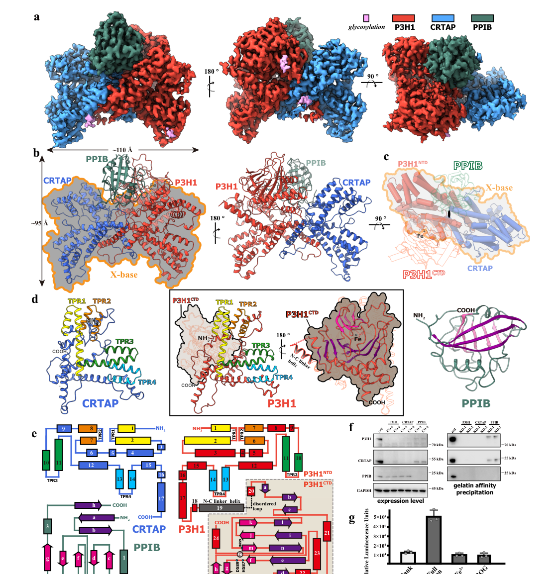
A major 2024 advance is a cryo-EM structural framework for the human P3H1/CRTAP/PPIB (PCP) complex (Nature Communications; publication month Sep 2024; URL/DOI below). The study reports:
- a 1:1:1 PCP complex, approximately 150 kDa and about 110 Γ… in length, resolved at ~3.17–3.75 Γ… in supplemented conditions,
- P3H1 and PPIB active sites positioned face-to-face, forming a proposed bifunctional reaction center consistent with coupled hydroxylation and prolyl isomerization,
- multiple collagen substrate binding sites (CBS1–CBS5) suggesting an extended substrate-interaction zone and processing path,
- an unexpected dual-ternary (hetero-hexameric) state, whose equilibrium can be altered by mutations in P3H1/PPIB active sites or PPIB inhibitors such as cyclosporin A.

These findings refine the current understanding from β€œthree proteins that modify/fold collagen” to a more spatially integrated model in which enzymatic hydroxylation (P3H1) and isomerization (PPIB) are structurally coordinated around collagen peptide binding. (li2024thestructuralbasis pages 1-2, li2024thestructuralbasis pages 7-8)

Publication details: Li et al., Nature Communications (Sep 2024). DOI: https://doi.org/10.1038/s41467-024-52321-6 (li2024thestructuralbasis pages 1-2)

4.2 2023: Mechanism-based classification perspective in OI

A 2023 authoritative review (Orphanet Journal of Rare Diseases) organizes osteogenesis imperfecta into mechanistic categories and explicitly classifies LEPRE1 (P3H1) as an autosomal recessive OI Type VIII gene under collagen post-translational modification/processing/folding defects, with severe-to-lethal phenotypes in its typology. (yu2023pathogenicmechanismsof pages 1-2)

Publication details: Yu et al., Orphanet J Rare Dis (Aug 2023). DOI: https://doi.org/10.1186/s13023-023-02849-5 (yu2023pathogenicmechanismsof pages 1-2)

5) Disease associations and mechanisms (human and model evidence)

5.1 Osteogenesis imperfecta type VIII (LEPRE1/P3H1)

Biallelic loss-of-function variants in LEPRE1/P3H1 cause autosomal recessive osteogenesis imperfecta type VIII, a severe bone fragility disorder. Molecularly, P3H1/CRTAP/PPIB deficiency causes loss of 3-hydroxylation at COL1A1 Pro986, delayed collagen folding, and collagen overmodification. (chang2010prolyl3hydroxylase1 pages 1-2, cabral2014recessiveosteogenesisimperfecta pages 109-113)

5.2 Cellular pathophysiology: ER retention, UPR, and rescue concepts

Patient fibroblast studies across recessive OI types due to the collagen prolyl 3-hydroxylation complex show that intracellular retention of overmodified type I collagen can cause ER enlargement, protein aggregates, activation of the PERK branch of the unfolded protein response (UPR), and apoptosis. The chemical chaperone 4-phenylbutyrate (4-PBA) alleviated cellular stress markers and supported ER proteostasis in these experimental settings, supporting a model where complex dysfunction produces a combined enzymatic + proteostasis pathology. (besio2019cellularstressdue pages 1-2)

Publication details: Besio et al., Disease Models & Mechanisms (Jun 2019). DOI: https://doi.org/10.1242/dmm.038521 (besio2019cellularstressdue pages 1-2)

6) Applications and real-world implementations

6.1 Clinical genetics and diagnosis

Mechanism-based and gene-based classification frameworks for OI explicitly incorporate LEPRE1/P3H1 as an etiologic gene for severe autosomal recessive OI (type VIII), supporting the routine inclusion of LEPRE1 in clinical sequencing panels and variant interpretation workflows for bone fragility disorders. (yu2023pathogenicmechanismsof pages 1-2)

6.2 Treatment outcomes in recent clinical cohorts

A 2024 retrospective cohort of rare bone fragility disorders treated with bisphosphonates included 3 patients with pathogenic P3H1 variants (mean age 9.3 years, range 5–15). Across the treated cohort (n=18), fracture rate decreased from 3.9 in the year prior to treatment to 1.4 during treatment (p=0.02). One P3H1 patient had sustained fracture reduction after discontinuation. These data represent real-world management information relevant to P3H1-related bone fragility phenotypes, although the study is not limited to OI type VIII and includes multiple genotypes. (charpie2024clinicalspectrumof pages 4-5, charpie2024clinicalspectrumof pages 3-4)

Publication details: CharpiΓ© et al., European Journal of Human Genetics (Jun 2024). DOI: https://doi.org/10.1038/s41431-024-01645-4 (charpie2024clinicalspectrumof pages 4-5)

6.3 Population genetics and genetic counseling (founder variant example)

A recurring LEPRE1 splice-site variant (c.1080+1G>T) has been reported with carrier frequency ~0.40% in Mid-Atlantic African-Americans, with a predicted incidence of type VIII OI of approximately 1 in 250,000 African American births; in some West African cohorts, carrier frequency ~1.5% yields a much higher predicted incidence (reported as ~1 in 18,260 births in the cited summary). These kinds of estimates are used for population risk assessment and counseling in settings where founder alleles are common. (cabral2014recessiveosteogenesisimperfecta pages 180-185, cabral2014recessiveosteogenesisimperfecta pages 109-113)

7) Quantitative data highlights (selected)

7.1 Structural/biophysical (2024)

  • PCP complex size/scale: ~150 kDa, ~110 Γ… length. (li2024thestructuralbasis pages 1-2)
  • Cryo-EM resolution: ~3.17–3.75 Γ… for supplemented samples. (li2024thestructuralbasis pages 1-2)

7.2 Collagen biochemical consequences of P3H1 loss

Reported biochemical changes in P3H1/LEPRE1 loss-of-function contexts include:
- increased lysyl hydroxylation in patient collagen (32–35% vs 24% control in one summarized dataset),
- delayed secretion kinetics (15–20 min delay),
- and increased total collagen secretion per cell (20–50% increase) in the summarized report.
These data support a model where delayed folding increases dwell time for other ER modifying enzymes and perturbs secretion homeostasis. (cabral2014recessiveosteogenesisimperfecta pages 109-113)

8) Visual evidence from the primary 2024 structural study

The following cropped figures from the 2024 Nature Communications paper illustrate the architecture of the PCP complex and the collagen binding sites that underlie the β€œsubstrate interacting zone” model:
- overall PCP complex architecture/domain organization (li2024thestructuralbasis media 0746bcb1)
- collagen binding sites CBS1–CBS5 and the implied substrate interaction path (li2024thestructuralbasis media 707dfa86)

9) Concise evidence map (table)

Aspect Evidence-based statement Key supporting source
Identity Human P3H1 is LEPRE1/GROS1, encoding prolyl 3-hydroxylase 1 (UniProt Q32P28), the catalytic component of the ER collagen prolyl 3-hydroxylation machinery that modifies COL1A1 Pro986. (li2024thestructuralbasis pages 1-2, chang2010prolyl3hydroxylase1 pages 1-2) Li et al., 2024, 10.1038/s41467-024-52321-6, https://doi.org/10.1038/s41467-024-52321-6; Chang et al., 2010, 10.1093/hmg/ddp481, https://doi.org/10.1093/hmg/ddp481
Enzymatic reaction/cofactors P3H1 is a 2-oxoglutarate/Fe²⁺-dependent dioxygenase that catalyzes 3-hydroxylation of proline in collagen, requiring Fe²⁺, 2-oxoglutarate, Oβ‚‚, and ascorbate. (marini2007componentsofthe pages 4-5, li2024thestructuralbasis pages 1-2) Marini et al., 2007, 10.4161/cc.6.14.4474, https://doi.org/10.4161/cc.6.14.4474; Li et al., 2024, 10.1038/s41467-024-52321-6, https://doi.org/10.1038/s41467-024-52321-6
Substrate specificity The best-established substrate is type I collagen Ξ±1(I) Pro986; loss of P3H1 causes near-complete loss of 3-Hyp986 and collagen overmodification/delayed folding. Additional sites such as Ξ±2(I) Pro707 are modified to a lesser extent. (besio2019cellularstressdue pages 1-2, cabral2014recessiveosteogenesisimperfecta pages 35-39, hudson2013collagenprolyl3hydroxylation pages 1-2) Besio et al., 2019, 10.1242/dmm.038521, https://doi.org/10.1242/dmm.038521; Cabral, 2014; Hudson & Eyre, 2013, 10.3109/03008207.2013.800867, https://doi.org/10.3109/03008207.2013.800867
Complex partners/stoichiometry P3H1 forms a stable P3H1/CRTAP/PPIB (CyPB) complex in the ER with approximate 1:1:1 stoichiometry; CRTAP and P3H1 are mutually stabilizing, while PPIB provides prolyl cis-trans isomerase activity. (chang2010prolyl3hydroxylase1 pages 1-2, li2024thestructuralbasis pages 1-2) Chang et al., 2010, 10.1093/hmg/ddp481, https://doi.org/10.1093/hmg/ddp481; Li et al., 2024, 10.1038/s41467-024-52321-6, https://doi.org/10.1038/s41467-024-52321-6
Subcellular localization P3H1 enters the secretory pathway via an N-terminal signal peptide, contains a C-terminal ER-retention/retrieval motif (KDEL-like), and is experimentally localized to rough ER/ER-Golgi; immunostaining is strongest in fibrillar-collagen-rich tissues such as dermis, tendon, and cartilage. (fonsen2007prolylhydroxylasescloning pages 55-59, marini2007componentsofthe pages 5-5, cabral2014recessiveosteogenesisimperfecta pages 100-105, marini2007componentsofthe pages 4-5) FonsΓ©n, 2007; Marini et al., 2007, 10.4161/cc.6.14.4474, https://doi.org/10.4161/cc.6.14.4474; Cabral, 2014
Key 2024 structural insights Cryo-EM structures show a bifunctional reaction center with P3H1 and PPIB active sites face-to-face, multiple collagen-binding sites (CBS1–5), and an unexpected dual-ternary/hetero-hexameric state whose balance shifts with active-site mutations or cyclosporin A. (li2024thestructuralbasis pages 1-2, li2024thestructuralbasis pages 7-8, li2024thestructuralbasis media 0746bcb1) Li et al., 2024, 10.1038/s41467-024-52321-6, https://doi.org/10.1038/s41467-024-52321-6
Disease associations Biallelic P3H1/LEPRE1 variants cause recessive osteogenesis imperfecta type VIII, characterized by severe bone fragility, early fractures, collagen overmodification, ER stress, and impaired secretion/folding of type I collagen. (besio2019cellularstressdue pages 1-2, chang2010prolyl3hydroxylase1 pages 1-2, cabral2014recessiveosteogenesisimperfecta pages 109-113) Besio et al., 2019, 10.1242/dmm.038521, https://doi.org/10.1242/dmm.038521; Chang et al., 2010, 10.1093/hmg/ddp481, https://doi.org/10.1093/hmg/ddp481; Cabral, 2014
Quantitative stats Recent/representative numbers include: PCP complex ~150 kDa, ~110 Γ… long, cryo-EM ~3.17–3.75 Γ…; in a 2024 cohort, 3/18 treated rare bone-fragility patients had P3H1 variants, mean age 9.3 y (range 5–15), fracture rate fell from 3.9 to 1.4/year on bisphosphonates (p=0.02); older population data estimate the recurrent West African/African American splice variant carrier frequency at 0.4% in African Americans and ~1.5% in some West African cohorts. (li2024thestructuralbasis pages 1-2, charpie2024clinicalspectrumof pages 4-5, cabral2014recessiveosteogenesisimperfecta pages 180-185) Li et al., 2024, 10.1038/s41467-024-52321-6, https://doi.org/10.1038/s41467-024-52321-6; CharpiΓ© et al., 2024, 10.1038/s41431-024-01645-4, https://doi.org/10.1038/s41431-024-01645-4; Cabral, 2014

Table: This table summarizes the core functional annotation of human P3H1/LEPRE1 (UniProt Q32P28), including its enzymatic role, substrates, complex partners, localization, 2024 structural advances, disease relevance, and selected quantitative findings. It is useful as a concise evidence map for gene/protein annotation and interpretation.

10) Limitations and open questions (evidence-based)

Although the biochemical necessity of the P3H1/CRTAP/PPIB complex for COL1A1 Pro986 3-hydroxylation and normal collagen maturation is well supported, the precise in vivo contribution of the 3-Hyp mark itself versus broader chaperone/proteostasis functions of the complex remains discussed as not fully resolved in foundational synthesis: proposed roles for Pro986 3-hydroxylation include effects on fibril molecular packing, local helix behavior, or protein-binding interactions during fibrillogenesis. (hudson2013collagenprolyl3hydroxylation pages 1-2, cabral2014recessiveosteogenesisimperfecta pages 174-180)

References

  1. (li2024thestructuralbasis pages 1-2): Wenguo Li, Junjiang Peng, Deqiang Yao, Bing Rao, Ying Xia, Qian Wang, Shaobai Li, Mi Cao, Yafeng Shen, Peixiang Ma, Rijing Liao, An Qin, Jie Zhao, and Yu Cao. The structural basis for the collagen processing by human p3h1/crtap/ppib ternary complex. Nature Communications, Sep 2024. URL: https://doi.org/10.1038/s41467-024-52321-6, doi:10.1038/s41467-024-52321-6. This article has 17 citations and is from a highest quality peer-reviewed journal.

  2. (marini2007componentsofthe pages 4-5): Joan C. Marini, Wayne A. Cabral, Aileen M. Barnes, and Weizhong Chang. Components of the collagen prolyl 3-hydroxylation complex are crucial for normal bone development. Cell Cycle, 6:1675-1681, Jul 2007. URL: https://doi.org/10.4161/cc.6.14.4474, doi:10.4161/cc.6.14.4474. This article has 168 citations and is from a peer-reviewed journal.

  3. (besio2019cellularstressdue pages 1-2): Roberta Besio, Nadia Garibaldi, Laura Leoni, Lina Cipolla, Simone Sabbioneda, Marco Biggiogera, Monica Mottes, Mona Aglan, Ghada A. Otaify, Samia A. Temtamy, Antonio Rossi, and Antonella Forlino. Cellular stress due to impairment of collagen prolyl hydroxylation complex is rescued by the chaperone 4-phenylbutyrate. Disease Models & Mechanisms, Jun 2019. URL: https://doi.org/10.1242/dmm.038521, doi:10.1242/dmm.038521. This article has 66 citations and is from a domain leading peer-reviewed journal.

  4. (cabral2014recessiveosteogenesisimperfecta pages 35-39): WA Cabral. Recessive osteogenesis imperfecta: prevalence and pathophysiology of collagen prolyl-3-hydroxylation complex defects. Unknown journal, 2014.

  5. (hudson2013collagenprolyl3hydroxylation pages 1-2): David M. Hudson and David R. Eyre. Collagen prolyl 3-hydroxylation: a major role for a minor post-translational modification? Jun 2013. URL: https://doi.org/10.3109/03008207.2013.800867, doi:10.3109/03008207.2013.800867. This article has 133 citations and is from a peer-reviewed journal.

  6. (chang2010prolyl3hydroxylase1 pages 1-2): Weizhong Chang, Aileen M. Barnes, Wayne A. Cabral, Joann N. Bodurtha, and Joan C. Marini. Prolyl 3-hydroxylase 1 and crtap are mutually stabilizing in the endoplasmic reticulum collagen prolyl 3-hydroxylation complex. Human molecular genetics, 19 2:223-34, Jan 2010. URL: https://doi.org/10.1093/hmg/ddp481, doi:10.1093/hmg/ddp481. This article has 100 citations and is from a domain leading peer-reviewed journal.

  7. (fonsen2007prolylhydroxylasescloning pages 55-59): P FonsΓ©n. Prolyl hydroxylases: cloning and characterization of novel human and plant prolyl 4-hydroxylases, and three human prolyl 3-hydroxylases. Unknown journal, 2007.

  8. (marini2007componentsofthe pages 5-5): Joan C. Marini, Wayne A. Cabral, Aileen M. Barnes, and Weizhong Chang. Components of the collagen prolyl 3-hydroxylation complex are crucial for normal bone development. Cell Cycle, 6:1675-1681, Jul 2007. URL: https://doi.org/10.4161/cc.6.14.4474, doi:10.4161/cc.6.14.4474. This article has 168 citations and is from a peer-reviewed journal.

  9. (cabral2014recessiveosteogenesisimperfecta pages 100-105): WA Cabral. Recessive osteogenesis imperfecta: prevalence and pathophysiology of collagen prolyl-3-hydroxylation complex defects. Unknown journal, 2014.

  10. (li2024thestructuralbasis pages 7-8): Wenguo Li, Junjiang Peng, Deqiang Yao, Bing Rao, Ying Xia, Qian Wang, Shaobai Li, Mi Cao, Yafeng Shen, Peixiang Ma, Rijing Liao, An Qin, Jie Zhao, and Yu Cao. The structural basis for the collagen processing by human p3h1/crtap/ppib ternary complex. Nature Communications, Sep 2024. URL: https://doi.org/10.1038/s41467-024-52321-6, doi:10.1038/s41467-024-52321-6. This article has 17 citations and is from a highest quality peer-reviewed journal.

  11. (yu2023pathogenicmechanismsof pages 1-2): Hongjie Yu, Changrong Li, Huixiao Wu, Weibo Xia, Yanzhou Wang, Jiajun Zhao, and Chao Xu. Pathogenic mechanisms of osteogenesis imperfecta, evidence for classification. Orphanet Journal of Rare Diseases, Aug 2023. URL: https://doi.org/10.1186/s13023-023-02849-5, doi:10.1186/s13023-023-02849-5. This article has 36 citations and is from a peer-reviewed journal.

  12. (cabral2014recessiveosteogenesisimperfecta pages 109-113): WA Cabral. Recessive osteogenesis imperfecta: prevalence and pathophysiology of collagen prolyl-3-hydroxylation complex defects. Unknown journal, 2014.

  13. (charpie2024clinicalspectrumof pages 4-5): Maëlle Charpié, Perrine Brunelle, Geneviève Baujat, Caroline Michot, Julien Van Gils, Bruno Leheup, Élise Schaefer, Eugénie Koumakis, Zagorka Pejin, Graziella Pinto, Sophie Monnot, and Valérie Cormier-Daire. Clinical spectrum of rare bone fragility disorders and response to bisphosphonate treatment: a retrospective study. European journal of human genetics : EJHG, 32:1559-1566, Jun 2024. URL: https://doi.org/10.1038/s41431-024-01645-4, doi:10.1038/s41431-024-01645-4. This article has 5 citations.

  14. (charpie2024clinicalspectrumof pages 3-4): Maëlle Charpié, Perrine Brunelle, Geneviève Baujat, Caroline Michot, Julien Van Gils, Bruno Leheup, Élise Schaefer, Eugénie Koumakis, Zagorka Pejin, Graziella Pinto, Sophie Monnot, and Valérie Cormier-Daire. Clinical spectrum of rare bone fragility disorders and response to bisphosphonate treatment: a retrospective study. European journal of human genetics : EJHG, 32:1559-1566, Jun 2024. URL: https://doi.org/10.1038/s41431-024-01645-4, doi:10.1038/s41431-024-01645-4. This article has 5 citations.

  15. (cabral2014recessiveosteogenesisimperfecta pages 180-185): WA Cabral. Recessive osteogenesis imperfecta: prevalence and pathophysiology of collagen prolyl-3-hydroxylation complex defects. Unknown journal, 2014.

  16. (li2024thestructuralbasis media 0746bcb1): Wenguo Li, Junjiang Peng, Deqiang Yao, Bing Rao, Ying Xia, Qian Wang, Shaobai Li, Mi Cao, Yafeng Shen, Peixiang Ma, Rijing Liao, An Qin, Jie Zhao, and Yu Cao. The structural basis for the collagen processing by human p3h1/crtap/ppib ternary complex. Nature Communications, Sep 2024. URL: https://doi.org/10.1038/s41467-024-52321-6, doi:10.1038/s41467-024-52321-6. This article has 17 citations and is from a highest quality peer-reviewed journal.

  17. (li2024thestructuralbasis media 707dfa86): Wenguo Li, Junjiang Peng, Deqiang Yao, Bing Rao, Ying Xia, Qian Wang, Shaobai Li, Mi Cao, Yafeng Shen, Peixiang Ma, Rijing Liao, An Qin, Jie Zhao, and Yu Cao. The structural basis for the collagen processing by human p3h1/crtap/ppib ternary complex. Nature Communications, Sep 2024. URL: https://doi.org/10.1038/s41467-024-52321-6, doi:10.1038/s41467-024-52321-6. This article has 17 citations and is from a highest quality peer-reviewed journal.

  18. (cabral2014recessiveosteogenesisimperfecta pages 174-180): WA Cabral. Recessive osteogenesis imperfecta: prevalence and pathophysiology of collagen prolyl-3-hydroxylation complex defects. Unknown journal, 2014.

Artifacts

Citations

  1. li2024thestructuralbasis pages 1-2
  2. fonsen2007prolylhydroxylasescloning pages 55-59
  3. yu2023pathogenicmechanismsof pages 1-2
  4. besio2019cellularstressdue pages 1-2
  5. charpie2024clinicalspectrumof pages 4-5
  6. cabral2014recessiveosteogenesisimperfecta pages 109-113
  7. marini2007componentsofthe pages 4-5
  8. cabral2014recessiveosteogenesisimperfecta pages 35-39
  9. marini2007componentsofthe pages 5-5
  10. cabral2014recessiveosteogenesisimperfecta pages 100-105
  11. li2024thestructuralbasis pages 7-8
  12. charpie2024clinicalspectrumof pages 3-4
  13. cabral2014recessiveosteogenesisimperfecta pages 180-185
  14. cabral2014recessiveosteogenesisimperfecta pages 174-180
  15. https://doi.org/10.1038/s41467-024-52321-6
  16. https://doi.org/10.1186/s13023-023-02849-5
  17. https://doi.org/10.1242/dmm.038521
  18. https://doi.org/10.1038/s41431-024-01645-4
  19. https://doi.org/10.1038/s41467-024-52321-6;
  20. https://doi.org/10.1093/hmg/ddp481
  21. https://doi.org/10.4161/cc.6.14.4474;
  22. https://doi.org/10.1242/dmm.038521;
  23. https://doi.org/10.3109/03008207.2013.800867
  24. https://doi.org/10.1093/hmg/ddp481;
  25. https://doi.org/10.1038/s41431-024-01645-4;
  26. https://doi.org/10.1038/s41467-024-52321-6,
  27. https://doi.org/10.4161/cc.6.14.4474,
  28. https://doi.org/10.1242/dmm.038521,
  29. https://doi.org/10.3109/03008207.2013.800867,
  30. https://doi.org/10.1093/hmg/ddp481,
  31. https://doi.org/10.1186/s13023-023-02849-5,
  32. https://doi.org/10.1038/s41431-024-01645-4,

πŸ“š Additional Documentation

Notes

(P3H1-notes.md)

P3H1 (LEPRE1 / GROS1 / Leprecan-1) β€” gene review notes

UniProt: Q32P28 (P3H1_HUMAN), 736 aa precursor. HGNC:19316. Human.
EC 1.14.11.7 (procollagen-proline 3-dioxygenase / prolyl 3-hydroxylase).

Core identity and function

P3H1 is an ER-lumenal 2-oxoglutarate / Fe(II)-dependent dioxygenase that catalyzes
3-hydroxylation of specific proline residues in procollagen.

  • [file:human/P3H1/P3H1-uniprot.txt "Has prolyl 3-hydroxylase activity and catalyzes the post-"] / "translational formation of 3-hydroxyproline in -Xaa-Pro-Gly- sequences in pro-collagen chains, a critical step for the formation of mature collagen trimers (PubMed:39245686)."
  • Catalytic reaction (UniProt): "L-prolyl-[collagen] + 2-oxoglutarate + O2 = trans-3-hydroxy-L-prolyl-[collagen] + succinate + CO2 ... EC=1.14.11.7" supported by PMID:39245686.
  • Cofactors: Fe cation; L-ascorbate (vitamin C). UniProt COFACTOR section + keywords "Iron; Metal-binding; ... Vitamin C." Fe(3+) and 2-oxoglutarate binding residues His587, Asp589, His659, Arg669, etc., with mutagenesis (H587A, H659A, R669H = loss of activity).
  • Fe2OG dioxygenase domain 564-678; InterPro IPR005123 / IPR006620; member of leprecan family.

The specific physiological substrate residue is alpha1(I)Pro986 (and alpha1(II)Pro986).
- PMID:17277775
- PMID:22615817

The PCP / P3H1-CRTAP-PPIB complex

P3H1 is the catalytic core of the ER collagen prolyl 3-hydroxylation complex
("PCP complex") with CRTAP (O75718) and PPIB / cyclophilin B (P23284), in 1:1:1 stoichiometry.
- [file:human/P3H1/P3H1-uniprot.txt "Forms a ternary complex with PPIB (CYPB) and CRTAP, known as"] / "the PCP complex, in a 1:1:1 stoichiometric ratio; this complex binds unfolded collagen (PubMed:39245686)."
- Cryo-EM structures (8K0E/F/I/M/8K17/8KC9) of the ternary and dual-ternary complex with 2-OG and Fe(3+) and a collagen analog PMID:39245686.
- PMID:39245686
- The active sites of P3H1 and PPIB form a face-to-face bifunctional reaction center: "The active sites of P3H1 and PPIB form a face-to-face bifunctional reaction center, indicating a coupled modification mechanism." PMID:39245686
- P3H1 is the only complex component with a KDEL ER-retrieval signal (C-terminal "...KPKDEL", residues 733-736 "Prevents secretion from ER"). KDEL is essential for function in vivo PMID:22615817.

Mutual stabilization / chaperone role

CRTAP and P3H1 mutually stabilize each other in the ER; loss of either depletes both (proteasomal degradation of the orphan).
- PMID:19846465
- PMID:19846465
- The complex also has chaperone activity (inhibits citrate synthase aggregation, assists rhodanese refolding, collagen fibrillogenesis): PMID:19846465; PMID:22615817. This underpins protein folding / protein stabilization GO terms.

PPIB/CyPB loss does NOT abolish Pro986 3-hydroxylation PMID:20089953, i.e. P3H1 is the catalytic core; CyPB is the PPIase partner.

Localization

Isoform 1 (736 aa, KDEL) is ER-resident; this is the catalytically relevant location.
- [file:human/P3H1/P3H1-uniprot.txt "SUBCELLULAR LOCATION: [Isoform 1]: Endoplasmic reticulum"]
- ER lumen (Reactome TAS), ER (IDA PMID:19846465, PMID:20089953; EXP PMID:19088120).
- [PMID:19088120 "The affected splice form encodes a 736 amino acid (AA) protein with a \"KDEL\" endoplasmic reticulum retention signal."] β€” establishes isoform 1 / ER as the functional form.
- Secondary/secreted: as a CSPG, P3H1 (leprecan) can be secreted into ECM. UniProt: "Secreted, extracellular space, extracellular matrix" (by similarity to rat). This and proteomics localizations (exosome, membrane, ECM colocalization) are non-core / context-specific captures.

Disease

Osteogenesis imperfecta type 8 (OI8; MIM 610915), autosomal recessive, severe/lethal. Null LEPRE1 -> loss of Pro986 3-hydroxylation, collagen overmodification, delayed folding.
- PMID:17277775
- W675L variant: loss of catalytic activity (UniProt VARIANT + PMID:39245686).

"Growth suppressor 1" / proliferation

Original cloning as Gros1, a putative growth suppressor in fibroblasts (NIH3T3 overexpression slowed growth). This predates knowledge of its enzymatic role; NAS, weak/historical.
- PMID:10951563
- UniProt FUNCTION: "Has growth suppressive activity in fibroblasts." Keep as non-core (over-annotated relative to the well-established collagen-modifying enzyme role).

Annotation decisions summary

CORE:
- GO:0019797 procollagen-proline 3-dioxygenase activity (MF) β€” ACCEPT core (IDA PMID:39245686; IBA; ISS; IEA). EC 1.14.11.7.
- GO:0005788 ER lumen / GO:0005783 ER (CC) β€” ACCEPT (site of action).
- GO:0032963 collagen metabolic process (BP) β€” ACCEPT/core-ish (its biological role).
- GO:0032991 protein-containing complex part_of β€” ACCEPT (PCP complex membership).

Cofactor / catalytic-support MF (keep, but not bare core):
- GO:0005506 iron ion binding β€” ACCEPT (catalytic Fe(II) cofactor; structurally demonstrated).
- GO:0031418 L-ascorbic acid binding β€” KEEP_AS_NON_CORE / ACCEPT (vitamin C cofactor).
- GO:0016705 oxidoreductase activity (paired donors, O2) β€” parent of the specific dioxygenase MF; KEEP_AS_NON_CORE (generic; GO:0019797 is the informative term).

BP (complex chaperone / collagen biogenesis):
- GO:0006457 protein folding β€” ACCEPT/KEEP (complex chaperone function; IMP PMID:17277775, ISS PMID:15044469).
- GO:0050821 protein stabilization β€” ACCEPT (mutual stabilization, IMP PMID:19846465, PMID:22615817).
- GO:0060348 bone development β€” KEEP_AS_NON_CORE (downstream physiology; OI phenotype).
- GO:0050708 regulation of protein secretion β€” KEEP_AS_NON_CORE (collagen secretion delay in nulls).
- GO:1901874 negative regulation of post-translational protein modification β€” KEEP_AS_NON_CORE. This reflects that loss of P3H1 leads to OVER-modification (lysyl hydroxylation/glycosylation) of the helix; the wild-type complex limits over-modification by speeding folding. Slightly contorted but defensible from the IMP data (overmodification on P3H1 loss).

CC bare-binding / proteomics (non-core):
- GO:0005515 protein binding (IPI x3: PMID:30021884, PMID:33961781, PMID:39245686) β€” KEEP_AS_NON_CORE. Partners O75718=CRTAP and P23284=PPIB (in 39245686) are real complex partners; 30021884 (histone XL-MS) and 33961781 (BioPlex) capture CRTAP. Bare "protein binding" uninformative.
- GO:0005518 collagen binding contributes_to (ISS PMID:15044469) β€” ACCEPT/KEEP_AS_NON_CORE; the complex binds unfolded/denatured collagen substrate (consistent with structure & enzymology).
- GO:0070062 extracellular exosome (HDA PMID:19056867) β€” KEEP_AS_NON_CORE (urinary exosome proteomics; not the functional ER site).
- GO:0016020 membrane (HDA PMID:19946888) β€” KEEP_AS_NON_CORE (NK-cell membrane proteome; original immunoscreen was a "plasma membrane protein"; low-resolution).
- GO:0031012 extracellular matrix colocalizes_with (HDA PMID:28327460) β€” KEEP_AS_NON_CORE (consistent with secreted CSPG leprecan form; not the catalytic ER role).
- GO:0010976 positive regulation of neuron projection development (ISS GO_REF:0000024 from rat Q9R1J8) β€” MARK_AS_OVER_ANNOTATED (electronic ISS transfer from rat leprecan; no human evidence; tangential to collagen-modifying role).
- GO:0008285 negative regulation of cell population proliferation (NAS PMID:10951563) β€” KEEP_AS_NON_CORE (historical Gros1 growth-suppressor assertion; superseded by enzymatic characterization).

Falcon deep-research findings (incorporated 2026-06)

Falcon report largely corroborated the existing review; the PCP cryo-EM structural paper it foregrounds (Li et al. 2024, Nat Commun, DOI 10.1038/s41467-024-52321-6) is already in the review as PMID:39245686. Two additional verified references were added:

  • ER-stress / proteostasis pathophysiology of recessive OI complex defects: in primary fibroblasts from recessive OI patients (including P3H1/LEPRE1 deficiency), intracellular retention of overmodified type I collagen causes ER enlargement, protein aggregates, PERK-branch UPR activation and apoptosis; the chemical chaperone 4-phenylbutyrate (4-PBA) rescues ER proteostasis and cell survival PMID:31171565. Added as a MEDIUM-relevance reference (complex/disease-mechanism level, not P3H1-catalysis-specific). Not in publications cache.
  • Clinical/therapeutic cohort: a retrospective study of rare (non-COL1A1/COL1A2) bone-fragility disorders treated with bisphosphonates includes patients with pathogenic P3H1 variants; lumbar-spine BMD increased dose-dependently and fracture rate fell on treatment PMID:38926541. Added as a LOW-relevance reference (OI8 disease management context). Not in publications cache.
  • Falcon quantitative claims (e.g. PCP ~150 kDa / ~110 Γ… / 3.17–3.75 Γ…; bisphosphonate fracture-rate 3.9β†’1.4, p=0.02; founder splice variant c.1080+1G>T carrier frequencies) were noted but not added to YAML annotations; substrate/complex/localization facts were already fully covered by the existing review (alpha1(I)Pro986; 1:1:1 P3H1/CRTAP/PPIB complex; KDEL ER retention; bifunctional reaction center).
  • No action: changes made; reviews were already COMPLETE and these papers are downstream disease/mechanism context. Edits are additive (new references only).

Pn Notes

(P3H1-pn-notes.md)

P3H1 PN Consistency Notes

  • Generated: 2026-06-18
  • Project: PROTEOSTASIS
  • Scope: PN consistency rereview against local AIGR review and available deep-research artifacts
  • UniProt: Q32P28
  • AIGR review status: COMPLETE
  • Review batch: proteostasis-batch-2026-06-11
  • Batch change status: added

Source Files Checked

Deep Research Files

AIGR Review Snapshot

  • Description: P3H1 (prolyl 3-hydroxylase 1; gene LEPRE1, also known as leprecan-1 and growth suppressor 1) is an endoplasmic-reticulum-lumenal 2-oxoglutarate/Fe(II)-dependent dioxygenase (EC 1.14.11.7) of the leprecan family. It catalyzes the post-translational formation of 3-hydroxyproline at specific -Xaa-Pro-Gly- prolines in procollagen chains, most notably the alpha1(I)Pro986 residue of type I and type II collagen, a modification required for proper collagen triple-helix folding, assembly and stability. P3H1 is the catalytic core of the ER collagen prolyl 3-hydroxylation complex (the "PCP complex"), a 1:1:1 ternary assembly with cartilage-associated protein (CRTAP) and peptidyl-prolyl cis-trans isomerase B (PPIB/cyclophilin B). Within this complex P3H1 contributes prolyl 3-hydroxylase activity while PPIB provides cis-trans isomerase activity and CRTAP stabilizes the assembly and helps recruit collagen substrate; the complex also functions as a collagen chaperone. P3H1 carries a C-terminal KDEL ER-retrieval signal that retains it (and, with it, CRTAP) in the ER lumen. Catalysis requires a non-heme Fe(II) center, the co-substrate 2-oxoglutarate, molecular oxygen, and L-ascorbate (vitamin C) as cofactor. Loss-of-function mutations in LEPRE1 abolish alpha1(I)Pro986 3-hydroxylation, delay collagen folding and cause overmodification of the collagen helix, resulting in autosomal recessive osteogenesis imperfecta type 8 (OI8). A secreted chondroitin sulfate proteoglycan form of leprecan can also be deposited in the extracellular matrix.
  • Existing/core annotation action counts: ACCEPT: 22; KEEP_AS_NON_CORE: 14; MARK_AS_OVER_ANNOTATED: 2

PN Consistency Summary

  • Consistency: Deep research ↔ review ↔ PN annotation fully consistent. P3H1 is correctly the catalytic core (procollagen-proline 3-dioxygenase, EC 1.14.11.7, GO:0019797) of the ER PCP complex (with CRTAP + PPIB), required for collagen helix folding/stability; loss causes recessive OI8. PN "ER collagen processing and folding" matches precisely.
  • PN story / NEW pressure: No NEW GO pressure β€” catalytic, complex-membership, folding, stabilization and collagen-binding roles are all captured (GO:0019797, GO:0032963, GO:0032991, GO:0006457, GO:0050821, GO:0005518). Falcon added only disease/ER-stress context (PMID:31171565, 38926541). Conclude: already captured.
  • Evidence alignment: PN dossier is mapping-only (no titles). Review evidence is extensive and PubMed-verified (PMID:39245686 cryo-EM, 17277775, 19846465, 22615817, 15044469, 19088120). No bibliographic conflict or divergence.
  • Verdict: Review excellent and PN-consistent; the groupβ†’GO:0032964 mapping is sound and correctly branched (contrast with the over-broad lectin "N-glycosylation system" node). No changes needed.

Full Consistency Review

  • UniProt: Q32P28 (prolyl 3-hydroxylase 1 / LEPRE1 / leprecan-1) Β· batch: proteostasis-batch-2026-06-11 Β· review status: COMPLETE, very thorough (full annotation set + detailed notes + Falcon).
  • PN placement: ER proteostasis|Maturation and folding of specific substrates|ER collagen processing and folding ; PN-node mapping: group "ER collagen processing and folding" mappedβ†’GO:0032964 (collagen biosynthetic process, ok_for_propagation, more_specific_than_existing_goa); class/branch no_mapping.
  • Consistency: Deep research ↔ review ↔ PN annotation fully consistent. P3H1 is correctly the catalytic core (procollagen-proline 3-dioxygenase, EC 1.14.11.7, GO:0019797) of the ER PCP complex (with CRTAP + PPIB), required for collagen helix folding/stability; loss causes recessive OI8. PN "ER collagen processing and folding" matches precisely.
  • PN story / NEW pressure: No NEW GO pressure β€” catalytic, complex-membership, folding, stabilization and collagen-binding roles are all captured (GO:0019797, GO:0032963, GO:0032991, GO:0006457, GO:0050821, GO:0005518). Falcon added only disease/ER-stress context (PMID:31171565, 38926541). Conclude: already captured.
  • Mapping strategy: Groupβ†’GO:0032964 (collagen biosynthetic process) is defensible and verified real via OLS. It is broader than the review's accepted GO:0032963 (collagen metabolic process) only in being a biosynthesis-specific child β€” appropriate for the collagen-folding group and more_specific_than_existing_goa is reasonable (P3H1's GOA carries GO:0032963 metabolic, not the biosynthetic child). Unlike the lectin group node, this projection is the right branch (collagen biogenesis) for P3H1. Status/scope OK.
  • Evidence alignment: PN dossier is mapping-only (no titles). Review evidence is extensive and PubMed-verified (PMID:39245686 cryo-EM, 17277775, 19846465, 22615817, 15044469, 19088120). No bibliographic conflict or divergence.
  • Verdict: Review excellent and PN-consistent; the groupβ†’GO:0032964 mapping is sound and correctly branched (contrast with the over-broad lectin "N-glycosylation system" node). No changes needed.
    Recommended edits: None.

PN Dossier Context

  • review_batch: proteostasis-batch-2026-06-11
  • review_yaml: genes/human/P3H1/P3H1-ai-review.yaml
  • PN workbook rows: 1

PN row 1: ER proteostasis | Maturation and folding of specific substrates | ER collagen processing and folding

  • UniProt: Q32P28
  • In branches: ER
  • PN-node mapping records (path + ancestors):
    • [group] ER proteostasis|Maturation and folding of specific substrates|ER collagen processing and folding
      status=mapped scope=ok_for_propagation_to_go GO=[GO:0032964 collagen biosynthetic process]
      rationale: This PN group contains ER factors dedicated to collagen maturation, processing, and folding. Collagen biosynthetic process captures the shared substrate-specific pathway context.
    • [class] ER proteostasis|Maturation and folding of specific substrates
      status=no_mapping scope= GO=[]
      rationale: Reviewed as a broad PN category rather than a single GO class. The member genes span multiple activities, complexes, or contexts, so direct propagation from this node would overstate the shared biology.
    • [branch] ER proteostasis
      status=no_mapping scope= GO=[]
      rationale: Reviewed as a top-level PN branch. This is a systems/taxonomy umbrella, not a direct GO assertion; narrower child curations carry any propagating GO mappings.

Projected GO annotations (1)

  • GO:0032964 collagen biosynthetic process | scope=ok_for_propagation_to_go | goa_status=more_specific_than_existing_goa | from=ER proteostasis|Maturation and folding of specific substrates|ER collagen processing and folding

Note

This file is generated from the current PROTEOSTASIS phase-1 dossier and local gene-review artifacts. Edit the source review, PN mapping, or dossier rather than this generated note when correcting the underlying curation.

πŸ“„ View Raw YAML

id: Q32P28
gene_symbol: P3H1
product_type: PROTEIN
status: COMPLETE
taxon:
  id: NCBITaxon:9606
  label: Homo sapiens
description: P3H1 (prolyl 3-hydroxylase 1; gene LEPRE1, also known as leprecan-1 and growth
  suppressor 1) is an endoplasmic-reticulum-lumenal 2-oxoglutarate/Fe(II)-dependent dioxygenase
  (EC 1.14.11.7) of the leprecan family. It catalyzes the post-translational formation of
  3-hydroxyproline at specific -Xaa-Pro-Gly- prolines in procollagen chains, most notably the
  alpha1(I)Pro986 residue of type I and type II collagen, a modification required for proper
  collagen triple-helix folding, assembly and stability. P3H1 is the catalytic core of the ER
  collagen prolyl 3-hydroxylation complex (the "PCP complex"), a 1:1:1 ternary assembly with
  cartilage-associated protein (CRTAP) and peptidyl-prolyl cis-trans isomerase B (PPIB/cyclophilin
  B). Within this complex P3H1 contributes prolyl 3-hydroxylase activity while PPIB provides
  cis-trans isomerase activity and CRTAP stabilizes the assembly and helps recruit collagen
  substrate; the complex also functions as a collagen chaperone. P3H1 carries a C-terminal KDEL
  ER-retrieval signal that retains it (and, with it, CRTAP) in the ER lumen. Catalysis requires a
  non-heme Fe(II) center, the co-substrate 2-oxoglutarate, molecular oxygen, and L-ascorbate
  (vitamin C) as cofactor. Loss-of-function mutations in LEPRE1 abolish alpha1(I)Pro986
  3-hydroxylation, delay collagen folding and cause overmodification of the collagen helix,
  resulting in autosomal recessive osteogenesis imperfecta type 8 (OI8). A secreted chondroitin
  sulfate proteoglycan form of leprecan can also be deposited in the extracellular matrix.
alternative_products:
- name: 1 (GROS1-L, LEPREa, P3H1a)
  id: Q32P28-1
- name: 2 (GROS1-S)
  id: Q32P28-2
  sequence_note: VSP_019346, VSP_019347
- name: 3 (LEPREc)
  id: Q32P28-3
  sequence_note: VSP_019348
- name: 4 (LEPREb, P3H1b)
  id: Q32P28-4
  sequence_note: VSP_054864
existing_annotations:
- term:
    id: GO:0005783
    label: endoplasmic reticulum
  evidence_type: IBA
  original_reference_id: GO_REF:0000033
  qualifier: is_active_in
  review:
    summary: P3H1 acts in the ER lumen, where it 3-hydroxylates procollagen as part of the
      CRTAP/PPIB complex. The phylogenetic ER localization agrees with direct experimental
      evidence and the KDEL ER-retention signal.
    action: ACCEPT
    reason: Correct site of action; corroborated by EXP/IDA ER annotations and the KDEL retention
      motif retaining isoform 1 in the ER.
    supported_by:
    - reference_id: file:human/P3H1/P3H1-uniprot.txt
      supporting_text: 'SUBCELLULAR LOCATION: [Isoform 1]: Endoplasmic reticulum'
- term:
    id: GO:0019797
    label: procollagen-proline 3-dioxygenase activity
  evidence_type: IBA
  original_reference_id: GO_REF:0000033
  qualifier: enables
  review:
    summary: Core molecular function of P3H1, conserved across the leprecan/P3H family; it
      hydroxylates the 3-position of specific procollagen prolines.
    action: ACCEPT
    reason: Defining core MF; directly demonstrated experimentally (IDA, PMID:39245686, EC
      1.14.11.7) and conserved phylogenetically.
    supported_by:
    - reference_id: file:human/P3H1/P3H1-uniprot.txt
      supporting_text: Has prolyl 3-hydroxylase activity and catalyzes the post-
- term:
    id: GO:0032963
    label: collagen metabolic process
  evidence_type: IBA
  original_reference_id: GO_REF:0000033
  qualifier: involved_in
  review:
    summary: P3H1 participates in collagen post-translational modification/biosynthesis, a
      defining biological process for the gene.
    action: ACCEPT
    reason: Correct biological process; the enzyme is required for proper collagen biosynthesis,
      folding and assembly and is conserved across the family.
    supported_by:
    - reference_id: PMID:15044469
      supporting_text: The collagen prolyl hydroxylases are enzymes that are required for proper
- term:
    id: GO:0005506
    label: iron ion binding
  evidence_type: IEA
  original_reference_id: GO_REF:0000002
  qualifier: enables
  review:
    summary: P3H1 coordinates a catalytic non-heme Fe(II)/Fe(3+) ion in its Fe2OG dioxygenase
      domain (residues His587, Asp589, His659), a structural requirement for hydroxylation.
    action: KEEP_AS_NON_CORE
    reason: Accurate cofactor-binding attribute that supports the catalytic MF; structurally
      demonstrated, but subsidiary to the informative procollagen-proline 3-dioxygenase activity.
    supported_by:
    - reference_id: file:human/P3H1/P3H1-uniprot.txt
      supporting_text: Name=Fe cation; Xref=ChEBI:CHEBI:24875;
- term:
    id: GO:0005783
    label: endoplasmic reticulum
  evidence_type: IEA
  original_reference_id: GO_REF:0000044
  qualifier: located_in
  review:
    summary: Electronic transfer of ER localization from the UniProt subcellular location,
      consistent with stronger experimental evidence.
    action: ACCEPT
    reason: Correct compartment; redundant with EXP/IDA ER annotations.
    supported_by:
    - reference_id: file:human/P3H1/P3H1-uniprot.txt
      supporting_text: 'SUBCELLULAR LOCATION: [Isoform 1]: Endoplasmic reticulum'
- term:
    id: GO:0016705
    label: oxidoreductase activity, acting on paired donors, with incorporation or
      reduction of molecular oxygen
  evidence_type: IEA
  original_reference_id: GO_REF:0000002
  qualifier: enables
  review:
    summary: Parent molecular function describing the 2-oxoglutarate/Fe(II) dioxygenase chemistry
      that incorporates molecular oxygen during proline 3-hydroxylation.
    action: KEEP_AS_NON_CORE
    reason: Correct but generic; the specific GO:0019797 procollagen-proline 3-dioxygenase
      activity better captures the core function.
    supported_by:
    - reference_id: file:human/P3H1/P3H1-uniprot.txt
      supporting_text: 'Reaction=L-prolyl-[collagen] + 2-oxoglutarate + O2 = trans-3-hydroxy-L-'
- term:
    id: GO:0019797
    label: procollagen-proline 3-dioxygenase activity
  evidence_type: IEA
  original_reference_id: GO_REF:0000120
  qualifier: enables
  review:
    summary: Electronic assignment of the core dioxygenase activity from the EC/Rhea mapping (EC
      1.14.11.7; RHEA:22872), consistent with experimental evidence.
    action: ACCEPT
    reason: Correct core molecular function; redundant with IDA/IBA/ISS evidence.
    supported_by:
    - reference_id: file:human/P3H1/P3H1-uniprot.txt
      supporting_text: 'Reaction=L-prolyl-[collagen] + 2-oxoglutarate + O2 = trans-3-hydroxy-L-'
- term:
    id: GO:0031418
    label: L-ascorbic acid binding
  evidence_type: IEA
  original_reference_id: GO_REF:0000002
  qualifier: enables
  review:
    summary: P3H1, like other collagen hydroxylases, uses L-ascorbate (vitamin C) as a cofactor
      to maintain the catalytic iron in its reduced state.
    action: KEEP_AS_NON_CORE
    reason: Accurate cofactor-binding attribute supporting the catalytic MF; subsidiary to the
      core dioxygenase activity.
    supported_by:
    - reference_id: file:human/P3H1/P3H1-uniprot.txt
      supporting_text: Name=L-ascorbate; Xref=ChEBI:CHEBI:38290;
- term:
    id: GO:0032963
    label: collagen metabolic process
  evidence_type: IEA
  original_reference_id: GO_REF:0000002
  qualifier: involved_in
  review:
    summary: InterPro-based electronic assignment of the collagen metabolic process, consistent
      with the experimental and phylogenetic evidence.
    action: ACCEPT
    reason: Correct biological process; redundant with IBA/ISS.
    supported_by:
    - reference_id: file:human/P3H1/P3H1-uniprot.txt
      supporting_text: in pro-collagen chains, a critical step for the formation of mature
- term:
    id: GO:0005515
    label: protein binding
  evidence_type: IPI
  original_reference_id: PMID:30021884
  qualifier: enables
  review:
    summary: High-throughput crosslinking mass-spectrometry interactome capturing P3H1 with CRTAP
      (O75718). CRTAP is a genuine PCP complex partner, but the bare protein binding term is
      uninformative.
    action: KEEP_AS_NON_CORE
    reason: Records a real interaction with the complex partner CRTAP, but bare protein binding is
      uninformative; complex membership is better captured by GO:0032991 and the core catalytic MF.
    supported_by:
    - reference_id: file:human/P3H1/P3H1-uniprot.txt
      supporting_text: 'Q32P28; O75718: CRTAP;'
- term:
    id: GO:0005515
    label: protein binding
  evidence_type: IPI
  original_reference_id: PMID:33961781
  qualifier: enables
  review:
    summary: Proteome-scale (BioPlex) affinity-purification interactome capturing the P3H1-CRTAP
      (O75718) interaction. The partner is biologically real; the bare term is uninformative.
    action: KEEP_AS_NON_CORE
    reason: Real interaction with the complex partner CRTAP, but bare protein binding is
      uninformative and not elevated to core.
    supported_by:
    - reference_id: file:human/P3H1/P3H1-uniprot.txt
      supporting_text: 'Q32P28; O75718: CRTAP;'
- term:
    id: GO:0005515
    label: protein binding
  evidence_type: IPI
  original_reference_id: PMID:39245686
  qualifier: enables
  review:
    summary: Structural study establishing P3H1's direct interactions with CRTAP (O75718) and
      PPIB (P23284) within the PCP ternary complex. These are the genuine, biologically central
      complex partners, but the bare protein binding term is uninformative.
    action: KEEP_AS_NON_CORE
    reason: Partners CRTAP and PPIB are the real PCP complex components (demonstrated
      structurally), but bare protein binding does not convey the core function; captured better
      by GO:0032991 and GO:0019797.
    supported_by:
    - reference_id: PMID:39245686
      supporting_text: P3H1 and PPIB were identified as components of an ER-associated ternary
        complex consisting of P3H1/CRTAP/PPIB
- term:
    id: GO:0019797
    label: procollagen-proline 3-dioxygenase activity
  evidence_type: IDA
  original_reference_id: PMID:39245686
  qualifier: enables
  review:
    summary: Direct biochemical/structural demonstration that P3H1 is the core prolyl
      3-hydroxylase of the PCP complex, hydroxylating Pro986 of collagen alpha1(I); active-site
      mutants (H587A, D589A, H659A, R669) lose catalytic activity.
    action: ACCEPT
    reason: Core molecular function with direct experimental (IDA) support, EC 1.14.11.7 assigned,
      and mutagenesis confirming the catalytic residues.
    supported_by:
    - reference_id: PMID:39245686
      supporting_text: P3H1 is the core prolyl 3-hydroxylase, specially hydroxylating Pro986
- term:
    id: GO:0005783
    label: endoplasmic reticulum
  evidence_type: EXP
  original_reference_id: PMID:19088120
  qualifier: located_in
  review:
    summary: Experimental localization of the KDEL-bearing splice form (isoform 1) of P3H1 to the
      ER, the catalytically relevant compartment.
    action: ACCEPT
    reason: Experimentally supported ER localization consistent with the site of action.
    supported_by:
    - reference_id: file:human/P3H1/P3H1-uniprot.txt
      supporting_text: 'SUBCELLULAR LOCATION: [Isoform 1]: Endoplasmic reticulum'
- term:
    id: GO:0050821
    label: protein stabilization
  evidence_type: IMP
  original_reference_id: PMID:19846465
  qualifier: involved_in
  review:
    summary: P3H1 and CRTAP are mutually stabilizing within the ER prolyl 3-hydroxylation complex;
      loss of either leads to proteasomal degradation of the other. P3H1 thus stabilizes its
      partner CRTAP (and the complex stabilizes/chaperones collagen).
    action: ACCEPT
    reason: Directly supported by mutational analysis (null cells deplete both proteins; proteasome
      inhibitors partially rescue); reflects a genuine stabilization function of the complex.
    supported_by:
    - reference_id: PMID:19846465
      supporting_text: CRTAP and P3H1 are mutually stabilized in the collagen prolyl 3-hydroxylation
- term:
    id: GO:0050821
    label: protein stabilization
  evidence_type: IMP
  original_reference_id: PMID:22615817
  qualifier: involved_in
  review:
    summary: A KDEL-only mutation that prevents ER retention of P3H1 impairs its function,
      consistent with P3H1's role in maintaining the ER collagen-modifying complex and stabilizing
      its components/substrate.
    action: ACCEPT
    reason: Supported by patient mutation analysis; consistent with the genuine stabilization role
      of P3H1 within the ER collagen prolyl 3-hydroxylation complex.
    supported_by:
    - reference_id: PMID:22615817
      supporting_text: the KDEL ER- retrieval sequence is essential for P3H1 functionality
- term:
    id: GO:0031012
    label: extracellular matrix
  evidence_type: HDA
  original_reference_id: PMID:28327460
  qualifier: colocalizes_with
  review:
    summary: High-throughput proteomic detection of P3H1 in stem-cell-derived extracellular
      matrix. Consistent with the secreted chondroitin-sulfate proteoglycan (leprecan) form, but
      not the catalytic ER site of action.
    action: KEEP_AS_NON_CORE
    reason: Plausible given the documented secreted ECM proteoglycan form, but a weak
      colocalizes_with proteomics annotation peripheral to the core ER enzymatic role.
    supported_by:
    - reference_id: file:human/P3H1/P3H1-uniprot.txt
      supporting_text: 'Secreted, extracellular space, extracellular'
- term:
    id: GO:0005518
    label: collagen binding
  evidence_type: ISS
  original_reference_id: PMID:15044469
  qualifier: contributes_to
  review:
    summary: P3H1 specifically interacts with denatured/unfolded collagen, contributing to the
      PCP complex's substrate-binding (collagen recruitment) function.
    action: ACCEPT
    reason: Supported by enzyme characterization (specific interaction with denatured collagen)
      and the structurally demonstrated collagen-binding of the complex; an informative MF.
    supported_by:
    - reference_id: PMID:15044469
      supporting_text: specifically interact with denatured collagen
- term:
    id: GO:0006457
    label: protein folding
  evidence_type: ISS
  original_reference_id: PMID:15044469
  qualifier: involved_in
  review:
    summary: P3H1 (within the PCP complex) is required for proper collagen folding and assembly,
      and the complex has molecular chaperone activity.
    action: ACCEPT
    reason: Supported by enzyme characterization and the complex's documented chaperone role;
      loss of P3H1 delays collagen helix folding.
    supported_by:
    - reference_id: PMID:15044469
      supporting_text: required for proper
- term:
    id: GO:0010976
    label: positive regulation of neuron projection development
  evidence_type: ISS
  original_reference_id: GO_REF:0000024
  qualifier: involved_in
  review:
    summary: Electronic ISS transfer of a neuronal role from the rat leprecan ortholog (Q9R1J8).
      No human/primary evidence supports a neuron-projection function for P3H1, and it is tangential
      to its established collagen-modifying role.
    action: KEEP_AS_NON_CORE
    reason: Ortholog-based ISS with no supporting primary evidence in the P3H1 literature reviewed;
      peripheral and not corroborated, so retained only as non-core.
    supported_by:
    - reference_id: file:human/P3H1/P3H1-uniprot.txt
      supporting_text: Leucine- and proline-enriched proteoglycan 1
- term:
    id: GO:0016020
    label: membrane
  evidence_type: HDA
  original_reference_id: PMID:19946888
  qualifier: located_in
  review:
    summary: High-throughput NK-cell membrane proteome detection. P3H1 is an ER-lumenal/secreted
      protein; the generic "membrane" assignment most likely reflects co-fractionation rather than
      a genuine integral-membrane localization.
    action: MARK_AS_OVER_ANNOTATED
    reason: Generic, low-resolution proteomics localization inconsistent with the soluble
      ER-lumenal/secreted nature of P3H1; likely a contaminant/co-fractionation capture.
    supported_by:
    - reference_id: file:human/P3H1/P3H1-uniprot.txt
      supporting_text: 'SUBCELLULAR LOCATION: [Isoform 1]: Endoplasmic reticulum'
- term:
    id: GO:0005783
    label: endoplasmic reticulum
  evidence_type: IDA
  original_reference_id: PMID:20089953
  qualifier: located_in
  review:
    summary: Direct experimental ER localization of P3H1 from the cyclophilin B osteogenesis
      imperfecta study, consistent with the catalytic site of action.
    action: ACCEPT
    reason: IDA-supported ER localization agrees with the site of collagen hydroxylation.
    supported_by:
    - reference_id: file:human/P3H1/P3H1-uniprot.txt
      supporting_text: 'SUBCELLULAR LOCATION: [Isoform 1]: Endoplasmic reticulum'
- term:
    id: GO:0019797
    label: procollagen-proline 3-dioxygenase activity
  evidence_type: ISS
  original_reference_id: PMID:15044469
  qualifier: enables
  review:
    summary: Sequence-similarity assignment of the core prolyl 3-hydroxylase activity, transferred
      from the chick P3H1 ortholog and consistent with the direct human evidence.
    action: ACCEPT
    reason: Correct core molecular function; redundant with IDA/IBA/IEA support.
    supported_by:
    - reference_id: PMID:15044469
      supporting_text: required for proper
- term:
    id: GO:0032963
    label: collagen metabolic process
  evidence_type: ISS
  original_reference_id: PMID:15044469
  qualifier: involved_in
  review:
    summary: Sequence-similarity assignment of the collagen metabolic process, consistent with the
      enzyme's documented role in collagen biosynthesis and modification.
    action: ACCEPT
    reason: Correct biological process; redundant with IBA/IEA support.
    supported_by:
    - reference_id: PMID:15044469
      supporting_text: required for proper
- term:
    id: GO:0032991
    label: protein-containing complex
  evidence_type: ISS
  original_reference_id: PMID:15044469
  qualifier: part_of
  review:
    summary: P3H1 exists in a tight ER-resident complex (the PCP complex with CRTAP and PPIB).
      This captures complex membership, now established structurally.
    action: ACCEPT
    reason: Genuine and central complex membership; the generic parent term is the available CC
      capturing the PCP ternary complex.
    supported_by:
    - reference_id: file:human/P3H1/P3H1-uniprot.txt
      supporting_text: Forms a ternary complex with PPIB (CYPB) and CRTAP, known as
- term:
    id: GO:0060348
    label: bone development
  evidence_type: IMP
  original_reference_id: PMID:22615817
  qualifier: involved_in
  review:
    summary: Loss-of-function LEPRE1 mutations cause recessive osteogenesis imperfecta, reflecting
      P3H1's requirement for normal skeletal collagen and bone development.
    action: KEEP_AS_NON_CORE
    reason: Bone development is a downstream physiological consequence of the core collagen
      3-hydroxylation function; experimentally grounded in OI phenotype but not the core MF/BP.
    supported_by:
    - reference_id: PMID:17277775
      supporting_text: crucial for bone development and collagen helix formation
- term:
    id: GO:1901874
    label: negative regulation of post-translational protein modification
  evidence_type: IMP
  original_reference_id: PMID:22615817
  qualifier: involved_in
  review:
    summary: Loss of P3H1 delays collagen folding, leading to overmodification (excess lysyl
      hydroxylation/glycosylation) of the helix; functional P3H1 thereby limits this
      overmodification.
    action: KEEP_AS_NON_CORE
    reason: An indirect, downstream description (overmodification follows delayed folding); a
      defensible IMP but secondary to the core hydroxylation function.
    supported_by:
    - reference_id: PMID:17277775
      supporting_text: excess lysyl hydroxylation and
- term:
    id: GO:0005783
    label: endoplasmic reticulum
  evidence_type: IDA
  original_reference_id: PMID:19846465
  qualifier: located_in
  review:
    summary: Direct immunofluorescence/biochemical ER localization of P3H1 from the mutual
      stabilization study, consistent with its catalytic site.
    action: ACCEPT
    reason: IDA-supported ER localization agrees with the documented compartment.
    supported_by:
    - reference_id: file:human/P3H1/P3H1-uniprot.txt
      supporting_text: 'SUBCELLULAR LOCATION: [Isoform 1]: Endoplasmic reticulum'
- term:
    id: GO:0006457
    label: protein folding
  evidence_type: IMP
  original_reference_id: PMID:17277775
  qualifier: involved_in
  review:
    summary: P3H1 deficiency delays collagen helix folding; P3H1 (within the PCP complex) is
      required for proper, timely collagen folding.
    action: ACCEPT
    reason: Supported by patient/mutation analysis showing delayed collagen folding in P3H1-null
      cells; reflects the complex's chaperone/folding role.
    supported_by:
    - reference_id: PMID:17277775
      supporting_text: crucial for bone development and collagen helix formation
- term:
    id: GO:0050708
    label: regulation of protein secretion
  evidence_type: IMP
  original_reference_id: PMID:17277775
  qualifier: involved_in
  review:
    summary: P3H1-null proband cells show altered collagen secretion (moderately delayed secretion
      but increased total collagen secretion), indicating an influence on collagen secretion.
    action: KEEP_AS_NON_CORE
    reason: A downstream/indirect effect on collagen secretion observed in null cells; secondary
      to the core hydroxylation/folding function.
    supported_by:
    - reference_id: PMID:17277775
      supporting_text: moderately delayed, but total collagen secretion was increased
- term:
    id: GO:0060348
    label: bone development
  evidence_type: IMP
  original_reference_id: PMID:17277775
  qualifier: involved_in
  review:
    summary: LEPRE1 null alleles cause a recessive metabolic bone disorder resembling
      severe/lethal osteogenesis imperfecta, establishing P3H1 as crucial for bone development.
    action: KEEP_AS_NON_CORE
    reason: Downstream physiological role; experimentally grounded in the OI phenotype but not the
      core molecular function.
    supported_by:
    - reference_id: PMID:17277775
      supporting_text: crucial for bone development and collagen helix formation
- term:
    id: GO:1901874
    label: negative regulation of post-translational protein modification
  evidence_type: IMP
  original_reference_id: PMID:17277775
  qualifier: involved_in
  review:
    summary: P3H1 deficiency leads to excess lysyl hydroxylation and glycosylation of the collagen
      helix; functional P3H1 limits this overmodification by enabling normal-rate folding.
    action: KEEP_AS_NON_CORE
    reason: Indirect downstream consequence of delayed folding; a defensible IMP but secondary to
      the core function.
    supported_by:
    - reference_id: PMID:17277775
      supporting_text: excess lysyl hydroxylation and
- term:
    id: GO:0070062
    label: extracellular exosome
  evidence_type: HDA
  original_reference_id: PMID:19056867
  qualifier: located_in
  review:
    summary: High-throughput detection of P3H1 in urinary exosomes. Consistent with the secreted
      proteoglycan form, but not the functional ER compartment.
    action: KEEP_AS_NON_CORE
    reason: Proteomics localization peripheral to the core ER catalytic role; plausible given the
      documented secreted form but non-core.
    supported_by:
    - reference_id: file:human/P3H1/P3H1-uniprot.txt
      supporting_text: 'Secreted, extracellular space, extracellular'
- term:
    id: GO:0005788
    label: endoplasmic reticulum lumen
  evidence_type: TAS
  original_reference_id: Reactome:R-HSA-1980233
  qualifier: located_in
  review:
    summary: Reactome curation of P3H1 in the ER lumen, the soluble compartment where it catalyzes
      collagen prolyl 3-hydroxylation.
    action: ACCEPT
    reason: Correct, specific compartment for this soluble KDEL-bearing enzyme; consistent with
      experimental ER evidence.
    supported_by:
    - reference_id: file:human/P3H1/P3H1-uniprot.txt
      supporting_text: 'SUBCELLULAR LOCATION: [Isoform 1]: Endoplasmic reticulum'
- term:
    id: GO:0005788
    label: endoplasmic reticulum lumen
  evidence_type: TAS
  original_reference_id: Reactome:R-HSA-2022073
  qualifier: located_in
  review:
    summary: Reactome curation of P3H1 in the ER lumen during procollagen triple-helix formation.
    action: ACCEPT
    reason: Correct, specific compartment; redundant with experimental ER evidence.
    supported_by:
    - reference_id: file:human/P3H1/P3H1-uniprot.txt
      supporting_text: 'SUBCELLULAR LOCATION: [Isoform 1]: Endoplasmic reticulum'
- term:
    id: GO:0005788
    label: endoplasmic reticulum lumen
  evidence_type: TAS
  original_reference_id: Reactome:R-HSA-8948226
  qualifier: located_in
  review:
    summary: Reactome curation of P3H1 in the ER lumen (prolyl 3-hydroxylase complex reaction).
    action: ACCEPT
    reason: Correct, specific compartment; redundant with experimental ER evidence.
    supported_by:
    - reference_id: file:human/P3H1/P3H1-uniprot.txt
      supporting_text: 'SUBCELLULAR LOCATION: [Isoform 1]: Endoplasmic reticulum'
- term:
    id: GO:0005788
    label: endoplasmic reticulum lumen
  evidence_type: TAS
  original_reference_id: Reactome:R-HSA-8948230
  qualifier: located_in
  review:
    summary: Reactome curation of P3H1 in the ER lumen (binding of 4-Hyp collagen propeptides).
    action: ACCEPT
    reason: Correct, specific compartment; redundant with experimental ER evidence.
    supported_by:
    - reference_id: file:human/P3H1/P3H1-uniprot.txt
      supporting_text: 'SUBCELLULAR LOCATION: [Isoform 1]: Endoplasmic reticulum'
- term:
    id: GO:0008285
    label: negative regulation of cell population proliferation
  evidence_type: NAS
  original_reference_id: PMID:10951563
  qualifier: involved_in
  review:
    summary: Historical "growth suppressor 1" (Gros1) annotation from the original cloning paper,
      based on slowed growth of NIH3T3 cells overexpressing the cDNA. This predates and is not part
      of the established collagen prolyl 3-hydroxylase function.
    action: MARK_AS_OVER_ANNOTATED
    reason: NAS, historical assertion superseded by enzymatic characterization; no mechanistic link
      to P3H1's established function and not corroborated by later work.
    supported_by:
    - reference_id: PMID:10951563
      supporting_text: 85-kDa protein into NIH3T3 cells resulted in their slow growth and reduced
references:
- id: GO_REF:0000002
  title: Gene Ontology annotation through association of InterPro records with GO
    terms
  findings: []
- id: GO_REF:0000024
  title: Manual transfer of experimentally-verified manual GO annotation data to orthologs
    by curator judgment of sequence similarity
  findings: []
- id: GO_REF:0000033
  title: Annotation inferences using phylogenetic trees
  findings: []
- id: GO_REF:0000044
  title: Gene Ontology annotation based on UniProtKB/Swiss-Prot Subcellular Location
    vocabulary mapping, accompanied by conservative changes to GO terms applied by
    UniProt
  findings: []
- id: GO_REF:0000120
  title: Combined Automated Annotation using Multiple IEA Methods
  findings: []
- id: PMID:10951563
  title: 'Gros1, a potential growth suppressor on chromosome 1: its identity to basement
    membrane-associated proteoglycan, leprecan.'
  findings:
  - statement: Original cloning of leprecan/Gros1 as a putative growth suppressor; NIH3T3 cells
      overexpressing the cDNA showed slowed growth and reduced colony-forming efficiency.
    reference_section_type: ABSTRACT
  reference_review:
    relevance: LOW
    correctness: VERIFIED
    review_notes: Historical cloning/growth-suppressor paper predating the enzymatic
      characterization; source of the GO:0008285 NAS annotation and the "growth suppressor 1"
      synonym. Not relevant to the established collagen prolyl 3-hydroxylase function.
- id: PMID:15044469
  title: Prolyl 3-hydroxylase 1, enzyme characterization and identification of a novel
    family of enzymes.
  findings:
  - statement: P3H1 is an ER-resident 2-oxoglutarate/iron-dependent dioxygenase with prolyl
      3-hydroxylase activity on full-length procollagen, specifically interacts with denatured
      collagen, and exists in a tight complex with other ER-resident proteins.
    reference_section_type: ABSTRACT
  reference_review:
    relevance: HIGH
    correctness: VERIFIED
    review_notes: Founding enzyme characterization of P3H1; supports the dioxygenase MF, collagen
      binding, protein folding and complex-membership annotations (abstract-only in cache).
- id: PMID:17277775
  title: Prolyl 3-hydroxylase 1 deficiency causes a recessive metabolic bone disorder
    resembling lethal/severe osteogenesis imperfecta.
  findings:
  - statement: Null LEPRE1 alleles abolish alpha1(I)Pro986 3-hydroxylation, cause overmodification
      and delayed/altered collagen secretion, and produce recessive OI; P3H1 is crucial for bone
      development and collagen helix formation.
    reference_section_type: ABSTRACT
  reference_review:
    relevance: HIGH
    correctness: VERIFIED
    review_notes: Establishes the OI8 disease link and supports the bone development, protein
      folding, secretion regulation and negative regulation of PTM annotations.
- id: PMID:19056867
  title: Large-scale proteomics and phosphoproteomics of urinary exosomes.
  findings: []
  reference_review:
    relevance: LOW
    correctness: VERIFIED
    review_notes: High-throughput urinary exosome proteomics; source of the extracellular exosome
      HDA localization, peripheral to the core ER function.
- id: PMID:19088120
  title: 'Recessive osteogenesis imperfecta caused by LEPRE1 mutations: clinical documentation
    and identification of the splice form responsible for prolyl 3-hydroxylation.'
  findings:
  - statement: Identifies the 736-aa KDEL-bearing splice form (isoform 1) as the ER-resident form
      responsible for prolyl 3-hydroxylation; its loss severely reduces alpha1(I)Pro986
      3-hydroxylation.
    reference_section_type: ABSTRACT
  reference_review:
    relevance: HIGH
    correctness: VERIFIED
    review_notes: Supports the EXP ER localization and the functional relevance of the KDEL
      ER-retained isoform 1.
- id: PMID:19846465
  title: Prolyl 3-hydroxylase 1 and CRTAP are mutually stabilizing in the endoplasmic
    reticulum collagen prolyl 3-hydroxylation complex.
  findings:
  - statement: CRTAP and P3H1 are mutually stabilized in the ER collagen prolyl 3-hydroxylation
      complex; loss of one leads to proteasomal degradation of the other, and the complex has
      chaperone activity.
    reference_section_type: ABSTRACT
  reference_review:
    relevance: HIGH
    correctness: VERIFIED
    review_notes: Supports the protein stabilization annotation and the ER complex/chaperone role;
      full text available in cache.
- id: PMID:19946888
  title: Defining the membrane proteome of NK cells.
  findings: []
  reference_review:
    relevance: LOW
    correctness: VERIFIED
    review_notes: High-throughput NK-cell membrane proteome; source of the generic "membrane" HDA
      localization, likely a co-fractionation capture for this soluble ER-lumenal protein.
- id: PMID:20089953
  title: Lack of cyclophilin B in osteogenesis imperfecta with normal collagen folding.
  findings:
  - statement: CRTAP and P3H1 are the catalytically essential components of the collagen prolyl
      3-hydroxylation complex; provides ER localization (IDA) for P3H1.
    reference_section_type: ABSTRACT
  reference_review:
    relevance: MEDIUM
    correctness: VERIFIED
    review_notes: Cyclophilin B OI paper; cited for P3H1 ER localization and complex context.
- id: PMID:22615817
  title: A novel mutation in LEPRE1 that eliminates only the KDEL ER- retrieval sequence
    causes non-lethal osteogenesis imperfecta.
  findings:
  - statement: The C-terminal KDEL ER-retrieval sequence is essential for P3H1 functionality in
      vivo; the complex has prolyl 3-hydroxylase, PPIase and molecular chaperone activities.
    reference_section_type: ABSTRACT
  reference_review:
    relevance: HIGH
    correctness: VERIFIED
    review_notes: Supports the ER-retention requirement and the protein stabilization, bone
      development and negative-regulation-of-PTM annotations; full text available.
- id: PMID:31171565
  title: Cellular stress due to impairment of collagen prolyl hydroxylation complex
    is rescued by the chaperone 4-phenylbutyrate.
  findings:
  - statement: Primary fibroblasts from recessive OI patients (including P3H1/LEPRE1
      deficiency) retain overmodified type I collagen, causing ER enlargement, protein
      aggregates, PERK-branch unfolded protein response activation and apoptosis;
      4-phenylbutyrate (4-PBA) restores ER proteostasis and cell survival.
    reference_section_type: ABSTRACT
  reference_review:
    relevance: MEDIUM
    correctness: VERIFIED
    review_notes: PubMed-verified (DOI 10.1242/dmm.038521). Recessive-OI cohort spanning
      CRTAP/P3H1/PPIB complex defects; documents the downstream ER-stress/UPR/apoptosis
      pathophysiology and 4-PBA rescue. Supports the cellular consequences of P3H1
      loss-of-function (collagen overmodification, ER proteostasis) but is a
      complex-level/disease-mechanism study, not a P3H1-specific catalytic study.
      Not cached; no verbatim supporting_text added.
- id: PMID:38926541
  title: 'Clinical spectrum of rare bone fragility disorders and response to bisphosphonate
    treatment: a retrospective study.'
  findings:
  - statement: Retrospective cohort of bone-fragility patients with non-COL1A1/COL1A2
      variants (including pathogenic P3H1 variants) treated with bisphosphonates;
      lumbar-spine BMD increased dose-dependently and fracture rate decreased on treatment.
    reference_section_type: ABSTRACT
  reference_review:
    relevance: LOW
    correctness: VERIFIED
    review_notes: PubMed-verified (DOI 10.1038/s41431-024-01645-4). Clinical/therapeutic
      cohort that includes P3H1-variant patients among several rare-OI genotypes;
      relevant to the OI8 disease association and management, not to the molecular
      function. Not cached; no verbatim supporting_text added.
- id: PMID:28327460
  title: Comprehensive proteomic characterization of stem cell-derived extracellular
    matrices.
  findings: []
  reference_review:
    relevance: LOW
    correctness: VERIFIED
    review_notes: High-throughput ECM proteomics; source of the extracellular matrix
      colocalizes_with HDA annotation, consistent with the secreted proteoglycan form.
- id: PMID:30021884
  title: Histone Interaction Landscapes Visualized by Crosslinking Mass Spectrometry
    in Intact Cell Nuclei.
  findings: []
  reference_review:
    relevance: LOW
    correctness: VERIFIED
    review_notes: High-throughput crosslinking MS; source of a P3H1-CRTAP (O75718) IPI protein
      binding annotation. Partner is a real complex member; bare protein binding uninformative.
- id: PMID:33961781
  title: Dual proteome-scale networks reveal cell-specific remodeling of the human
    interactome.
  findings: []
  reference_review:
    relevance: LOW
    correctness: VERIFIED
    review_notes: BioPlex proteome-scale interactome; source of a P3H1-CRTAP (O75718) IPI protein
      binding annotation. Partner real; bare term uninformative.
- id: PMID:39245686
  title: The structural basis for the collagen processing by human P3H1/CRTAP/PPIB
    ternary complex.
  findings:
  - statement: Cryo-EM structures of the P3H1/CRTAP/PPIB (PCP) ternary and dual-ternary complexes,
      with 2-oxoglutarate and Fe, and with a collagen peptide; P3H1 is the core prolyl
      3-hydroxylase hydroxylating Pro986 of collagen alpha1(I).
    reference_section_type: ABSTRACT
  - statement: P3H1 and PPIB active sites form a face-to-face bifunctional reaction center;
      active-site mutations (H587A, D589A, H659A, R669) abolish/severely reduce 2OG-dependent
      oxygenase activity, confirming the catalytic residues.
    reference_section_type: RESULTS
  reference_review:
    relevance: HIGH
    correctness: VERIFIED
    review_notes: Definitive structural/biochemical study; establishes the PCP complex
      architecture, the catalytic activity (EC 1.14.11.7 assigned), and the CRTAP/PPIB interactions.
- id: Reactome:R-HSA-1980233
  title: Collagen prolyl 3-hydroxylase converts 4-Hyp collagen to 3,4-Hyp collagen
  findings: []
- id: Reactome:R-HSA-2022073
  title: Procollagen triple helix formation
  findings: []
- id: Reactome:R-HSA-8948226
  title: Prolyl 3-hydroxylases:Fe2+:3,4-Hyp collagen propeptides dissociates
  findings: []
- id: Reactome:R-HSA-8948230
  title: P3HB binds 4-Hyp-collagen propeptides
  findings: []
- id: file:human/P3H1/P3H1-uniprot.txt
  title: UniProt entry Q32P28 (P3H1_HUMAN), prolyl 3-hydroxylase 1
  findings:
  - statement: ER-lumenal 2-oxoglutarate/Fe(II)-dependent dioxygenase (EC 1.14.11.7) that
      3-hydroxylates -Xaa-Pro-Gly- prolines in procollagen; forms a 1:1:1 PCP ternary complex with
      CRTAP and PPIB; uses Fe and L-ascorbate cofactors; KDEL-retained isoform 1 in the ER;
      loss-of-function causes OI8.
    reference_section_type: OTHER
core_functions:
- description: ER-lumenal procollagen-proline 3-dioxygenase that catalyzes 3-hydroxylation of
    specific procollagen prolines (notably alpha1(I)Pro986) using a non-heme Fe(II) center,
    2-oxoglutarate, O2 and L-ascorbate, a modification required for proper collagen folding.
  molecular_function:
    id: GO:0019797
    label: procollagen-proline 3-dioxygenase activity
  locations:
  - id: GO:0005788
    label: endoplasmic reticulum lumen
  supported_by:
  - reference_id: PMID:39245686
    supporting_text: P3H1 is the core prolyl 3-hydroxylase, specially hydroxylating Pro986
  - reference_id: file:human/P3H1/P3H1-uniprot.txt
    supporting_text: Has prolyl 3-hydroxylase activity and catalyzes the post-
- description: Catalytic core of the ER collagen prolyl 3-hydroxylation (PCP) complex with CRTAP
    and PPIB/cyclophilin B, which binds, modifies and chaperones unfolded collagen and mutually
    stabilizes its components in the ER lumen.
  molecular_function:
    id: GO:0019797
    label: procollagen-proline 3-dioxygenase activity
  locations:
  - id: GO:0005788
    label: endoplasmic reticulum lumen
  supported_by:
  - reference_id: file:human/P3H1/P3H1-uniprot.txt
    supporting_text: Forms a ternary complex with PPIB (CYPB) and CRTAP, known as
  - reference_id: PMID:19846465
    supporting_text: CRTAP and P3H1 are mutually stabilized in the collagen prolyl 3-hydroxylation
  directly_involved_in:
  - id: GO:0032963
    label: collagen metabolic process
proposed_new_terms: []
suggested_questions:
- question: Does P3H1 have any biologically meaningful function (e.g., growth suppression) outside
    the PCP collagen-modifying complex, or are the historical "growth suppressor 1" and secreted
    proteoglycan observations independent of its enzymatic role?
- question: What determines the strict substrate specificity of P3H1 for alpha1(I)Pro986 among the
    many Pro-containing triplets in collagen, and how is this encoded by the collagen-binding sites
    of the PCP complex?
suggested_experiments:
- description: Reconstitute the purified P3H1/CRTAP/PPIB complex with a full-length procollagen
    substrate and use mass spectrometry to map the complete set of 3-hydroxyproline sites and the
    kinetics conferred by each complex component.
- description: Generate catalytically dead (e.g., H587A/H659A) versus complex-assembly-deficient
    P3H1 knock-in cells and compare collagen 3-hydroxylation, folding kinetics, overmodification
    and secretion to separate the enzymatic from the chaperone/stabilization contributions.