EMC3

UniProt ID: Q9P0I2
Organism: Homo sapiens
Review Status: COMPLETE
πŸ“ Provide Detailed Feedback

Gene Description

EMC3 (ER membrane protein complex subunit 3; also TMEM111) is a 261-residue polytopic ER membrane protein with three transmembrane helices and a lumenal N-terminus, and is the catalytic insertase subunit of the ER membrane protein complex (EMC), a conserved nine- to ten-subunit transmembrane-domain insertase and membrane-protein chaperone of the endoplasmic reticulum. EMC3 belongs to the Oxa1/YidC/Get1 insertase superfamily and is a distant homolog of the tail-anchored-protein insertase Get1; together with the small subunit EMC6 it forms the membrane-embedded hydrophilic vestibule through which substrate transmembrane domains are inserted. A methionine-rich cytosolic loop of EMC3 is required for substrate engagement, and structure-guided mutations of EMC3 residues lining the vestibule (e.g. Arg-31, the Met-rich loop, Arg-180) reduce client insertion without disrupting complex assembly, demonstrating that EMC3 provides the substrate-conducting active site. As part of the EMC, EMC3 enables the energy-independent insertion of newly synthesized membrane proteins into the ER membrane, with a preference for transmembrane domains that are weakly hydrophobic or carry destabilizing charged or aromatic residues. It mediates post-translational insertion of tail-anchored proteins and cotranslational insertion and N-exo topogenesis of multipass membrane proteins, including the first transmembrane domain of G protein-coupled receptors, in cooperation with the Sec61 translocon. EMC3 is broadly expressed and resides in the ER membrane.

Existing Annotations Review

GO Term Evidence Action Reason
GO:0032977 membrane insertase activity
IBA
GO_REF:0000033
ACCEPT
Summary: Phylogenetic (PAN-GO) assignment of membrane insertase activity across the EMC3/Oxa1-YidC family. EMC3 forms the substrate-conducting catalytic vestibule of the EMC with EMC6, so membrane insertase activity is the core molecular function; the contributes_to qualifier reflects that EMC3 acts within the multi-subunit complex.
Reason: Core molecular function; EMC3 is the catalytic insertase subunit (YidC/Oxa1/Get1 superfamily) and provides the membrane vestibule, with mutagenesis separating its insertion role from complex assembly.
Supporting Evidence:
file:human/EMC3/EMC3-uniprot.txt
No effect on EMC assembly but decreased
GO:0071816 tail-anchored membrane protein insertion into ER membrane
IBA
GO_REF:0000033
ACCEPT
Summary: Phylogenetic propagation of the EMC's tail-anchored protein insertion role across the EMC3 family, consistent with direct experimental and structural evidence. Core EMC process executed at the EMC3/EMC6 vestibule.
Reason: Core EMC-mediated process; the EMC post-translationally inserts tail-anchored proteins through the EMC3-containing vestibule.
Supporting Evidence:
file:human/EMC3/EMC3-uniprot.txt
post-translational insertion of tail-anchored/TA proteins in
GO:0072546 EMC complex
IBA
GO_REF:0000033
ACCEPT
Summary: Phylogenetic assignment of EMC complex membership across the EMC3 family, matching direct experimental and structural evidence. Core structural identity of EMC3.
Reason: EMC complex membership is a core cellular-component identity of EMC3 and is supported by IDA, cryo-EM structures, and the conserved EMC3 family.
Supporting Evidence:
file:human/EMC3/EMC3-uniprot.txt
Component of the ER membrane protein complex (EMC).
GO:0005789 endoplasmic reticulum membrane
IEA
GO_REF:0000044
ACCEPT
Summary: Electronic transfer of the ER membrane subcellular location from UniProt; the correct and core compartment for the multipass ER membrane subunit EMC3.
Reason: Correct core location; redundant with experimental EXP/IDA evidence.
Supporting Evidence:
file:human/EMC3/EMC3-uniprot.txt
SUBCELLULAR LOCATION: Endoplasmic reticulum membrane
GO:0016020 membrane
IEA
GO_REF:0000002
KEEP AS NON CORE
Summary: InterPro-based generic membrane assignment, a parent of the specific ER membrane localization.
Reason: Correct but generic; the specific ER membrane term captures the informative localization.
Supporting Evidence:
file:human/EMC3/EMC3-uniprot.txt
Multi-pass membrane protein
GO:0005515 protein binding
IPI
PMID:26496610
A human interactome in three quantitative dimensions organiz...
KEEP AS NON CORE
Summary: Quantitative interactome capture of EMC3 with the EMC cytosolic scaffold EMC2 (Q15006). A bona fide intra-complex partnership, but bare protein binding is uninformative.
Reason: Genuine EMC subunit interaction (EMC2); bare protein binding is uninformative and not core.
Supporting Evidence:
file:human/EMC3/EMC3-uniprot.txt
Q9P0I2; Q15006: EMC2
GO:0005515 protein binding
IPI
PMID:32296183
A reference map of the human binary protein interactome.
KEEP AS NON CORE
Summary: High-throughput binary (HuRI) interactome capture of EMC3 with a non-EMC partner (Q13126). Bare protein binding is uninformative and the partner is most plausibly an incidental high-throughput hit or a membrane-protein client.
Reason: High-throughput binary interaction; bare protein binding is uninformative and not core.
Supporting Evidence:
file:human/EMC3/EMC3-uniprot.txt
Component of the ER membrane protein complex (EMC).
GO:0005515 protein binding
IPI
PMID:33961781
Dual proteome-scale networks reveal cell-specific remodeling...
KEEP AS NON CORE
Summary: BioPlex affinity-MS interactome capture of EMC3 with the EMC scaffold EMC2 (Q15006). Genuine EMC partner but bare protein binding is uninformative.
Reason: Genuine EMC subunit interaction (EMC2); bare protein binding is uninformative and not core.
Supporting Evidence:
file:human/EMC3/EMC3-uniprot.txt
Q9P0I2; Q15006: EMC2
GO:0005515 protein binding
IPI
PMID:35271311
OpenCell: Endogenous tagging for the cartography of human ce...
KEEP AS NON CORE
Summary: OpenCell endogenous-tagging interactome capture of EMC3 with the EMC scaffold EMC2 (Q15006). Genuine EMC partner but bare protein binding is uninformative.
Reason: Genuine EMC subunit interaction (EMC2); bare protein binding is uninformative and not core.
Supporting Evidence:
file:human/EMC3/EMC3-uniprot.txt
Q9P0I2; Q15006: EMC2
GO:0005515 protein binding
IPI
PMID:40205054
Multimodal cell maps as a foundation for structural and func...
KEEP AS NON CORE
Summary: Multimodal cell-map interactome capture of EMC3 with the EMC scaffold EMC2 (Q15006). Genuine EMC partner but bare protein binding is uninformative.
Reason: Genuine EMC subunit interaction (EMC2); bare protein binding is uninformative and not core.
Supporting Evidence:
file:human/EMC3/EMC3-uniprot.txt
Q9P0I2; Q15006: EMC2
GO:0005789 endoplasmic reticulum membrane
EXP
PMID:22119785
Defining human ERAD networks through an integrative mapping ...
ACCEPT
Summary: Experimental ER membrane localization from the foundational ERAD-network mapping study that first identified the EMC. Core compartment.
Reason: Experimentally supported core location.
Supporting Evidence:
file:human/EMC3/EMC3-uniprot.txt
SUBCELLULAR LOCATION: Endoplasmic reticulum membrane
GO:0005789 endoplasmic reticulum membrane
NAS
PMID:29242231
The ER membrane protein complex is a transmembrane domain in...
ACCEPT
Summary: ComplexPortal NAS annotation of ER membrane localization for the EMC, consistent with the experimental evidence and core compartment of EMC3.
Reason: Correct core location; consistent with EXP/IDA evidence.
Supporting Evidence:
file:human/EMC3/EMC3-uniprot.txt
SUBCELLULAR LOCATION: Endoplasmic reticulum membrane
GO:0045050 protein insertion into ER membrane by stop-transfer membrane-anchor sequence
IDA
PMID:29242231
The ER membrane protein complex is a transmembrane domain in...
ACCEPT
Summary: The EMC inserts transmembrane domains, including stop-transfer membrane-anchor sequences of multipass clients; EMC3 forms the insertase vestibule. Core EMC process.
Reason: Core EMC-mediated process; EMC3 provides the substrate-conducting active site of the insertase.
Supporting Evidence:
PMID:29242231
transmembrane domain insertase
GO:0071816 tail-anchored membrane protein insertion into ER membrane
IDA
PMID:29242231
The ER membrane protein complex is a transmembrane domain in...
ACCEPT
Summary: The EMC mediates post-translational insertion of tail-anchored proteins with moderately hydrophobic TMDs, demonstrated directly in this study; insertion occurs at the EMC3/EMC6 vestibule. Core EMC process.
Reason: Core EMC-mediated process; directly demonstrated, executed at the EMC3-containing vestibule.
Supporting Evidence:
PMID:29242231
tail-anchored membrane proteins with moderately hydrophobic transmembrane
GO:0072546 EMC complex
IPI
PMID:32439656
Structural basis for membrane insertion by the human ER memb...
ACCEPT
Summary: ComplexPortal IPI assignment of EMC complex membership based on the cryo-EM structure of the human EMC, which places EMC3 at the catalytic insertion vestibule. Core structural identity.
Reason: Structurally demonstrated core EMC membership.
Supporting Evidence:
PMID:32439656
formed by the subunits EMC3 and EMC6
GO:0032977 membrane insertase activity
IMP
PMID:29809151
The ER membrane protein complex interacts cotranslationally ...
ACCEPT
Summary: IMP evidence (cotranslational multipass biogenesis study) that the EMC has membrane insertase activity; EMC3 provides the catalytic vestibule. Core MF.
Reason: Core MF; EMC3 is the catalytic insertase subunit, with activity separable from assembly by mutagenesis.
Supporting Evidence:
file:human/EMC3/EMC3-uniprot.txt
No effect on EMC assembly but decreased
GO:0032977 membrane insertase activity
IMP
PMID:30415835
EMC Is Required to Initiate Accurate Membrane Protein Topoge...
ACCEPT
Summary: IMP evidence (topogenesis study) supporting the EMC's membrane insertase activity; EMC3 forms the substrate-conducting vestibule. Core MF.
Reason: Core MF; EMC3 provides the catalytic insertase active site.
Supporting Evidence:
file:human/EMC3/EMC3-uniprot.txt
No effect on EMC assembly but decreased
GO:0045050 protein insertion into ER membrane by stop-transfer membrane-anchor sequence
IMP
PMID:29809151
The ER membrane protein complex interacts cotranslationally ...
ACCEPT
Summary: The EMC is required for cotranslational insertion of multipass proteins in which stop-transfer membrane-anchor sequences become membrane-spanning helices; EMC3 is the catalytic subunit. Core EMC process.
Reason: Core EMC-mediated process; supported by IMP of EMC subunits.
Supporting Evidence:
file:human/EMC3/EMC3-uniprot.txt
stop-transfer membrane-anchor sequences become ER membrane spanning
GO:0005789 endoplasmic reticulum membrane
IDA
PMID:32439656
Structural basis for membrane insertion by the human ER memb...
ACCEPT
Summary: Direct (cryo-EM structural) evidence placing EMC3 in the ER membrane as a multipass subunit at the insertase vestibule. Core compartment.
Reason: Experimentally supported core location.
Supporting Evidence:
file:human/EMC3/EMC3-uniprot.txt
SUBCELLULAR LOCATION: Endoplasmic reticulum membrane
GO:0045050 protein insertion into ER membrane by stop-transfer membrane-anchor sequence
IMP
PMID:30415835
EMC Is Required to Initiate Accurate Membrane Protein Topoge...
ACCEPT
Summary: IMP evidence (topogenesis study) that the EMC inserts stop-transfer membrane-anchor sequences and sets the N-exo topology of multipass clients such as GPCRs; EMC3 is the catalytic subunit. Core EMC process.
Reason: Core EMC-mediated process.
Supporting Evidence:
PMID:30415835
G protein-coupled receptors
GO:0016020 membrane
IDA
PMID:22119785
Defining human ERAD networks through an integrative mapping ...
KEEP AS NON CORE
Summary: Direct generic membrane localization from the EMC-discovery study; a parent of the specific ER membrane term.
Reason: Correct but generic; the ER membrane term captures the informative localization.
Supporting Evidence:
file:human/EMC3/EMC3-uniprot.txt
SUBCELLULAR LOCATION: Endoplasmic reticulum membrane
GO:0072546 EMC complex
IDA
PMID:22119785
Defining human ERAD networks through an integrative mapping ...
ACCEPT
Summary: Direct experimental identification of EMC3 in the EMC by the foundational ERAD-network mapping study. Core structural identity.
Reason: Core EMC membership; directly demonstrated.
Supporting Evidence:
file:human/EMC3/EMC3-uniprot.txt
Component of the ER membrane protein complex (EMC).

Core Functions

Catalytic insertase subunit of the EMC (Oxa1/YidC/Get1 superfamily); together with EMC6 forms the membrane-embedded hydrophilic vestibule that provides the substrate-conducting active site for energy-independent insertion of transmembrane domains into the ER membrane.

Molecular Function:
membrane insertase activity
In Complex:
EMC complex
Supporting Evidence:
  • file:human/EMC3/EMC3-uniprot.txt
    No effect on EMC assembly but decreased
  • PMID:32439656
    formed by the subunits EMC3 and EMC6
  • PMID:37199759
    Positively charged residues at the entrance to the vestibule function as a selectivity filter

As the catalytic core of the EMC, mediates post-translational insertion of tail-anchored proteins and cotranslational insertion and N-exo topogenesis of multipass membrane proteins (including GPCRs) at the ER membrane.

Supporting Evidence:
  • file:human/EMC3/EMC3-uniprot.txt
    post-translational insertion of tail-anchored/TA proteins in
  • PMID:37957425
    TMDs near the carboxyl terminus of mammalian multipass proteins are inserted post-translationally by the endoplasmic reticulum membrane protein complex (EMC)

References

The architecture of EMC reveals a path for membrane protein insertion.
Gene Ontology annotation through association of InterPro records with GO terms
Annotation inferences using phylogenetic trees
Gene Ontology annotation based on UniProtKB/Swiss-Prot Subcellular Location vocabulary mapping, accompanied by conservative changes to GO terms applied by UniProt
Defining human ERAD networks through an integrative mapping strategy.
  • Affinity-MS ERAD-network mapping that first identified the EMC (including EMC3) in human cells and localized it to the ER membrane.
A human interactome in three quantitative dimensions organized by stoichiometries and abundances.
The ER membrane protein complex is a transmembrane domain insertase.
  • EMC is a transmembrane domain insertase that post-translationally inserts tail-anchored membrane proteins; EMC3 is a distant homolog of the Get1 insertase.
The ER membrane protein complex interacts cotranslationally to enable biogenesis of multipass membrane proteins.
  • The EMC engages multipass membrane protein clients cotranslationally to enable their biogenesis.
EMC Is Required to Initiate Accurate Membrane Protein Topogenesis.
  • The EMC sets the N-exo topology of the first TMD of GPCRs and other multipass proteins, initiating accurate topogenesis in cooperation with Sec61.
A reference map of the human binary protein interactome.
Structural basis for membrane insertion by the human ER membrane protein complex.
  • Cryo-EM structure of the human EMC; substrate insertion occurs via an enclosed hydrophilic vestibule formed by the subunits EMC3 and EMC6 and requires a methionine-rich cytosolic loop.
Dual proteome-scale networks reveal cell-specific remodeling of the human interactome.
OpenCell: Endogenous tagging for the cartography of human cellular organization.
Multimodal cell maps as a foundation for structural and functional genomics.
file:human/EMC3/EMC3-uniprot.txt
UniProt entry Q9P0I2 (EMC3_HUMAN), ER membrane protein complex subunit 3
  • Multipass ER membrane catalytic insertase subunit of the EMC (YidC/Oxa1/Get1 superfamily); with EMC6 forms the insertase vestibule; EMC3 vestibule/Met-rich-loop mutations reduce client insertion without affecting assembly.
A selectivity filter in the ER membrane protein complex limits protein misinsertion at the ER.
  • Two conserved positively charged EMC3 residues (R31 and R180) at the hydrophilic-vestibule entrance form a charge-repulsion selectivity filter that rejects mitochondrial tail-anchored proteins and enforces the positive-inside topology rule; introducing negative charge into the vestibule increases ER misinsertion, defining EMC3 as a fidelity determinant of insertion.
EMC rectifies the topology of multipass membrane proteins.
  • The EMC post-translationally inserts C-terminal transmembrane domains of multipass membrane proteins to rectify topology after ribosome release; the EMC cytosol-facing hydrophilic vestibule (formed by EMC3/EMC6) is adjacent to the pre-translocated C-terminal tail, and this mechanism may apply to ~250 diverse multipass proteins.
Structural insights into human EMC and its interaction with VDAC.
  • Cryo-EM of human EMC in apo and VDAC-bound states identifies a gating plug within the EMC3/EMC6 hydrophilic vestibule (substrate-binding pocket); conformational changes of the gating plug between states suggest the EMC is not insertion-competent in the VDAC1-bound state, indicating state-dependent regulation of the insertase vestibule.
ER complex proteins are required for rhodopsin biosynthesis and photoreceptor survival in Drosophila and mice.
  • Loss-of-function of emc3 (and emc5, emc6) in Drosophila causes defective phototransduction and photoreceptor degeneration with reduced rhodopsin, independent of ERAD; conditional Emc3 knockout in mice causes rhodopsin mislocalization and death of rod and cone photoreceptors, establishing a conserved in vivo requirement for EMC3 in rhodopsin (a multipass GPCR-like client) biogenesis.

Suggested Questions for Experts

Q: What is the precise reaction trajectory of a substrate transmembrane domain through the EMC3/EMC6 hydrophilic vestibule, and how do the Met-rich cytosolic loop and the vestibule arginines (R31, R180) lower the energetic barrier to insertion?

Q: How does EMC3 discriminate moderately hydrophobic or charge-bearing client TMDs from highly hydrophobic TMDs that are instead handled by the Sec61 translocon?

Suggested Experiments

Experiment: Reconstitute insertion of model tail-anchored and multipass substrates into proteoliposomes containing wild-type versus vestibule-mutant EMC3 (R31A, Met-loop mutants, R180A) to quantify the residue-specific contribution of EMC3 to insertion efficiency independent of complex assembly.

Experiment: Use site-specific crosslinking or time-resolved cryo-EM to capture substrate TMDs engaged at the EMC3/EMC6 vestibule during insertion and define the path of the translocating segment.

Deep Research

Falcon

(EMC3-deep-research-falcon.md)
Research Report: Human **EMC3** (UniProt **Q9P0I2**, gene **EMC3**, synonym **TMEM111**) β€” Functional Annotation Falcon Edison Scientific Literature 44 citations 2 artifacts 2026-06-12T01:36:08.932737

The research report should be a detailed narrative explaining the function, biological processes, and localization of the gene product. Citations should be given for all claims.

You should prioritize authoritative reviews and primary scientific literature when conducting research. You can supplement
this with annotations you find in gene/protein databases, but these can be outdated or inaccurate.

We are specifically interested in the primary function of the gene - for enzymes, what reaction is catalyzed, and what is the substrate specificity? For transporters, what is the substrate? For structural proteins or adapters, what is the broader structural role? For signaling molecules, what is the role in the pathway.

We are interested in where in or outside the cell the gene product carries out its function.

We are also interested in the signaling or biochemical pathways in which the gene functions. We are less interested in broad pleiotropic effects, except where these elucidate the precise role.

Include evidence where possible. We are interested in both experimental evidence as well as inference from structure, evolution, or bioinformatic analysis. Precise studies should be prioritized over high-throughput, where available.

Research Report: Human EMC3 (UniProt Q9P0I2, gene EMC3, synonym TMEM111) β€” Functional Annotation

1) Gene/protein identity verification (mandatory)

The literature retrieved and synthesized here matches the UniProt identity provided: Homo sapiens EMC3 (Q9P0I2), annotated as ER membrane protein complex subunit 3, also referred to as TMEM111, and described as an evolutionarily conserved membrane protein in the Oxa1/YidC/Alb3 (Oxa1) superfamily that forms the conserved core of the ER membrane protein complex (EMC) together with EMC6. This identity is consistently stated in authoritative structural and review sources describing human EMC and its EMC3–EMC6 core insertase module. (hegde2022thefunctionstructure pages 1-2, pleiner2020structuralbasisfor pages 1-3, odonnell2020thearchitectureof pages 1-2)

2) Key concepts, definitions, and current understanding

2.1 What is the ER membrane protein complex (EMC)?

The EMC is a conserved multi-subunit assembly in the endoplasmic reticulum (ER) that functions in membrane protein biogenesis. A central conclusion from review and structural work is that EMC has an established role as a co- and post-translational transmembrane-domain (TMD) insertase for a subset of membrane proteins, with additional roles proposed in later folding/assembly steps for some clients. (hegde2022thefunctionstructure pages 1-2, hegde2022thefunctionstructure pages 20-22)

2.2 What is EMC3’s primary molecular function?

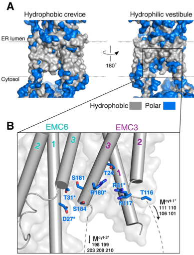
EMC3 is not an enzyme; rather, it is a core membrane insertase-like subunit that contributes directly to the EMC’s insertion pathway. In the human EMC structure, substrate insertion occurs via a membrane-embedded, enclosed β€œhydrophilic vestibule” formed by EMC3 and EMC6, and the complex uses local membrane thinning and a positively charged patch to reduce the energetic barrier of inserting challenging TMDs. (pleiner2020structuralbasisfor pages 1-3)

A key architectural/mechanistic framing is that EMC presents a cytosolic vestibule for initial TMD binding and a lipid-exposed intramembrane groove enabling energy-independent insertion following a hydrophobicity gradient from vestibule to membrane. (odonnell2020thearchitectureof pages 1-2, odonnell2020thearchitectureof pages 14-15)

2.3 Structural/biophysical determinants contributed by EMC3

Across human and yeast EMC structures and interpretation in authoritative reviews, EMC3:
- is structurally related to bacterial YidC (Oxa1 superfamily), consistent with insertase activity; (bai2020structureofthe pages 2-4, hegde2022thefunctionstructure pages 13-14)
- contains conserved basic residues within/near the vestibule whose mutation impairs insertion; (hegde2022thefunctionstructure pages 14-16)
- includes a methionine-rich cytosolic loop implicated in substrate capture/transfer into the vestibule; (pleiner2020structuralbasisfor pages 1-3)
- forms a three-helix domain (unusually extended vs many Oxa1-family proteins) that binds the cytosolic EMC scaffold subunit EMC2; (hegde2022thefunctionstructure pages 14-16)
These features support EMC3 being a principal functional element of the insertase machinery rather than a peripheral structural component.

3) Recent developments and latest research (prioritizing 2023–2024)

3.1 2023: A charge-based selectivity filter centered on EMC3 prevents misinsertion and enforces topology

Pleiner et al. (Journal of Cell Biology; May 2023; https://doi.org/10.1083/jcb.202212007) proposed and tested a selectivity filter in the EMC that protects ER proteome integrity by using charge repulsion at the vestibule entrance. Their model emphasizes that a substrate’s TMD is transiently captured while its adjacent polar tail β€œprobes” a positively charged hydrophilic vestibule; positively charged tails are repelled, increasing the likelihood of substrate rejection and reducing inappropriate ER insertion. (pleiner2023aselectivityfilter pages 10-11, pleiner2023aselectivityfilter pages 8-10)

Mechanistically and specifically for EMC3, they identify two conserved positively charged EMC3 residues (R31 and R180) as forming the charge barrier at the vestibule entrance (in the Oxa1 superfamily context), and show that changing vestibule charge (e.g., introducing negative charge) increases ER misinsertion of mitochondrial tail-anchored proteins and incorrect topology in multipass substrates. (pleiner2023aselectivityfilter pages 10-11, pleiner2023aselectivityfilter pages 8-10, pleiner2023aselectivityfilter pages 4-6)

This 2023 framework advances EMC3 annotation from β€œinsertase subunit” to an active fidelity determinant: EMC3’s vestibule electrostatics contribute to client discrimination, TA protein sorting (ER vs mitochondria), and positive-inside topology enforcement. (pleiner2023aselectivityfilter pages 10-11, pleiner2023aselectivityfilter pages 8-10)

3.2 2024: EMC-mediated post-translational β€œtopology rectification” expands EMC client scope

Wu et al. (Nature Structural & Molecular Biology; Nov 2024; https://doi.org/10.1038/s41594-023-01120-6) report that, in mammalian cells, some terminal TMDs near the C-terminus of multipass proteins are not fully inserted co-translationally and instead require a post-translational insertion step mediated by EMC to β€œrectify” topology after ribosome release. (wu2024emcrectifiesthe pages 1-2, wu2024emcrectifiesthe pages 7-9)

They estimate this sequential co-/post-translational mechanism may apply to roughly ~250 multipass proteins (new putative EMC-sensitive substrates), broadening the functional scope of EMC beyond classical TA insertion and selected co-translational events. (wu2024emcrectifiesthe pages 1-2, wu2024emcrectifiesthe pages 7-9)

The work also provides mechanistic constraints: EMC cannot access substrates very near a Sec61-bound ribosome exit tunnel, implying EMC acts after diffusion/partial release, consistent with an assembly/rectification role at late biogenesis steps for specific membrane-protein regions. (wu2024emcrectifiesthe pages 7-9)

4) Molecular function in pathways and cellular context

4.1 Pathway context: ER membrane protein biogenesis and quality control

EMC3 functions in the ER membrane biogenesis network that includes Sec61-mediated translocation/insertion. The EMC is positioned as a parallel/auxiliary insertase for β€œdifficult” TMDs (e.g., with polar/charged residues) and may also intersect with quality control pathways by preventing misinsertion (2023 selectivity filter) and helping complete topogenesis of multipass proteins (2024 rectification). (odonnell2020thearchitectureof pages 1-2, pleiner2023aselectivityfilter pages 8-10, wu2024emcrectifiesthe pages 1-2)

4.2 Substrates/clients: what types of proteins depend on EMC (and thus EMC3)

A major empirical theme is that EMC clients often contain at least one TMD with polar/charged residues, and altering these residues can shift EMC dependence.

  • In an unbiased quantitative proteomics study (Cell Reports; Sep 2019; https://doi.org/10.1016/j.celrep.2019.08.006), Tian et al. identified 36 EMC-dependent membrane proteins and 171 EMC-independent membrane proteins using stringent reproducibility criteria; EMC-dependent proteins were enriched among multipass proteins and were characterized by polar/charged residues in TMDs. (tian2019proteomicanalysisidentifies pages 1-3, tian2019proteomicanalysisidentifies pages 5-6)
  • Representative EMC-dependent proteins in that study include FDFT1/SQS (tail-anchored-like topology with low hydrophobicity), FZD6, ATP6V0A1, and SLC43A3; proteasome inhibition could rescue levels of some EMC-dependent proteins in EMC-deficient backgrounds, indicating failure of biogenesis leads to degradation. (tian2019proteomicanalysisidentifies pages 3-5, tian2019proteomicanalysisidentifies pages 5-6)
  • In structural/functional assays of human EMC, reporter substrates used to separate EMC-dependent insertion include SQS/FDFT1, VAMP2 (comparatively EMC-independent), and multipass reporters such as OPRK1 and TRAM2. (pleiner2020structuralbasisfor pages 7-11, pleiner2023aselectivityfilter pages 8-10)

5) Current applications and real-world implementations

5.1 Biomedical relevance via membrane-protein drug targets

Because a large fraction of therapeutics target integral membrane proteins, mechanisms governing membrane-protein biogenesis (including EMC3-dependent insertion) are relevant to broad biomedical contexts. The cryo-EM structure paper frames membrane protein biogenesis defects as underlying diverse human diseases and highlights the biomedical importance of understanding EMC-mediated insertion. (pleiner2020structuralbasisfor pages 7-11)

5.2 In vivo functional relevance demonstrated in mammalian retina

Although the report’s focus is human EMC3, mammalian in vivo models provide strong functional evidence for EMC3-dependent biogenesis of critical neural membrane proteins:
- Conditional loss of Emc3 in mouse photoreceptors leads to rhodopsin mislocalization and death of rod and cone photoreceptors, consistent with EMC3’s role in early biosynthesis/handling of a key multipass GPCR-like membrane protein in vivo. (xiong2020ercomplexproteins pages 1-2, xiong2020ercomplexproteins pages 10-13)
- Bipolar-cell conditional knockout shows progressive dysfunction and degeneration with quantitative electrophysiology and histology changes (below), offering a β€œreal-world” physiological implementation of EMC3-dependent membrane-protein maintenance in a defined neural circuit. (zhu2020lossofthe pages 4-6)

5.3 Limitations of evidence for direct clinical implementation

Within the retrieved evidence set, there were no direct, clinically deployed therapies that specifically target EMC3 or EMC insertase activity, and no human clinical trials were identified in this run. Translational relevance is therefore best supported currently through (i) mechanistic understanding of membrane-protein biogenesis and (ii) disease/phenotype models consistent with failure of EMC-dependent membrane-protein homeostasis.

6) Expert opinions and authoritative synthesis

The Annual Review of Biochemistry article by Hegde (Annual Review of Biochemistry; Jun 2022; https://doi.org/10.1146/annurev-biochem-032620-104553) provides an authoritative synthesis emphasizing:
- EMC is built around a conserved EMC3–EMC6 core; EMC3 is an Oxa1 superfamily member; (hegde2022thefunctionstructure pages 1-2)
- EMC has an established role in TMD insertion with additional less well understood roles in later folding/assembly; (hegde2022thefunctionstructure pages 1-2)
- key mechanistic questions remain about how EMC’s multiple putative substrate-handling modes relate (insertase versus chaperone-like effects), motivating careful substrate-specific topology/folding assays. (hegde2022thefunctionstructure pages 20-22)

This review perspective aligns with the trajectory of 2023–2024 primary research: EMC3 has clearly defined insertion and fidelity roles (2023), and EMC’s role in post-translational topology completion suggests an expanded functional repertoire consistent with β€œmultifunctional molecular machine” models. (pleiner2023aselectivityfilter pages 8-10, wu2024emcrectifiesthe pages 1-2)

7) Relevant statistics and quantitative data (recent studies emphasized where available)

7.1 Structural/biophysical quantitative measures

  • The human EMC cryo-EM structure was determined at ~3.4 Γ… overall resolution and includes a membrane region with 12 transmembrane helices (nine forming an ordered core) and a cytosolic basket (EMC2/EMC8). (pleiner2020structuralbasisfor pages 1-3)

7.2 Proteomics-defined EMC client set size

Tian et al. (Cell Reports; Sep 2019) quantified 5,570 proteins total; 4,446 were quantified with β‰₯2 peptides; 3,188 were membrane-associated by GO-CC; 971 carried UniProt β€œTransmembrane” annotation; among these, 36 were classified as EMC-dependent versus 171 as EMC-independent. (tian2019proteomicanalysisidentifies pages 3-5, tian2019proteomicanalysisidentifies pages 5-6)

7.3 2024 client-scope estimate for topology rectification

Wu et al. (NSMB; Nov 2024) estimated their sequential co-/post-translational rectification mechanism may apply to ~250 multipass proteins (new putative EMC substrates). (wu2024emcrectifiesthe pages 1-2, wu2024emcrectifiesthe pages 7-9)

7.4 Quantitative in vivo phenotype data linked to EMC3 loss

In a bipolar-cell-specific Emc3 conditional knockout in mice (PLoS ONE; Sep 2020; https://doi.org/10.1371/journal.pone.0238435):
- recombination affected ~75% of bipolar cells, with Emc3 expression reduced ~40–45% by RT-PCR; (zhu2020lossofthe pages 4-6)
- at 12 months, scotopic ERG b-wave amplitudes decreased ~50% and photopic b-waves ~30%, while a-waves were unchanged (post-receptoral defect); (zhu2020lossofthe pages 4-6)
- oscillatory potentials decreased to ~51–62% depending on flash intensity; (zhu2020lossofthe pages 4-6)
- ~50% loss of PKCΞ±+ rod bipolar cells and INL thickness reduced to ~55% of control. (zhu2020lossofthe pages 4-6)

8) Visual structural evidence (EMC3/EMC6 vestibule)

Cryo-EM figures from Pleiner et al. (Science; Jul 2020; https://doi.org/10.1126/science.abb5008) depict the EMC membrane region and highlight the hydrophilic vestibule formed by EMC3 and EMC6, including a cutaway/space-filling view of the conduit spanning the membraneβ€”directly supporting the mechanistic annotation of EMC3 as part of an intramembrane insertion vestibule. (pleiner2020structuralbasisfor media a4fc15a7, pleiner2020structuralbasisfor media 72e4d409)

9) Summary of functional annotation (concise)

Human EMC3 (Q9P0I2; TMEM111) is an ER-resident, Oxa1/YidC-like core subunit of the EMC insertase. Its primary function is to participate directly in selective insertion and topogenesis of a subset of membrane proteins by forming (with EMC6) a hydrophilic intramembrane vestibule; EMC3’s cytosolic loops mediate substrate capture, and its conserved vestibule charges enforce selectivity to prevent misinsertion and help implement the positive-inside topology rule. Recent work indicates EMC (and thus EMC3) also supports post-translational topology rectification for a substantial set of multipass proteins, consistent with a broader role in late-stage membrane-protein biogenesis and proteostasis. (pleiner2020structuralbasisfor pages 1-3, pleiner2023aselectivityfilter pages 8-10, pleiner2023aselectivityfilter pages 4-6, wu2024emcrectifiesthe pages 1-2)


Evidence summary table

Aspect Evidence Key sources (with year) URL
Identity/domains Human EMC3 corresponds to UniProt Q9P0I2, also called ER membrane protein complex subunit 3/TMEM111; it is a core transmembrane subunit of the ER membrane protein complex and belongs to the Oxa1/YidC/Alb3 insertase superfamily, consistent with EMC3-family/domain annotation (hegde2022thefunctionstructure pages 1-2, pleiner2020structuralbasisfor pages 1-3, odonnell2020thearchitectureof pages 1-2) Hegde 2022; Pleiner et al. 2020; O'Donnell et al. 2020 https://doi.org/10.1146/annurev-biochem-032620-104553 ; https://doi.org/10.1126/science.abb5008 ; https://doi.org/10.7554/elife.57887
Localization EMC3 is an ER membrane protein within the multi-subunit EMC; structurally it sits in the membrane core with EMC6 and contributes to the membrane-embedded insertase. In Drosophila retina, EMC3 co-localizes with the ER marker calnexin, supporting ER localization in vivo (hegde2022thefunctionstructure pages 13-14, pleiner2020structuralbasisfor pages 7-11, xiong2020ercomplexproteins pages 7-10) Pleiner et al. 2020; Hegde 2022; Xiong et al. 2020 https://doi.org/10.1126/science.abb5008 ; https://doi.org/10.1146/annurev-biochem-032620-104553 ; https://doi.org/10.1038/s41418-019-0378-6
Molecular function/mechanism EMC3 is a core insertase subunit that, together with EMC6, forms a hydrophilic intramembrane vestibule for insertion of selected transmembrane domains, especially tail-anchored proteins and some N-terminal or terminal helices of multipass proteins. EMC-mediated insertion is energy-independent and aided by membrane thinning and a positively charged patch that lower the energetic barrier for insertion (hegde2022thefunctionstructure pages 14-16, pleiner2020structuralbasisfor pages 7-11, odonnell2020thearchitectureof pages 1-2, pleiner2020structuralbasisfor pages 1-3) Pleiner et al. 2020; O'Donnell et al. 2020; Hegde 2022 https://doi.org/10.1126/science.abb5008 ; https://doi.org/10.7554/elife.57887 ; https://doi.org/10.1146/annurev-biochem-032620-104553
EMC3-specific structural elements EMC3 is a three-transmembrane, YidC-like bundle that abuts EMC6 at the complex midline; it has a methionine-rich cytosolic loop required for substrate capture/insertion, a conserved hydrophilic vestibule with key basic residues, lumenal-plane helices that may remodel bilayer properties, and a C-terminal three-helix bundle that binds the cytosolic scaffold EMC2 (hegde2022thefunctionstructure pages 13-14, hegde2022thefunctionstructure pages 14-16, pleiner2020structuralbasisfor pages 1-3, pleiner2023aselectivityfilter pages 4-6) Pleiner et al. 2020; Hegde 2022; Pleiner et al. 2023 https://doi.org/10.1126/science.abb5008 ; https://doi.org/10.1146/annurev-biochem-032620-104553 ; https://doi.org/10.1083/jcb.202212007
Client/substrate examples EMC-dependent clients include tail-anchored SQS/FDFT1 and multipass proteins such as OPRK1, TRAM2, ATP6V0A1, FZD6, SLC43A3, CD9, and SEC61A1; EMC dependence correlates with polar/charged residues in at least one transmembrane domain, and mutagenesis can switch proteins between EMC-dependent and EMC-independent states (pleiner2020structuralbasisfor pages 7-11, tian2019proteomicanalysisidentifies pages 1-3, tian2019proteomicanalysisidentifies pages 3-5, tian2019proteomicanalysisidentifies pages 5-6, tian2019proteomicanalysisidentifies pages 8-10) Pleiner et al. 2020; Tian et al. 2019 https://doi.org/10.1126/science.abb5008 ; https://doi.org/10.1016/j.celrep.2019.08.006
2023-2024 key advances 2023 work showed that EMC selectivity is enforced by a vestibule-entry charge filter centered on conserved EMC3 residues R31 and R180, which repel positively charged tails to reduce ER misinsertion of mitochondrial TA proteins and help enforce the positive-inside rule. 2024 work showed EMC can post-translationally rectify topology of multipass proteins by inserting C-terminal TMDs after ribosome release, expanding EMC function beyond earlier co-/post-translational insertion models (pleiner2023aselectivityfilter pages 10-11, pleiner2023aselectivityfilter pages 8-10, pleiner2023aselectivityfilter pages 2-4, pleiner2023aselectivityfilter pages 4-6, wu2024emcrectifiesthe pages 1-2, wu2024emcrectifiesthe pages 7-9, wu2024emcrectifiesthe pages 2-3) Pleiner et al. 2023; Wu et al. 2024 https://doi.org/10.1083/jcb.202212007 ; https://doi.org/10.1038/s41594-023-01120-6
Phenotypes/disease relevance In vivo loss of Emc3 impairs rhodopsin biogenesis and photoreceptor survival across species and causes retinal degeneration phenotypes. In mouse photoreceptors, Emc3 deletion caused rhodopsin mislocalization and rod/cone death; in bipolar-cell conditional knockout mice, Emc3 loss caused age-dependent bipolar-cell degeneration, synaptic disorganization, reduced ERG b-waves, and reactive gliosis, indicating dependence of neural membrane-protein homeostasis on EMC3 (xiong2020ercomplexproteins pages 1-2, zhu2020lossofthe pages 6-9, xiong2020ercomplexproteins pages 10-13, zhu2020lossofthe pages 1-2, zhu2020lossofthe pages 9-11, zhu2020lossofthe pages 4-6) Xiong et al. 2020; Zhu et al. 2020 https://doi.org/10.1038/s41418-019-0378-6 ; https://doi.org/10.1371/journal.pone.0238435
Quantitative statistics Structural studies resolved human EMC at 3.4 Γ… overall resolution with ~12 TMs in the membrane region; proteomics identified 5,570 proteins total, 4,446 quantified, 3,188 membrane-associated, 971 with transmembrane annotation, and 36 EMC-dependent membrane proteins versus 171 EMC-independent ones. Recent topology work estimated ~250 new putative EMC-sensitive multipass substrates. In bipolar-cell cKO mice, Emc3 deletion affected ~75% of BCs, reduced Emc3 expression by ~40-45%, lowered scotopic b-waves by ~50%, photopic b-waves by ~30%, OPs to ~51-62% of control, caused ~50% loss of PKCΞ±+ rod bipolar cells, and reduced INL thickness to ~55% of control at 12 months (pleiner2020structuralbasisfor pages 1-3, tian2019proteomicanalysisidentifies pages 3-5, tian2019proteomicanalysisidentifies pages 5-6, wu2024emcrectifiesthe pages 7-9, zhu2020lossofthe pages 9-11, zhu2020lossofthe pages 4-6) Pleiner et al. 2020; Tian et al. 2019; Wu et al. 2024; Zhu et al. 2020 https://doi.org/10.1126/science.abb5008 ; https://doi.org/10.1016/j.celrep.2019.08.006 ; https://doi.org/10.1038/s41594-023-01120-6 ; https://doi.org/10.1371/journal.pone.0238435

Table: This table summarizes verified identity, mechanism, localization, key recent advances, representative clients, phenotypic relevance, and quantitative evidence for human EMC3 (UniProt Q9P0I2). It is useful as a compact evidence-based reference for functional annotation.

References

  1. (hegde2022thefunctionstructure pages 1-2): Ramanujan S. Hegde. The function, structure, and origins of the er membrane protein complex. Annual Review of Biochemistry, 91:651-678, Jun 2022. URL: https://doi.org/10.1146/annurev-biochem-032620-104553, doi:10.1146/annurev-biochem-032620-104553. This article has 65 citations and is from a domain leading peer-reviewed journal.

  2. (pleiner2020structuralbasisfor pages 1-3): Tino Pleiner, Giovani Pinton Tomaleri, Kurt Januszyk, Alison J. Inglis, Masami Hazu, and Rebecca M. Voorhees. Structural basis for membrane insertion by the human er membrane protein complex. Jul 2020. URL: https://doi.org/10.1126/science.abb5008, doi:10.1126/science.abb5008. This article has 192 citations and is from a highest quality peer-reviewed journal.

  3. (odonnell2020thearchitectureof pages 1-2): John P O'Donnell, Ben P Phillips, Yuichi Yagita, Szymon Juszkiewicz, Armin Wagner, Duccio Malinverni, Robert J Keenan, Elizabeth A Miller, and Ramanujan S Hegde. The architecture of emc reveals a path for membrane protein insertion. May 2020. URL: https://doi.org/10.7554/elife.57887, doi:10.7554/elife.57887. This article has 121 citations and is from a domain leading peer-reviewed journal.

  4. (hegde2022thefunctionstructure pages 20-22): Ramanujan S. Hegde. The function, structure, and origins of the er membrane protein complex. Annual Review of Biochemistry, 91:651-678, Jun 2022. URL: https://doi.org/10.1146/annurev-biochem-032620-104553, doi:10.1146/annurev-biochem-032620-104553. This article has 65 citations and is from a domain leading peer-reviewed journal.

  5. (odonnell2020thearchitectureof pages 14-15): John P O'Donnell, Ben P Phillips, Yuichi Yagita, Szymon Juszkiewicz, Armin Wagner, Duccio Malinverni, Robert J Keenan, Elizabeth A Miller, and Ramanujan S Hegde. The architecture of emc reveals a path for membrane protein insertion. May 2020. URL: https://doi.org/10.7554/elife.57887, doi:10.7554/elife.57887. This article has 121 citations and is from a domain leading peer-reviewed journal.

  6. (bai2020structureofthe pages 2-4): Lin Bai, Qinglong You, Xiang Feng, Amanda Kovach, and Huilin Li. Structure of the er membrane complex, a transmembrane-domain insertase. Jun 2020. URL: https://doi.org/10.1038/s41586-020-2389-3, doi:10.1038/s41586-020-2389-3. This article has 164 citations and is from a highest quality peer-reviewed journal.

  7. (hegde2022thefunctionstructure pages 13-14): Ramanujan S. Hegde. The function, structure, and origins of the er membrane protein complex. Annual Review of Biochemistry, 91:651-678, Jun 2022. URL: https://doi.org/10.1146/annurev-biochem-032620-104553, doi:10.1146/annurev-biochem-032620-104553. This article has 65 citations and is from a domain leading peer-reviewed journal.

  8. (hegde2022thefunctionstructure pages 14-16): Ramanujan S. Hegde. The function, structure, and origins of the er membrane protein complex. Annual Review of Biochemistry, 91:651-678, Jun 2022. URL: https://doi.org/10.1146/annurev-biochem-032620-104553, doi:10.1146/annurev-biochem-032620-104553. This article has 65 citations and is from a domain leading peer-reviewed journal.

  9. (pleiner2023aselectivityfilter pages 10-11): Tino Pleiner, Masami Hazu, Giovani Pinton Tomaleri, Vy N. Nguyen, Kurt Januszyk, and Rebecca M. Voorhees. A selectivity filter in the er membrane protein complex limits protein misinsertion at the er. The Journal of Cell Biology, May 2023. URL: https://doi.org/10.1083/jcb.202212007, doi:10.1083/jcb.202212007. This article has 28 citations.

  10. (pleiner2023aselectivityfilter pages 8-10): Tino Pleiner, Masami Hazu, Giovani Pinton Tomaleri, Vy N. Nguyen, Kurt Januszyk, and Rebecca M. Voorhees. A selectivity filter in the er membrane protein complex limits protein misinsertion at the er. The Journal of Cell Biology, May 2023. URL: https://doi.org/10.1083/jcb.202212007, doi:10.1083/jcb.202212007. This article has 28 citations.

  11. (pleiner2023aselectivityfilter pages 4-6): Tino Pleiner, Masami Hazu, Giovani Pinton Tomaleri, Vy N. Nguyen, Kurt Januszyk, and Rebecca M. Voorhees. A selectivity filter in the er membrane protein complex limits protein misinsertion at the er. The Journal of Cell Biology, May 2023. URL: https://doi.org/10.1083/jcb.202212007, doi:10.1083/jcb.202212007. This article has 28 citations.

  12. (wu2024emcrectifiesthe pages 1-2): Haoxi Wu, Luka SmalinskaitΔ—, and Ramanujan S. Hegde. Emc rectifies the topology of multipass membrane proteins. Nature Structural & Molecular Biology, 31:32-41, Nov 2024. URL: https://doi.org/10.1038/s41594-023-01120-6, doi:10.1038/s41594-023-01120-6. This article has 41 citations and is from a highest quality peer-reviewed journal.

  13. (wu2024emcrectifiesthe pages 7-9): Haoxi Wu, Luka SmalinskaitΔ—, and Ramanujan S. Hegde. Emc rectifies the topology of multipass membrane proteins. Nature Structural & Molecular Biology, 31:32-41, Nov 2024. URL: https://doi.org/10.1038/s41594-023-01120-6, doi:10.1038/s41594-023-01120-6. This article has 41 citations and is from a highest quality peer-reviewed journal.

  14. (tian2019proteomicanalysisidentifies pages 1-3): Songhai Tian, Quan Wu, Bo Zhou, Mei Yuk Choi, Bo Ding, Wei Yang, and Min Dong. Proteomic analysis identifies membrane proteins dependent on the er membrane protein complex. Cell reports, 28:2517-2526.e5, Sep 2019. URL: https://doi.org/10.1016/j.celrep.2019.08.006, doi:10.1016/j.celrep.2019.08.006. This article has 79 citations and is from a highest quality peer-reviewed journal.

  15. (tian2019proteomicanalysisidentifies pages 5-6): Songhai Tian, Quan Wu, Bo Zhou, Mei Yuk Choi, Bo Ding, Wei Yang, and Min Dong. Proteomic analysis identifies membrane proteins dependent on the er membrane protein complex. Cell reports, 28:2517-2526.e5, Sep 2019. URL: https://doi.org/10.1016/j.celrep.2019.08.006, doi:10.1016/j.celrep.2019.08.006. This article has 79 citations and is from a highest quality peer-reviewed journal.

  16. (tian2019proteomicanalysisidentifies pages 3-5): Songhai Tian, Quan Wu, Bo Zhou, Mei Yuk Choi, Bo Ding, Wei Yang, and Min Dong. Proteomic analysis identifies membrane proteins dependent on the er membrane protein complex. Cell reports, 28:2517-2526.e5, Sep 2019. URL: https://doi.org/10.1016/j.celrep.2019.08.006, doi:10.1016/j.celrep.2019.08.006. This article has 79 citations and is from a highest quality peer-reviewed journal.

  17. (pleiner2020structuralbasisfor pages 7-11): Tino Pleiner, Giovani Pinton Tomaleri, Kurt Januszyk, Alison J. Inglis, Masami Hazu, and Rebecca M. Voorhees. Structural basis for membrane insertion by the human er membrane protein complex. Jul 2020. URL: https://doi.org/10.1126/science.abb5008, doi:10.1126/science.abb5008. This article has 192 citations and is from a highest quality peer-reviewed journal.

  18. (xiong2020ercomplexproteins pages 1-2): Liangyao Xiong, Lin Zhang, Yeming Yang, Na Li, Wenjia Lai, Fengchao Wang, Xianjun Zhu, and Tao Wang. Er complex proteins are required for rhodopsin biosynthesis and photoreceptor survival in drosophila and mice. Cell Death and Differentiation, 27:646-661, Jul 2020. URL: https://doi.org/10.1038/s41418-019-0378-6, doi:10.1038/s41418-019-0378-6. This article has 45 citations and is from a domain leading peer-reviewed journal.

  19. (xiong2020ercomplexproteins pages 10-13): Liangyao Xiong, Lin Zhang, Yeming Yang, Na Li, Wenjia Lai, Fengchao Wang, Xianjun Zhu, and Tao Wang. Er complex proteins are required for rhodopsin biosynthesis and photoreceptor survival in drosophila and mice. Cell Death and Differentiation, 27:646-661, Jul 2020. URL: https://doi.org/10.1038/s41418-019-0378-6, doi:10.1038/s41418-019-0378-6. This article has 45 citations and is from a domain leading peer-reviewed journal.

  20. (zhu2020lossofthe pages 4-6): Xiong Zhu, Xin Qi, Yeming Yang, Wanli Tian, Wenjing Liu, Zhilin Jiang, Shuzhen Li, and Xianjun Zhu. Loss of the er membrane protein complex subunit emc3 leads to retinal bipolar cell degeneration in aged mice. PLoS ONE, 15:e0238435, Sep 2020. URL: https://doi.org/10.1371/journal.pone.0238435, doi:10.1371/journal.pone.0238435. This article has 16 citations and is from a peer-reviewed journal.

  21. (pleiner2020structuralbasisfor media a4fc15a7): Tino Pleiner, Giovani Pinton Tomaleri, Kurt Januszyk, Alison J. Inglis, Masami Hazu, and Rebecca M. Voorhees. Structural basis for membrane insertion by the human er membrane protein complex. Jul 2020. URL: https://doi.org/10.1126/science.abb5008, doi:10.1126/science.abb5008. This article has 192 citations and is from a highest quality peer-reviewed journal.

  22. (pleiner2020structuralbasisfor media 72e4d409): Tino Pleiner, Giovani Pinton Tomaleri, Kurt Januszyk, Alison J. Inglis, Masami Hazu, and Rebecca M. Voorhees. Structural basis for membrane insertion by the human er membrane protein complex. Jul 2020. URL: https://doi.org/10.1126/science.abb5008, doi:10.1126/science.abb5008. This article has 192 citations and is from a highest quality peer-reviewed journal.

  23. (xiong2020ercomplexproteins pages 7-10): Liangyao Xiong, Lin Zhang, Yeming Yang, Na Li, Wenjia Lai, Fengchao Wang, Xianjun Zhu, and Tao Wang. Er complex proteins are required for rhodopsin biosynthesis and photoreceptor survival in drosophila and mice. Cell Death and Differentiation, 27:646-661, Jul 2020. URL: https://doi.org/10.1038/s41418-019-0378-6, doi:10.1038/s41418-019-0378-6. This article has 45 citations and is from a domain leading peer-reviewed journal.

  24. (tian2019proteomicanalysisidentifies pages 8-10): Songhai Tian, Quan Wu, Bo Zhou, Mei Yuk Choi, Bo Ding, Wei Yang, and Min Dong. Proteomic analysis identifies membrane proteins dependent on the er membrane protein complex. Cell reports, 28:2517-2526.e5, Sep 2019. URL: https://doi.org/10.1016/j.celrep.2019.08.006, doi:10.1016/j.celrep.2019.08.006. This article has 79 citations and is from a highest quality peer-reviewed journal.

  25. (pleiner2023aselectivityfilter pages 2-4): Tino Pleiner, Masami Hazu, Giovani Pinton Tomaleri, Vy N. Nguyen, Kurt Januszyk, and Rebecca M. Voorhees. A selectivity filter in the er membrane protein complex limits protein misinsertion at the er. The Journal of Cell Biology, May 2023. URL: https://doi.org/10.1083/jcb.202212007, doi:10.1083/jcb.202212007. This article has 28 citations.

  26. (wu2024emcrectifiesthe pages 2-3): Haoxi Wu, Luka SmalinskaitΔ—, and Ramanujan S. Hegde. Emc rectifies the topology of multipass membrane proteins. Nature Structural & Molecular Biology, 31:32-41, Nov 2024. URL: https://doi.org/10.1038/s41594-023-01120-6, doi:10.1038/s41594-023-01120-6. This article has 41 citations and is from a highest quality peer-reviewed journal.

  27. (zhu2020lossofthe pages 6-9): Xiong Zhu, Xin Qi, Yeming Yang, Wanli Tian, Wenjing Liu, Zhilin Jiang, Shuzhen Li, and Xianjun Zhu. Loss of the er membrane protein complex subunit emc3 leads to retinal bipolar cell degeneration in aged mice. PLoS ONE, 15:e0238435, Sep 2020. URL: https://doi.org/10.1371/journal.pone.0238435, doi:10.1371/journal.pone.0238435. This article has 16 citations and is from a peer-reviewed journal.

  28. (zhu2020lossofthe pages 1-2): Xiong Zhu, Xin Qi, Yeming Yang, Wanli Tian, Wenjing Liu, Zhilin Jiang, Shuzhen Li, and Xianjun Zhu. Loss of the er membrane protein complex subunit emc3 leads to retinal bipolar cell degeneration in aged mice. PLoS ONE, 15:e0238435, Sep 2020. URL: https://doi.org/10.1371/journal.pone.0238435, doi:10.1371/journal.pone.0238435. This article has 16 citations and is from a peer-reviewed journal.

  29. (zhu2020lossofthe pages 9-11): Xiong Zhu, Xin Qi, Yeming Yang, Wanli Tian, Wenjing Liu, Zhilin Jiang, Shuzhen Li, and Xianjun Zhu. Loss of the er membrane protein complex subunit emc3 leads to retinal bipolar cell degeneration in aged mice. PLoS ONE, 15:e0238435, Sep 2020. URL: https://doi.org/10.1371/journal.pone.0238435, doi:10.1371/journal.pone.0238435. This article has 16 citations and is from a peer-reviewed journal.

Artifacts

Citations

  1. pleiner2020structuralbasisfor pages 1-3
  2. hegde2022thefunctionstructure pages 14-16
  3. wu2024emcrectifiesthe pages 7-9
  4. pleiner2020structuralbasisfor pages 7-11
  5. zhu2020lossofthe pages 4-6
  6. hegde2022thefunctionstructure pages 1-2
  7. hegde2022thefunctionstructure pages 20-22
  8. odonnell2020thearchitectureof pages 1-2
  9. odonnell2020thearchitectureof pages 14-15
  10. bai2020structureofthe pages 2-4
  11. hegde2022thefunctionstructure pages 13-14
  12. pleiner2023aselectivityfilter pages 10-11
  13. pleiner2023aselectivityfilter pages 8-10
  14. pleiner2023aselectivityfilter pages 4-6
  15. wu2024emcrectifiesthe pages 1-2
  16. tian2019proteomicanalysisidentifies pages 1-3
  17. tian2019proteomicanalysisidentifies pages 5-6
  18. tian2019proteomicanalysisidentifies pages 3-5
  19. xiong2020ercomplexproteins pages 1-2
  20. xiong2020ercomplexproteins pages 10-13
  21. xiong2020ercomplexproteins pages 7-10
  22. tian2019proteomicanalysisidentifies pages 8-10
  23. pleiner2023aselectivityfilter pages 2-4
  24. wu2024emcrectifiesthe pages 2-3
  25. zhu2020lossofthe pages 6-9
  26. zhu2020lossofthe pages 1-2
  27. zhu2020lossofthe pages 9-11
  28. https://doi.org/10.1083/jcb.202212007
  29. https://doi.org/10.1038/s41594-023-01120-6
  30. https://doi.org/10.1016/j.celrep.2019.08.006
  31. https://doi.org/10.1146/annurev-biochem-032620-104553
  32. https://doi.org/10.1371/journal.pone.0238435
  33. https://doi.org/10.1126/science.abb5008
  34. https://doi.org/10.7554/elife.57887
  35. https://doi.org/10.1038/s41418-019-0378-6
  36. https://doi.org/10.1146/annurev-biochem-032620-104553,
  37. https://doi.org/10.1126/science.abb5008,
  38. https://doi.org/10.7554/elife.57887,
  39. https://doi.org/10.1038/s41586-020-2389-3,
  40. https://doi.org/10.1083/jcb.202212007,
  41. https://doi.org/10.1038/s41594-023-01120-6,
  42. https://doi.org/10.1016/j.celrep.2019.08.006,
  43. https://doi.org/10.1038/s41418-019-0378-6,
  44. https://doi.org/10.1371/journal.pone.0238435,

πŸ“š Additional Documentation

Notes

(EMC3-notes.md)

EMC3 (human, Q9P0I2) review notes

Identity / architecture

  • EMC3 = ER membrane protein complex subunit 3 (synonym TMEM111). 261 aa, multi-pass ER membrane protein with 3 TM helices (TM1 15-38, TM2 115-130, TM3 169-187; N-terminus lumenal) [file:human/EMC3/EMC3-uniprot.txt TOPO_DOM/TRANSMEM features].
  • Oxa1/YidC-superfamily insertase fold; Pfam PF01956 EMC3_TMCO1; distant homolog of Get1/YidC: "EMC3 is a distant homolog of Get1" PMID:29242231, both "evolved from an ancestral prokaryotic insertase of the YidC family".
  • Belongs to the EMC3 family [file:human/EMC3/EMC3-uniprot.txt "Belongs to the EMC3 family"].

EMC complex membership

  • Identified biochemically in the EMC during ERAD network mapping [PMID:22119785; UniProt "Component of the ER membrane protein complex (EMC)."].
  • Strongest verified binary partner is EMC2: UniProt INTERACTION line "Q9P0I2; Q15006: EMC2; NbExp=12" β€” EMC2 is the cytosolic scaffold that cradles EMC3.

Function β€” EMC is a TMD insertase; EMC3 is the catalytic core

  • EMC catalyzes energy-independent insertion of newly synthesized membrane proteins into the ER membrane [file:human/EMC3/EMC3-uniprot.txt "(EMC) that enables the energy-independent insertion into endoplasmic"].
  • Demonstrated as a transmembrane-domain insertase, reconstituted in liposomes: "EMC is a transmembrane domain insertase, a function" PMID:29242231; "EMC as an ER-resident insertase for moderately hydrophobic TMDs" PMID:29242231.
  • Post-translational insertion of tail-anchored (TA) proteins (e.g. SQS/squalene synthase) [PMID:29242231; UniProt "post-translational insertion of tail-anchored/TA proteins in"].
  • Cotranslational role for multipass proteins: "EMC binds to and promotes the biogenesis of a range of multipass transmembrane proteins" and "EMC engages clients cotranslationally and immediately following" clusters of charged TMDs PMID:29809151.
  • Initiates accurate topogenesis by inserting the first TMD (N-exo) of GPCRs; cooperates with Sec61: "EMC inserts TMDs co-translationally and cooperates with the Sec61 translocon to ensure accurate topogenesis" PMID:30415835; "Sec61 translocon was necessary for insertion of the next TMD" PMID:30415835. This = stop-transfer membrane-anchor insertion (GO:0045050).

EMC3 is the substrate-conducting active site (insertase core)

  • Cryo-EM structure: substrate insertion occurs via "an enclosed hydrophilic vestibule within the membrane formed by the subunits EMC3 and EMC6" [PMID:32439656 abstract]; requires "a methionine-rich cytosolic loop".
  • Structure-guided MUTAGEN of EMC3 residues R31, M101/M106/M110/M111, R180 (the Met-rich loop and the hydrophilic vestibule): "No effect on EMC assembly but decreased membrane insertion of hydrophobic transmembrane helices-containing proteins by the EMC" [file:human/EMC3/EMC3-uniprot.txt MUTAGEN 31/101/106/110/111/180]. Controls F173 and conservative R->K substitutions had no effect. => EMC3 forms the substrate-conducting insertase active site; activity separable from assembly.
  • Hence GO:0032977 membrane insertase activity with qualifier contributes_to is the CORE molecular function (EMC3 is the catalytic subunit but acts within the complex).

Curation decisions (summary)

  • CORE: GO:0032977 (insertase activity, contributes_to), GO:0071816 (TA insertion), GO:0045050 (stop-transfer insertion), GO:0072546 (EMC complex), GO:0005789 (ER membrane). All ACCEPT.
  • GO:0016020 membrane (generic parent of ER membrane) -> KEEP_AS_NON_CORE.
  • GO:0005515 protein binding IPIs (PMID:26496610/32296183/33961781/35271311/40205054, mostly the EMC2 interaction) -> KEEP_AS_NON_CORE (real high-throughput interactions but uninformative bare term).
  • No REMOVE/MODIFY/UNDECIDED needed; experimental insertase/process annotations are well supported by full-text and the EMC3-specific mutagenesis.

Verification pass (2026-06-11)

  • All 22 PENDING annotations reviewed and filled; status COMPLETE.
  • Verbatim supporting substrings confirmed present: uniprot "Component of the ER membrane protein complex (EMC).", "SUBCELLULAR LOCATION: Endoplasmic reticulum membrane", "Multi-pass membrane protein", "Q9P0I2; Q15006: EMC2", "No effect on EMC assembly but decreased" (EMC3 vestibule/Met-loop mutagenesis), "post-translational insertion of tail-anchored/TA proteins in", "stop-transfer membrane-anchor sequences become ER membrane spanning"; PMID:29242231 "transmembrane domain insertase" / "tail-anchored membrane proteins with moderately hydrophobic transmembrane" / "EMC3 is a distant homolog of Get1"; PMID:32439656 "formed by the subunits EMC3 and EMC6" / "enclosed hydrophilic" / "methionine-rich cytosolic loop"; PMID:30415835 "G protein-coupled receptors".
  • IPI partners: PMID:26496610/33961781/35271311/40205054 -> EMC2 (Q15006); PMID:32296183 -> Q13126 (non-EMC, likely incidental/client). All bare protein binding -> KEEP_AS_NON_CORE.
  • CORE molecular function GO:0032977 (contributes_to) is the catalytic insertase activity; ACCEPT. No REMOVE/MODIFY/UNDECIDED needed.

Falcon deep-research findings (incorporated 2026-06)

New recent (2020-2024) papers identified by Falcon deep research and verified against PubMed; added to the review references. EMC3-specific / EMC3-vestibule findings not previously captured:

  • EMC3 selectivity filter (EMC3-specific mechanism): two conserved positively charged EMC3 residues, R31 and R180, at the hydrophilic-vestibule entrance form a charge-repulsion filter that rejects mitochondrial tail-anchored proteins and enforces the positive-inside rule; making the vestibule negative increases ER misinsertion PMID:37199759. Advances EMC3 from "insertase subunit" to an active client-discrimination/topology-fidelity determinant (added to insertase core_function supported_by).
  • Post-translational topology rectification at the EMC3/EMC6 vestibule: EMC inserts C-terminal TMDs of multipass proteins (e.g. SOAT1) after ribosome release; the EMC cytosol-facing hydrophilic vestibule is adjacent to the pre-translocated C-tail; ~250 putative multipass clients PMID:37957425. Expands the EMC3 catalytic vestibule's role (added to process core_function supported_by).
  • Gating plug regulates the EMC3 vestibule: human EMC cryo-EM (apo vs VDAC-bound) reveals a gating plug inside the hydrophilic vestibule (the substrate-binding pocket); its conformational change suggests the EMC is not insertion-competent in the VDAC1-bound state, i.e. state-dependent regulation of the EMC3/EMC6 insertion pocket PMID:38517390.
  • In vivo EMC3 requirement (disease/physiology): emc3 loss in Drosophila causes defective phototransduction and photoreceptor degeneration with reduced rhodopsin (ERAD-independent); conditional Emc3 knockout in mice causes rhodopsin mislocalization and rod/cone photoreceptor death PMID:31263175. Conserved in vivo evidence that EMC3 is required for biogenesis of rhodopsin, a multipass GPCR-like client.
  • Note (not added to YAML): Falcon also cited Zhu et al. 2020 (PLoS ONE, DOI:10.1371/journal.pone.0238435), a mouse bipolar-cell Emc3 conditional-KO retinal degeneration study; a PMID could not be resolved via PubMed search in this pass, so it is recorded here only by DOI. The closely related, fully PubMed-verified Xiong 2020 paper (PMID:31263175) covers the core EMC3 retina in-vivo finding and was added instead.

Pn Notes

(EMC3-pn-notes.md)

EMC3 PN Consistency Notes

  • Generated: 2026-06-18
  • Project: PROTEOSTASIS
  • Scope: PN consistency rereview against local AIGR review and available deep-research artifacts
  • UniProt: Q9P0I2
  • AIGR review status: COMPLETE
  • Review batch: proteostasis-batch-2026-06-11
  • Batch change status: added

Source Files Checked

Deep Research Files

AIGR Review Snapshot

  • Description: EMC3 (ER membrane protein complex subunit 3; also TMEM111) is a 261-residue polytopic ER membrane protein with three transmembrane helices and a lumenal N-terminus, and is the catalytic insertase subunit of the ER membrane protein complex (EMC), a conserved nine- to ten-subunit transmembrane-domain insertase and membrane-protein chaperone of the endoplasmic reticulum. EMC3 belongs to the Oxa1/YidC/Get1 insertase superfamily and is a distant homolog of the tail-anchored-protein insertase Get1; together with the small subunit EMC6 it forms the membrane-embedded hydrophilic vestibule through which substrate transmembrane domains are inserted. A methionine-rich cytosolic loop of EMC3 is required for substrate engagement, and structure-guided mutations of EMC3 residues lining the vestibule (e.g. Arg-31, the Met-rich loop, Arg-180) reduce client insertion without disrupting complex assembly, demonstrating that EMC3 provides the substrate-conducting active site. As part of the EMC, EMC3 enables the energy-independent insertion of newly synthesized membrane proteins into the ER membrane, with a preference for transmembrane domains that are weakly hydrophobic or carry destabilizing charged or aromatic residues. It mediates post-translational insertion of tail-anchored proteins and cotranslational insertion and N-exo topogenesis of multipass membrane proteins, including the first transmembrane domain of G protein-coupled receptors, in cooperation with the Sec61 translocon. EMC3 is broadly expressed and resides in the ER membrane.
  • Existing/core annotation action counts: ACCEPT: 15; KEEP_AS_NON_CORE: 7

PN Consistency Summary

  • Consistency: Strong agreement, and notably EMC3 is the catalytic insertase subunit (Oxa1/YidC/Get1 superfamily) forming the EMC3/EMC6 vestibule. Deep research, review, and PN annotation are aligned. Review correctly elevates GO:0032977 membrane insertase activity to core (contributes_to, but distinguished from accessory subunits by vestibule mutagenesis R31/R180, Met-rich loop). GO:0072546, ER membrane, and insertion BPs all captured. No contradictions.
  • PN story / NEW pressure: PN ("transmembrane protein import" / "EMC complex component") asserts nothing beyond GO coverage. EMC3's catalytic/selectivity-filter role (PMID:37199759) is captured by GO:0032977 + insertion BPs. No new GO term warranted. Conclusion: already captured.
  • Evidence alignment: Excellent overlap (PMID:22119785, PMID:29242231, PMID:32439656, PMID:30415835). Review adds EMC3-specific mechanistic depth: selectivity filter PMID:37199759, topology rectification PMID:37957425, in vivo rhodopsin/photoreceptor requirement PMID:31263175. PN cites no row-1 titles; no divergence.
  • Verdict: Consistent (EMC3 = catalytic core, correctly handled); PN adds no NEW pressure; projected group/class terms broader than review (no mapping change warranted).

Full Consistency Review

  • UniProt: Q9P0I2 Β· batch: proteostasis-batch-2026-06-11 Β· review status: COMPLETE (thorough; all annotations adjudicated)
  • PN placement: ER proteostasis | Protein transport | Transmembrane protein import | EMC complex component; PN-node mapping: type=mapped/ok_for_propagation β†’ GO:0072546 EMC complex (already_in_goa_exact); groupβ†’GO:0044743, classβ†’GO:0015031 (new_to_goa); branch=no_mapping.
  • Consistency: Strong agreement, and notably EMC3 is the catalytic insertase subunit (Oxa1/YidC/Get1 superfamily) forming the EMC3/EMC6 vestibule. Deep research, review, and PN annotation are aligned. Review correctly elevates GO:0032977 membrane insertase activity to core (contributes_to, but distinguished from accessory subunits by vestibule mutagenesis R31/R180, Met-rich loop). GO:0072546, ER membrane, and insertion BPs all captured. No contradictions.
  • PN story / NEW pressure: PN ("transmembrane protein import" / "EMC complex component") asserts nothing beyond GO coverage. EMC3's catalytic/selectivity-filter role (PMID:37199759) is captured by GO:0032977 + insertion BPs. No new GO term warranted. Conclusion: already captured.
  • Mapping strategy: EMC3 does not change the node. typeβ†’GO:0072546 is correct/exact for complex membership; importantly the EMC complex CC term (membership) is correctly distinguished from EMC3's catalytic insertase MF β€” the PN node maps only membership, which is right. Projected group/class terms (GO:0044743, GO:0015031) are broader than the review's specific terms β€” broader-ancestor pattern (cf. TOMM20/HSPA8/RAB7A). No mapping change warranted.
  • Evidence alignment: Excellent overlap (PMID:22119785, PMID:29242231, PMID:32439656, PMID:30415835). Review adds EMC3-specific mechanistic depth: selectivity filter PMID:37199759, topology rectification PMID:37957425, in vivo rhodopsin/photoreceptor requirement PMID:31263175. PN cites no row-1 titles; no divergence.
  • Verdict: Consistent (EMC3 = catalytic core, correctly handled); PN adds no NEW pressure; projected group/class terms broader than review (no mapping change warranted).

PN Dossier Context

  • review_batch: proteostasis-batch-2026-06-11
  • review_yaml: genes/human/EMC3/EMC3-ai-review.yaml
  • PN workbook rows: 1

PN row 1: ER proteostasis | Protein transport | Transmembrane protein import | EMC complex component

  • UniProt: Q9P0I2
  • In branches: ER
  • PN-node mapping records (path + ancestors):
    • [type] ER proteostasis|Protein transport|Transmembrane protein import|EMC complex component
      status=mapped scope=ok_for_propagation_to_go GO=[GO:0072546 EMC complex]
      rationale: This PN type denotes ER membrane protein complex components. The GO EMC complex cellular-component term is the direct target.
    • [group] ER proteostasis|Protein transport|Transmembrane protein import
      status=mapped scope=ok_for_propagation_to_go GO=[GO:0044743 protein transmembrane import into intracellular organelle]
      rationale: This PN group covers ER transmembrane-protein insertion/import systems such as EMC- and PAT-related pathways. The local GO cache does not expose an ER-specific matching term, so the broader intracellular-organelle transmembrane-import process is the best supported propagation target.
    • [class] ER proteostasis|Protein transport
      status=mapped scope=ok_for_propagation_to_go GO=[GO:0015031 protein transport]
      rationale: The PN ER Protein transport class groups ER-targeting and ER-insertion pathways. GO protein transport is the appropriate propagation target, while the source class remains ER-specific and broader than any single GO transport subtype.
    • [branch] ER proteostasis
      status=no_mapping scope= GO=[]
      rationale: Reviewed as a top-level PN branch. This is a systems/taxonomy umbrella, not a direct GO assertion; narrower child curations carry any propagating GO mappings.

Projected GO annotations (3)

  • GO:0015031 protein transport | scope=ok_for_propagation_to_go | goa_status=new_to_goa | from=ER proteostasis|Protein transport
  • GO:0044743 protein transmembrane import into intracellular organelle | scope=ok_for_propagation_to_go | goa_status=new_to_goa | from=ER proteostasis|Protein transport|Transmembrane protein import
  • GO:0072546 EMC complex | scope=ok_for_propagation_to_go | goa_status=already_in_goa_exact | from=ER proteostasis|Protein transport|Transmembrane protein import|EMC complex component

Note

This file is generated from the current PROTEOSTASIS phase-1 dossier and local gene-review artifacts. Edit the source review, PN mapping, or dossier rather than this generated note when correcting the underlying curation.

πŸ“„ View Raw YAML

id: Q9P0I2
gene_symbol: EMC3
product_type: PROTEIN
status: COMPLETE
taxon:
  id: NCBITaxon:9606
  label: Homo sapiens
description: EMC3 (ER membrane protein complex subunit 3; also TMEM111) is a 261-residue polytopic ER membrane protein with three transmembrane helices and a lumenal N-terminus, and is the catalytic insertase subunit of the ER membrane protein complex (EMC), a conserved nine- to ten-subunit transmembrane-domain insertase and membrane-protein chaperone of the endoplasmic reticulum. EMC3 belongs to the Oxa1/YidC/Get1 insertase superfamily and is a distant homolog of the tail-anchored-protein insertase Get1; together with the small subunit EMC6 it forms the membrane-embedded hydrophilic vestibule through which substrate transmembrane domains are inserted. A methionine-rich cytosolic loop of EMC3 is required for substrate engagement, and structure-guided mutations of EMC3 residues lining the vestibule (e.g. Arg-31, the Met-rich loop, Arg-180) reduce client insertion without disrupting complex assembly, demonstrating that EMC3 provides the substrate-conducting active site. As part of the EMC, EMC3 enables the energy-independent insertion of newly synthesized membrane proteins into the ER membrane, with a preference for transmembrane domains that are weakly hydrophobic or carry destabilizing charged or aromatic residues. It mediates post-translational insertion of tail-anchored proteins and cotranslational insertion and N-exo topogenesis of multipass membrane proteins, including the first transmembrane domain of G protein-coupled receptors, in cooperation with the Sec61 translocon. EMC3 is broadly expressed and resides in the ER membrane.
alternative_products:
- name: '1'
  id: Q9P0I2-1
- name: '2'
  id: Q9P0I2-2
  sequence_note: VSP_014886
existing_annotations:
- term:
    id: GO:0032977
    label: membrane insertase activity
  evidence_type: IBA
  original_reference_id: GO_REF:0000033
  qualifier: contributes_to
  review:
    summary: Phylogenetic (PAN-GO) assignment of membrane insertase activity across the EMC3/Oxa1-YidC family. EMC3 forms the substrate-conducting catalytic vestibule of the EMC with EMC6, so membrane insertase activity is the core molecular function; the contributes_to qualifier reflects that EMC3 acts within the multi-subunit complex.
    action: ACCEPT
    reason: Core molecular function; EMC3 is the catalytic insertase subunit (YidC/Oxa1/Get1 superfamily) and provides the membrane vestibule, with mutagenesis separating its insertion role from complex assembly.
    supported_by:
    - reference_id: file:human/EMC3/EMC3-uniprot.txt
      supporting_text: No effect on EMC assembly but decreased
- term:
    id: GO:0071816
    label: tail-anchored membrane protein insertion into ER membrane
  evidence_type: IBA
  original_reference_id: GO_REF:0000033
  qualifier: involved_in
  review:
    summary: Phylogenetic propagation of the EMC's tail-anchored protein insertion role across the EMC3 family, consistent with direct experimental and structural evidence. Core EMC process executed at the EMC3/EMC6 vestibule.
    action: ACCEPT
    reason: Core EMC-mediated process; the EMC post-translationally inserts tail-anchored proteins through the EMC3-containing vestibule.
    supported_by:
    - reference_id: file:human/EMC3/EMC3-uniprot.txt
      supporting_text: post-translational insertion of tail-anchored/TA proteins in
- term:
    id: GO:0072546
    label: EMC complex
  evidence_type: IBA
  original_reference_id: GO_REF:0000033
  qualifier: part_of
  review:
    summary: Phylogenetic assignment of EMC complex membership across the EMC3 family, matching direct experimental and structural evidence. Core structural identity of EMC3.
    action: ACCEPT
    reason: EMC complex membership is a core cellular-component identity of EMC3 and is supported by IDA, cryo-EM structures, and the conserved EMC3 family.
    supported_by:
    - reference_id: file:human/EMC3/EMC3-uniprot.txt
      supporting_text: Component of the ER membrane protein complex (EMC).
- term:
    id: GO:0005789
    label: endoplasmic reticulum membrane
  evidence_type: IEA
  original_reference_id: GO_REF:0000044
  qualifier: located_in
  review:
    summary: Electronic transfer of the ER membrane subcellular location from UniProt; the correct and core compartment for the multipass ER membrane subunit EMC3.
    action: ACCEPT
    reason: Correct core location; redundant with experimental EXP/IDA evidence.
    supported_by:
    - reference_id: file:human/EMC3/EMC3-uniprot.txt
      supporting_text: 'SUBCELLULAR LOCATION: Endoplasmic reticulum membrane'
- term:
    id: GO:0016020
    label: membrane
  evidence_type: IEA
  original_reference_id: GO_REF:0000002
  qualifier: located_in
  review:
    summary: InterPro-based generic membrane assignment, a parent of the specific ER membrane localization.
    action: KEEP_AS_NON_CORE
    reason: Correct but generic; the specific ER membrane term captures the informative localization.
    supported_by:
    - reference_id: file:human/EMC3/EMC3-uniprot.txt
      supporting_text: Multi-pass membrane protein
- term:
    id: GO:0005515
    label: protein binding
  evidence_type: IPI
  original_reference_id: PMID:26496610
  qualifier: enables
  review:
    summary: Quantitative interactome capture of EMC3 with the EMC cytosolic scaffold EMC2 (Q15006). A bona fide intra-complex partnership, but bare protein binding is uninformative.
    action: KEEP_AS_NON_CORE
    reason: Genuine EMC subunit interaction (EMC2); bare protein binding is uninformative and not core.
    supported_by:
    - reference_id: file:human/EMC3/EMC3-uniprot.txt
      supporting_text: 'Q9P0I2; Q15006: EMC2'
- term:
    id: GO:0005515
    label: protein binding
  evidence_type: IPI
  original_reference_id: PMID:32296183
  qualifier: enables
  review:
    summary: High-throughput binary (HuRI) interactome capture of EMC3 with a non-EMC partner (Q13126). Bare protein binding is uninformative and the partner is most plausibly an incidental high-throughput hit or a membrane-protein client.
    action: KEEP_AS_NON_CORE
    reason: High-throughput binary interaction; bare protein binding is uninformative and not core.
    supported_by:
    - reference_id: file:human/EMC3/EMC3-uniprot.txt
      supporting_text: Component of the ER membrane protein complex (EMC).
- term:
    id: GO:0005515
    label: protein binding
  evidence_type: IPI
  original_reference_id: PMID:33961781
  qualifier: enables
  review:
    summary: BioPlex affinity-MS interactome capture of EMC3 with the EMC scaffold EMC2 (Q15006). Genuine EMC partner but bare protein binding is uninformative.
    action: KEEP_AS_NON_CORE
    reason: Genuine EMC subunit interaction (EMC2); bare protein binding is uninformative and not core.
    supported_by:
    - reference_id: file:human/EMC3/EMC3-uniprot.txt
      supporting_text: 'Q9P0I2; Q15006: EMC2'
- term:
    id: GO:0005515
    label: protein binding
  evidence_type: IPI
  original_reference_id: PMID:35271311
  qualifier: enables
  review:
    summary: OpenCell endogenous-tagging interactome capture of EMC3 with the EMC scaffold EMC2 (Q15006). Genuine EMC partner but bare protein binding is uninformative.
    action: KEEP_AS_NON_CORE
    reason: Genuine EMC subunit interaction (EMC2); bare protein binding is uninformative and not core.
    supported_by:
    - reference_id: file:human/EMC3/EMC3-uniprot.txt
      supporting_text: 'Q9P0I2; Q15006: EMC2'
- term:
    id: GO:0005515
    label: protein binding
  evidence_type: IPI
  original_reference_id: PMID:40205054
  qualifier: enables
  review:
    summary: Multimodal cell-map interactome capture of EMC3 with the EMC scaffold EMC2 (Q15006). Genuine EMC partner but bare protein binding is uninformative.
    action: KEEP_AS_NON_CORE
    reason: Genuine EMC subunit interaction (EMC2); bare protein binding is uninformative and not core.
    supported_by:
    - reference_id: file:human/EMC3/EMC3-uniprot.txt
      supporting_text: 'Q9P0I2; Q15006: EMC2'
- term:
    id: GO:0005789
    label: endoplasmic reticulum membrane
  evidence_type: EXP
  original_reference_id: PMID:22119785
  qualifier: located_in
  review:
    summary: Experimental ER membrane localization from the foundational ERAD-network mapping study that first identified the EMC. Core compartment.
    action: ACCEPT
    reason: Experimentally supported core location.
    supported_by:
    - reference_id: file:human/EMC3/EMC3-uniprot.txt
      supporting_text: 'SUBCELLULAR LOCATION: Endoplasmic reticulum membrane'
- term:
    id: GO:0005789
    label: endoplasmic reticulum membrane
  evidence_type: NAS
  original_reference_id: PMID:29242231
  qualifier: located_in
  review:
    summary: ComplexPortal NAS annotation of ER membrane localization for the EMC, consistent with the experimental evidence and core compartment of EMC3.
    action: ACCEPT
    reason: Correct core location; consistent with EXP/IDA evidence.
    supported_by:
    - reference_id: file:human/EMC3/EMC3-uniprot.txt
      supporting_text: 'SUBCELLULAR LOCATION: Endoplasmic reticulum membrane'
- term:
    id: GO:0045050
    label: protein insertion into ER membrane by stop-transfer membrane-anchor sequence
  evidence_type: IDA
  original_reference_id: PMID:29242231
  qualifier: involved_in
  review:
    summary: The EMC inserts transmembrane domains, including stop-transfer membrane-anchor sequences of multipass clients; EMC3 forms the insertase vestibule. Core EMC process.
    action: ACCEPT
    reason: Core EMC-mediated process; EMC3 provides the substrate-conducting active site of the insertase.
    supported_by:
    - reference_id: PMID:29242231
      supporting_text: transmembrane domain insertase
- term:
    id: GO:0071816
    label: tail-anchored membrane protein insertion into ER membrane
  evidence_type: IDA
  original_reference_id: PMID:29242231
  qualifier: involved_in
  review:
    summary: The EMC mediates post-translational insertion of tail-anchored proteins with moderately hydrophobic TMDs, demonstrated directly in this study; insertion occurs at the EMC3/EMC6 vestibule. Core EMC process.
    action: ACCEPT
    reason: Core EMC-mediated process; directly demonstrated, executed at the EMC3-containing vestibule.
    supported_by:
    - reference_id: PMID:29242231
      supporting_text: tail-anchored membrane proteins with moderately hydrophobic transmembrane
- term:
    id: GO:0072546
    label: EMC complex
  evidence_type: IPI
  original_reference_id: PMID:32439656
  qualifier: part_of
  review:
    summary: ComplexPortal IPI assignment of EMC complex membership based on the cryo-EM structure of the human EMC, which places EMC3 at the catalytic insertion vestibule. Core structural identity.
    action: ACCEPT
    reason: Structurally demonstrated core EMC membership.
    supported_by:
    - reference_id: PMID:32439656
      supporting_text: formed by the subunits EMC3 and EMC6
- term:
    id: GO:0032977
    label: membrane insertase activity
  evidence_type: IMP
  original_reference_id: PMID:29809151
  qualifier: contributes_to
  review:
    summary: IMP evidence (cotranslational multipass biogenesis study) that the EMC has membrane insertase activity; EMC3 provides the catalytic vestibule. Core MF.
    action: ACCEPT
    reason: Core MF; EMC3 is the catalytic insertase subunit, with activity separable from assembly by mutagenesis.
    supported_by:
    - reference_id: file:human/EMC3/EMC3-uniprot.txt
      supporting_text: No effect on EMC assembly but decreased
- term:
    id: GO:0032977
    label: membrane insertase activity
  evidence_type: IMP
  original_reference_id: PMID:30415835
  qualifier: contributes_to
  review:
    summary: IMP evidence (topogenesis study) supporting the EMC's membrane insertase activity; EMC3 forms the substrate-conducting vestibule. Core MF.
    action: ACCEPT
    reason: Core MF; EMC3 provides the catalytic insertase active site.
    supported_by:
    - reference_id: file:human/EMC3/EMC3-uniprot.txt
      supporting_text: No effect on EMC assembly but decreased
- term:
    id: GO:0045050
    label: protein insertion into ER membrane by stop-transfer membrane-anchor sequence
  evidence_type: IMP
  original_reference_id: PMID:29809151
  qualifier: involved_in
  review:
    summary: The EMC is required for cotranslational insertion of multipass proteins in which stop-transfer membrane-anchor sequences become membrane-spanning helices; EMC3 is the catalytic subunit. Core EMC process.
    action: ACCEPT
    reason: Core EMC-mediated process; supported by IMP of EMC subunits.
    supported_by:
    - reference_id: file:human/EMC3/EMC3-uniprot.txt
      supporting_text: stop-transfer membrane-anchor sequences become ER membrane spanning
- term:
    id: GO:0005789
    label: endoplasmic reticulum membrane
  evidence_type: IDA
  original_reference_id: PMID:32439656
  qualifier: located_in
  review:
    summary: Direct (cryo-EM structural) evidence placing EMC3 in the ER membrane as a multipass subunit at the insertase vestibule. Core compartment.
    action: ACCEPT
    reason: Experimentally supported core location.
    supported_by:
    - reference_id: file:human/EMC3/EMC3-uniprot.txt
      supporting_text: 'SUBCELLULAR LOCATION: Endoplasmic reticulum membrane'
- term:
    id: GO:0045050
    label: protein insertion into ER membrane by stop-transfer membrane-anchor sequence
  evidence_type: IMP
  original_reference_id: PMID:30415835
  qualifier: involved_in
  review:
    summary: IMP evidence (topogenesis study) that the EMC inserts stop-transfer membrane-anchor sequences and sets the N-exo topology of multipass clients such as GPCRs; EMC3 is the catalytic subunit. Core EMC process.
    action: ACCEPT
    reason: Core EMC-mediated process.
    supported_by:
    - reference_id: PMID:30415835
      supporting_text: G protein-coupled receptors
- term:
    id: GO:0016020
    label: membrane
  evidence_type: IDA
  original_reference_id: PMID:22119785
  qualifier: located_in
  review:
    summary: Direct generic membrane localization from the EMC-discovery study; a parent of the specific ER membrane term.
    action: KEEP_AS_NON_CORE
    reason: Correct but generic; the ER membrane term captures the informative localization.
    supported_by:
    - reference_id: file:human/EMC3/EMC3-uniprot.txt
      supporting_text: 'SUBCELLULAR LOCATION: Endoplasmic reticulum membrane'
- term:
    id: GO:0072546
    label: EMC complex
  evidence_type: IDA
  original_reference_id: PMID:22119785
  qualifier: part_of
  review:
    summary: Direct experimental identification of EMC3 in the EMC by the foundational ERAD-network mapping study. Core structural identity.
    action: ACCEPT
    reason: Core EMC membership; directly demonstrated.
    supported_by:
    - reference_id: file:human/EMC3/EMC3-uniprot.txt
      supporting_text: Component of the ER membrane protein complex (EMC).
core_functions:
- description: Catalytic insertase subunit of the EMC (Oxa1/YidC/Get1 superfamily); together with EMC6 forms the membrane-embedded hydrophilic vestibule that provides the substrate-conducting active site for energy-independent insertion of transmembrane domains into the ER membrane.
  molecular_function:
    id: GO:0032977
    label: membrane insertase activity
  in_complex:
    id: GO:0072546
    label: EMC complex
  locations:
  - id: GO:0005789
    label: endoplasmic reticulum membrane
  supported_by:
  - reference_id: file:human/EMC3/EMC3-uniprot.txt
    supporting_text: No effect on EMC assembly but decreased
  - reference_id: PMID:32439656
    supporting_text: formed by the subunits EMC3 and EMC6
  - reference_id: PMID:37199759
    supporting_text: Positively charged residues at the entrance to the vestibule function as a selectivity filter
- description: As the catalytic core of the EMC, mediates post-translational insertion of tail-anchored proteins and cotranslational insertion and N-exo topogenesis of multipass membrane proteins (including GPCRs) at the ER membrane.
  molecular_function:
    id: GO:0032977
    label: membrane insertase activity
  locations:
  - id: GO:0005789
    label: endoplasmic reticulum membrane
  supported_by:
  - reference_id: file:human/EMC3/EMC3-uniprot.txt
    supporting_text: post-translational insertion of tail-anchored/TA proteins in
  - reference_id: PMID:37957425
    supporting_text: TMDs near the carboxyl terminus of mammalian multipass proteins are inserted post-translationally by the endoplasmic reticulum membrane protein complex (EMC)
  directly_involved_in:
  - id: GO:0071816
    label: tail-anchored membrane protein insertion into ER membrane
  - id: GO:0045050
    label: protein insertion into ER membrane by stop-transfer membrane-anchor sequence
proposed_new_terms: []
references:
- id: PMID:32459176
  title: The architecture of EMC reveals a path for membrane protein insertion.
  reference_review:
    relevance: HIGH
    correctness: VERIFIED
    review_notes: 'O''Donnell et al. 2020 (eLife). Cryo-EM architecture of the human EMC,
      establishing the overall complex organization and subunit topology relevant to
      EMC3 as a constitutive EMC subunit.'
- id: GO_REF:0000002
  title: Gene Ontology annotation through association of InterPro records with GO terms
  findings: []
- id: GO_REF:0000033
  title: Annotation inferences using phylogenetic trees
  findings: []
- id: GO_REF:0000044
  title: Gene Ontology annotation based on UniProtKB/Swiss-Prot Subcellular Location vocabulary mapping, accompanied by conservative changes to GO terms applied by UniProt
  findings: []
- id: PMID:22119785
  title: Defining human ERAD networks through an integrative mapping strategy.
  findings:
  - statement: Affinity-MS ERAD-network mapping that first identified the EMC (including EMC3) in human cells and localized it to the ER membrane.
    reference_section_type: ABSTRACT
  reference_review:
    relevance: HIGH
    correctness: VERIFIED
    review_notes: Foundational identification of the human EMC; source of EMC complex membership and ER membrane localization for EMC3.
- id: PMID:26496610
  title: A human interactome in three quantitative dimensions organized by stoichiometries and abundances.
  findings: []
  reference_review:
    relevance: LOW
    correctness: VERIFIED
    review_notes: Quantitative interactome; source of an IPI protein-binding annotation with the EMC scaffold EMC2.
- id: PMID:29242231
  title: The ER membrane protein complex is a transmembrane domain insertase.
  findings:
  - statement: EMC is a transmembrane domain insertase that post-translationally inserts tail-anchored membrane proteins; EMC3 is a distant homolog of the Get1 insertase.
    reference_section_type: ABSTRACT
  reference_review:
    relevance: HIGH
    correctness: VERIFIED
    review_notes: Establishes the insertase function of the EMC and the YidC/Get1 ancestry of EMC3; basis for the insertion BP/MF annotations.
- id: PMID:29809151
  title: The ER membrane protein complex interacts cotranslationally to enable biogenesis of multipass membrane proteins.
  findings:
  - statement: The EMC engages multipass membrane protein clients cotranslationally to enable their biogenesis.
    reference_section_type: ABSTRACT
  reference_review:
    relevance: HIGH
    correctness: VERIFIED
    review_notes: Cotranslational multipass biogenesis role of the EMC; basis for the IMP MF/BP annotations.
- id: PMID:30415835
  title: EMC Is Required to Initiate Accurate Membrane Protein Topogenesis.
  findings:
  - statement: The EMC sets the N-exo topology of the first TMD of GPCRs and other multipass proteins, initiating accurate topogenesis in cooperation with Sec61.
    reference_section_type: ABSTRACT
  reference_review:
    relevance: HIGH
    correctness: VERIFIED
    review_notes: Topogenesis/orientation role of the EMC; GPCR clients; basis for IMP MF/BP annotations.
- id: PMID:32296183
  title: A reference map of the human binary protein interactome.
  findings: []
  reference_review:
    relevance: LOW
    correctness: VERIFIED
    review_notes: High-throughput HuRI binary interactome; source of an IPI protein-binding annotation with a non-EMC partner (likely incidental or a client).
- id: PMID:32439656
  title: Structural basis for membrane insertion by the human ER membrane protein complex.
  findings:
  - statement: Cryo-EM structure of the human EMC; substrate insertion occurs via an enclosed hydrophilic vestibule formed by the subunits EMC3 and EMC6 and requires a methionine-rich cytosolic loop.
    reference_section_type: ABSTRACT
  reference_review:
    relevance: HIGH
    correctness: VERIFIED
    review_notes: Structural basis for the EMC3/EMC6 insertase vestibule; identifies EMC3 as the catalytic core. Abstract-only in cache.
- id: PMID:33961781
  title: Dual proteome-scale networks reveal cell-specific remodeling of the human interactome.
  findings: []
  reference_review:
    relevance: LOW
    correctness: VERIFIED
    review_notes: BioPlex affinity-MS interactome; source of an IPI protein-binding annotation with the EMC scaffold EMC2.
- id: PMID:35271311
  title: 'OpenCell: Endogenous tagging for the cartography of human cellular organization.'
  findings: []
  reference_review:
    relevance: LOW
    correctness: VERIFIED
    review_notes: OpenCell endogenous-tagging interactome; source of an IPI protein-binding annotation with the EMC scaffold EMC2.
- id: PMID:40205054
  title: Multimodal cell maps as a foundation for structural and functional genomics.
  findings: []
  reference_review:
    relevance: LOW
    correctness: VERIFIED
    review_notes: Multimodal cell-map interactome; source of an IPI protein-binding annotation with the EMC scaffold EMC2.
- id: file:human/EMC3/EMC3-uniprot.txt
  title: UniProt entry Q9P0I2 (EMC3_HUMAN), ER membrane protein complex subunit 3
  findings:
  - statement: Multipass ER membrane catalytic insertase subunit of the EMC (YidC/Oxa1/Get1 superfamily); with EMC6 forms the insertase vestibule; EMC3 vestibule/Met-rich-loop mutations reduce client insertion without affecting assembly.
    reference_section_type: OTHER
- id: PMID:37199759
  title: A selectivity filter in the ER membrane protein complex limits protein misinsertion
    at the ER.
  findings:
  - statement: Two conserved positively charged EMC3 residues (R31 and R180) at the hydrophilic-vestibule entrance form a charge-repulsion selectivity filter that rejects mitochondrial tail-anchored proteins and enforces the positive-inside topology rule; introducing negative charge into the vestibule increases ER misinsertion, defining EMC3 as a fidelity determinant of insertion.
    reference_section_type: RESULTS
  reference_review:
    relevance: HIGH
    correctness: VERIFIED
    review_notes: PubMed-verified (J Cell Biol 2023, 222:e202212007). EMC3-specific mechanism (vestibule arginines R31/R180 as selectivity filter); advances EMC3 from insertase subunit to active client-discrimination/topology-fidelity determinant.
- id: PMID:37957425
  title: EMC rectifies the topology of multipass membrane proteins.
  findings:
  - statement: The EMC post-translationally inserts C-terminal transmembrane domains of multipass membrane proteins to rectify topology after ribosome release; the EMC cytosol-facing hydrophilic vestibule (formed by EMC3/EMC6) is adjacent to the pre-translocated C-terminal tail, and this mechanism may apply to ~250 diverse multipass proteins.
    reference_section_type: ABSTRACT
  reference_review:
    relevance: HIGH
    correctness: VERIFIED
    review_notes: PubMed-verified (Nat Struct Mol Biol 2024, 31:32-41). Expands the EMC3/EMC6 vestibule's role to post-translational topology rectification of multipass clients; directly relevant to EMC3's catalytic insertase function.
- id: PMID:38517390
  title: Structural insights into human EMC and its interaction with VDAC.
  findings:
  - statement: Cryo-EM of human EMC in apo and VDAC-bound states identifies a gating plug within the EMC3/EMC6 hydrophilic vestibule (substrate-binding pocket); conformational changes of the gating plug between states suggest the EMC is not insertion-competent in the VDAC1-bound state, indicating state-dependent regulation of the insertase vestibule.
    reference_section_type: ABSTRACT
  reference_review:
    relevance: MEDIUM
    correctness: VERIFIED
    review_notes: PubMed-verified (Aging (Albany NY) 2024, 16:5501-5525). Identifies a gating plug regulating the EMC3-containing insertion vestibule; structural context directly relevant to EMC3's catalytic core.
- id: PMID:31263175
  title: ER complex proteins are required for rhodopsin biosynthesis and photoreceptor
    survival in Drosophila and mice.
  findings:
  - statement: Loss-of-function of emc3 (and emc5, emc6) in Drosophila causes defective phototransduction and photoreceptor degeneration with reduced rhodopsin, independent of ERAD; conditional Emc3 knockout in mice causes rhodopsin mislocalization and death of rod and cone photoreceptors, establishing a conserved in vivo requirement for EMC3 in rhodopsin (a multipass GPCR-like client) biogenesis.
    reference_section_type: ABSTRACT
  reference_review:
    relevance: HIGH
    correctness: VERIFIED
    review_notes: PubMed-verified (Cell Death Differ 2020, 27:646-661). In vivo (fly and mouse) evidence that EMC3 is required for rhodopsin biosynthesis and photoreceptor survival; supports EMC3's physiological role in multipass membrane-protein biogenesis.
suggested_questions:
- question: What is the precise reaction trajectory of a substrate transmembrane domain through the EMC3/EMC6 hydrophilic vestibule, and how do the Met-rich cytosolic loop and the vestibule arginines (R31, R180) lower the energetic barrier to insertion?
- question: How does EMC3 discriminate moderately hydrophobic or charge-bearing client TMDs from highly hydrophobic TMDs that are instead handled by the Sec61 translocon?
suggested_experiments:
- description: Reconstitute insertion of model tail-anchored and multipass substrates into proteoliposomes containing wild-type versus vestibule-mutant EMC3 (R31A, Met-loop mutants, R180A) to quantify the residue-specific contribution of EMC3 to insertion efficiency independent of complex assembly.
- description: Use site-specific crosslinking or time-resolved cryo-EM to capture substrate TMDs engaged at the EMC3/EMC6 vestibule during insertion and define the path of the translocating segment.