ATAD1 is a conserved single-pass AAA+ ATPase anchored mainly in the mitochondrial outer membrane, with additional evidence for peroxisomal membrane localization. It functions as an ATP-dependent membrane protein dislocase that extracts mistargeted tail-anchored membrane proteins from the mitochondrial outer membrane so that they can be cleared, thereby protecting mitochondrial integrity. In neurons, ATAD1/Thorase has a separate disease-relevant role in AMPA receptor complex disassembly and postsynaptic receptor trafficking, but the conserved molecular activity remains ATP-driven membrane protein extraction.
| GO Term | Evidence | Action | Reason |
|---|---|---|---|
|
GO:0140570
extraction of mislocalized protein from mitochondrial outer membrane
|
IBA
GO_REF:0000033 |
ACCEPT |
Summary: This is the most specific existing biological-process annotation for the conserved ATAD1/Msp1 pathway.
Reason: The IBA call matches direct human/yeast evidence that ATAD1/Msp1 limits accumulation of mistargeted tail-anchored proteins on mitochondria and promotes their extraction and degradation. This should be retained as a core function and is more precise than the PN-projected parent process.
Supporting Evidence:
PMID:24843043
human ATAD1 limits the mitochondrial mislocalization of PEX26 and GOS28
PMID:24843043
conserved members of the mitochondrial protein quality control system that might promote the extraction and degradation of mislocalized TA proteins
PMID:35550246
removes mislocalized membrane proteins, as well as stuck import substrates from the mitochondrial outer membrane, facilitating their re-insertion into their cognate organelles and maintaining mitochondria's protein import capacity. In doing so, it helps to maintain proteostasis in mitochondria
|
|
GO:0005741
mitochondrial outer membrane
|
IBA
GO_REF:0000033 |
ACCEPT |
Summary: ATAD1 is an outer-mitochondrial-membrane anchored AAA+ dislocase.
Reason: The mitochondrial outer membrane is the active location for ATAD1-mediated removal of mistargeted tail-anchored proteins.
Supporting Evidence:
file:human/ATAD1/ATAD1-uniprot.txt
SUBCELLULAR LOCATION: Mitochondrion outer membrane
PMID:24843043
Msp1 limits the accumulation of mislocalized TA proteins on mitochondria
|
|
GO:0005524
ATP binding
|
IEA
GO_REF:0000002 |
KEEP AS NON CORE |
Summary: ATAD1 has a canonical AAA ATPase domain and predicted ATP-binding residues.
Reason: ATP binding is accurate but less informative than ATP hydrolysis activity and membrane protein dislocase activity, which capture the functional mechanism.
Supporting Evidence:
file:human/ATAD1/ATAD1-uniprot.txt
/ligand="ATP"
|
|
GO:0005741
mitochondrial outer membrane
|
IEA
GO_REF:0000044 |
ACCEPT |
Summary: UniProt subcellular-location mapping correctly places ATAD1 in the mitochondrial outer membrane.
Reason: This is the core membrane location for the dislocase function.
Supporting Evidence:
file:human/ATAD1/ATAD1-uniprot.txt
SUBCELLULAR LOCATION: Mitochondrion outer membrane
|
|
GO:0005778
peroxisomal membrane
|
IEA
GO_REF:0000044 |
KEEP AS NON CORE |
Summary: UniProt reports peroxisomal membrane localization, but the reviewed core function is mitochondrial outer-membrane protein extraction.
Reason: Keep this localization as supported non-core context. Current evidence does not establish peroxisomal membrane extraction as ATAD1's main conserved function.
Supporting Evidence:
file:human/ATAD1/ATAD1-uniprot.txt
Peroxisome membrane {ECO:0000269|PubMed:24843043}
|
|
GO:0016020
membrane
|
IEA
GO_REF:0000117 |
MODIFY |
Summary: ATAD1 is a membrane protein, but the generic term loses the informative mitochondrial outer-membrane and peroxisomal-membrane localizations.
Reason: Replace the generic membrane annotation with the specific experimentally supported membrane locations.
Proposed replacements:
mitochondrial outer membrane
peroxisomal membrane
Supporting Evidence:
file:human/ATAD1/ATAD1-uniprot.txt
Mitochondrion outer membrane
file:human/ATAD1/ATAD1-uniprot.txt
Peroxisome membrane
|
|
GO:0016887
ATP hydrolysis activity
|
IEA
GO_REF:0000120 |
ACCEPT |
Summary: ATP hydrolysis is the enzymatic activity that powers ATAD1 dislocase function. The cryo-EM structures of human ATAD1 (PDB 7UPR with ATP/Mg; 7UPT with ADP+ATP/Mg) capture the hexameric AAA+ assembly engaging a peptide substrate, consistent with ATP-hydrolysis-driven substrate translocation through the central pore.
Reason: Retain this molecular-function annotation as a core activity of the AAA+ ATPase.
Supporting Evidence:
file:human/ATAD1/ATAD1-uniprot.txt
ATAD1-catalyzed ATP hydrolysis
PMID:35550246
extract hydrophobic membrane proteins from the lipid bilayer...utilization of multiple aromatic amino acids to firmly grip the substrate in the central pore
|
|
GO:0045211
postsynaptic membrane
|
IEA
GO_REF:0000120 |
KEEP AS NON CORE |
Summary: Postsynaptic localization is transferred from mouse Thorase/Atad1 biology and is relevant to the AMPA receptor trafficking phenotype.
Reason: Keep as non-core neuronal context. The conserved core activity is membrane protein extraction at the mitochondrial outer membrane.
Supporting Evidence:
file:human/ATAD1/ATAD1-uniprot.txt
Postsynaptic cell membrane
PMID:29659736
ATAD1 encephalopathy and stiff baby syndrome
|
|
GO:0140567
membrane protein dislocase activity
|
IEA
GO_REF:0000116 |
ACCEPT |
Summary: Membrane protein dislocase activity captures the core molecular function of ATAD1 more informatively than ATP binding alone. The cryo-EM structures of human ATAD1 (PDB 7UPR/7UPT) bound to a peptide substrate show a hexameric AAA+ spiral that grips the substrate in its central pore via conserved aromatic pore-loop 1 residues, directly visualizing the extraction/dislocase mechanism.
Reason: UniProt describes ATAD1 as a dislocase that mediates ATP-dependent extraction of mistargeted tail-anchored transmembrane proteins; the Rhea mapping is therefore biologically appropriate.
Supporting Evidence:
file:human/ATAD1/ATAD1-uniprot.txt
acts as a dislocase that mediates the ATP-dependent extraction of mistargeted tail-anchored transmembrane proteins
PMID:35550246
removes mislocalized membrane proteins, as well as stuck import substrates from the mitochondrial outer membrane...utilization of multiple aromatic amino acids to firmly grip the substrate in the central pore...both aromatic amino acids in pore-loop 1 are required for ATAD1's function and cannot be substituted by aliphatic amino acids
|
|
GO:0002092
positive regulation of receptor internalization
|
IEA
GO_REF:0000107 |
KEEP AS NON CORE |
Summary: This automated transfer reflects AMPA receptor internalization biology in the Thorase/Atad1 literature.
Reason: Retain as a non-core neuronal receptor-trafficking process. It is not the primary conserved proteostasis function emphasized by the direct ATAD1 mitochondrial evidence.
Supporting Evidence:
file:human/ATAD1/ATAD1-uniprot.txt
Required for NMDA-stimulated AMPAR internalization
|
|
GO:0007612
learning
|
IEA
GO_REF:0000107 |
MARK AS OVER ANNOTATED |
Summary: Learning is a high-level organismal phenotype transferred from mouse Thorase/Atad1 studies.
Reason: The term is too far downstream for a human ATAD1 gene-function review. The mechanistic neuronal annotations should be retained instead of treating learning as a core ATAD1 function.
Supporting Evidence:
file:human/ATAD1/ATAD1-uniprot.txt
thereby regulating synaptic plasticity and learning
|
|
GO:0007613
memory
|
IEA
GO_REF:0000107 |
MARK AS OVER ANNOTATED |
Summary: Memory is a high-level behavioral consequence inferred from mouse Thorase/Atad1 studies.
Reason: This phenotype-level term is too indirect for the core human annotation set. AMPAR receptor trafficking terms are more mechanistically useful.
Supporting Evidence:
file:human/ATAD1/ATAD1-uniprot.txt
learning and memory (By similarity)
|
|
GO:0051967
negative regulation of synaptic transmission, glutamatergic
|
IEA
GO_REF:0000107 |
KEEP AS NON CORE |
Summary: This term summarizes the inferred synaptic consequence of ATAD1-dependent AMPAR trafficking.
Reason: The annotation is plausible for neuronal ATAD1/Thorase biology but is secondary to the conserved mitochondrial dislocase/protein-quality-control function.
Supporting Evidence:
file:human/ATAD1/ATAD1-uniprot.txt
regulating synaptic plasticity
|
|
GO:0098794
postsynapse
|
IEA
GO_REF:0000107 |
KEEP AS NON CORE |
Summary: Postsynapse is a transferred neuronal location consistent with the AMPAR-trafficking model.
Reason: Keep as non-core neuronal context; it should not displace the mitochondrial outer membrane as the principal active location.
Supporting Evidence:
file:human/ATAD1/ATAD1-uniprot.txt
Postsynaptic cell membrane
|
|
GO:0098978
glutamatergic synapse
|
IEA
GO_REF:0000107 |
KEEP AS NON CORE |
Summary: Glutamatergic synapse is a transferred neuronal location for the AMPAR-trafficking role.
Reason: Keep as non-core context because the mechanistic evidence is by similarity and disease context rather than direct human localization.
Supporting Evidence:
file:human/ATAD1/ATAD1-uniprot.txt
Required for NMDA-stimulated AMPAR internalization
|
|
GO:0099149
regulation of postsynaptic neurotransmitter receptor internalization
|
IEA
GO_REF:0000107 |
KEEP AS NON CORE |
Summary: This is the most specific of the transferred receptor-internalization annotations.
Reason: Retain as non-core neuronal context. It is mechanistically more appropriate than learning or memory but remains secondary to ATAD1's conserved membrane protein dislocase role.
Supporting Evidence:
file:human/ATAD1/ATAD1-uniprot.txt
Required for NMDA-stimulated AMPAR internalization
|
|
GO:0045211
postsynaptic membrane
|
ISS
GO_REF:0000024 |
KEEP AS NON CORE |
Summary: The manual transfer from mouse supports a neuronal postsynaptic membrane context.
Reason: Keep as non-core because ATAD1's best-supported conserved location is the mitochondrial outer membrane.
Supporting Evidence:
file:human/ATAD1/ATAD1-uniprot.txt
Postsynaptic cell membrane
|
|
GO:0005739
mitochondrion
|
HTP
PMID:34800366 Quantitative high-confidence human mitochondrial proteome an... |
MODIFY |
Summary: The MitoCoP proteomics study supports mitochondrial assignment, but the more precise location for ATAD1 is the mitochondrial outer membrane.
Reason: Replace the broad mitochondrion term with mitochondrial outer membrane when representing ATAD1's active localization.
Proposed replacements:
mitochondrial outer membrane
Supporting Evidence:
PMID:34800366
mitochondrial high-confidence proteome of >1,100 proteins
file:human/ATAD1/ATAD1-uniprot.txt
Mitochondrion outer membrane
|
|
GO:0016887
ATP hydrolysis activity
|
ISS
GO_REF:0000024 |
ACCEPT |
Summary: ATP hydrolysis activity is conserved across ATAD1/Msp1 orthologs and powers dislocation/extraction.
Reason: Retain as a core molecular function of the AAA+ ATPase.
Supporting Evidence:
file:human/ATAD1/ATAD1-uniprot.txt
ATAD1-catalyzed ATP hydrolysis
|
|
GO:0005741
mitochondrial outer membrane
|
IDA
PMID:24843043 Msp1/ATAD1 maintains mitochondrial function by facilitating ... |
ACCEPT |
Summary: Direct ATAD1 work supports mitochondrial localization for the protein quality-control function.
Reason: The mitochondrial outer membrane is the site from which ATAD1 extracts mistargeted tail-anchored proteins.
Supporting Evidence:
PMID:24843043
Msp1 limits the accumulation of mislocalized TA proteins on mitochondria
file:human/ATAD1/ATAD1-uniprot.txt
Mitochondrion outer membrane
|
|
GO:0005778
peroxisomal membrane
|
IDA
PMID:24843043 Msp1/ATAD1 maintains mitochondrial function by facilitating ... |
KEEP AS NON CORE |
Summary: Peroxisomal membrane localization is supported, but the direct functional evidence in this paper centers on mitochondrial mislocalization and mitochondrial quality control.
Reason: Keep the location as supported non-core context. Do not infer an equivalent peroxisomal extraction function without direct evidence.
Supporting Evidence:
file:human/ATAD1/ATAD1-uniprot.txt
Peroxisome membrane {ECO:0000269|PubMed:24843043}
|
|
GO:0140570
extraction of mislocalized protein from mitochondrial outer membrane
|
IDA
PMID:24843043 Msp1/ATAD1 maintains mitochondrial function by facilitating ... |
ACCEPT |
Summary: Direct ATAD1/Msp1 evidence supports extraction of mislocalized tail-anchored proteins from mitochondria.
Reason: This term captures the direct, specific ATAD1 biological process and is the best annotation for the PN-relevant mitochondrial quality-control role.
Supporting Evidence:
PMID:24843043
human ATAD1 limits the mitochondrial mislocalization of PEX26 and GOS28
PMID:24843043
promote the extraction and degradation of mislocalized TA proteins
|
|
GO:0005778
peroxisomal membrane
|
TAS
Reactome:R-HSA-9603775 |
KEEP AS NON CORE |
Summary: Reactome treats ATAD1 as a class I peroxisomal membrane protein in the PEX19/PEX3 import pathway.
Reason: Keep as non-core localization/pathway context. This does not change the core function from mitochondrial outer-membrane dislocation.
Supporting Evidence:
Reactome:R-HSA-9603804
Human class I peroxisomal membrane proteins that are bound by PEX19 include
Reactome:R-HSA-9603804
ATAD1 (Liu et al. 2016)
|
|
GO:0005829
cytosol
|
TAS
Reactome:R-HSA-9603775 |
REMOVE |
Summary: The cytosol localization appears to come from the PEX19 cytosolic step in peroxisomal membrane protein import rather than ATAD1 itself.
Reason: ATAD1 is a single-pass membrane protein with mitochondrial outer membrane, peroxisomal membrane, and postsynaptic membrane annotations; cytosol is not an appropriate cellular-component annotation for the gene product.
Supporting Evidence:
Reactome:R-HSA-9603804
In the cytosol, PEX19 binds newly synthesized class I peroxisomal membrane proteins
file:human/ATAD1/ATAD1-uniprot.txt
Single-pass membrane protein
|
|
GO:0005829
cytosol
|
TAS
Reactome:R-HSA-9603784 |
REMOVE |
Summary: This Reactome cytosol annotation reflects cytosolic PEX19-cargo handling, not a soluble ATAD1 pool.
Reason: Cytosol is inappropriate for ATAD1 because the protein is membrane anchored. The reaction can remain pathway context, but not as an ATAD1 cellular-component annotation.
Supporting Evidence:
Reactome:R-HSA-9603784
Cytosolic PEX19 bound to a peroxisomal membrane protein
file:human/ATAD1/ATAD1-uniprot.txt
Single-pass membrane protein
|
|
GO:0005829
cytosol
|
TAS
Reactome:R-HSA-9603804 |
REMOVE |
Summary: Reactome explicitly places PEX19 in the cytosol; ATAD1 is one of the membrane protein cargos listed in the same pathway context.
Reason: The cytosolic reaction context should not be propagated as ATAD1 cytosolic localization.
Supporting Evidence:
Reactome:R-HSA-9603804
In the cytosol, PEX19 binds newly synthesized class I peroxisomal membrane proteins
Reactome:R-HSA-9603804
ATAD1 (Liu et al. 2016)
|
|
GO:0016020
membrane
|
HDA
PMID:19946888 Defining the membrane proteome of NK cells. |
MODIFY |
Summary: The high-throughput NK-cell membrane proteome annotation supports ATAD1 as membrane-associated but is less specific than curated subcellular locations.
Reason: Replace the broad membrane term with mitochondrial outer membrane and peroxisomal membrane where relevant.
Proposed replacements:
mitochondrial outer membrane
peroxisomal membrane
Supporting Evidence:
PMID:19946888
approximately 40% of the identified proteins were predicted as plausible membrane proteins
file:human/ATAD1/ATAD1-uniprot.txt
Mitochondrion outer membrane
|
|
GO:0005778
peroxisomal membrane
|
HDA
PMID:21525035 PEX14 is required for microtubule-based peroxisome motility ... |
KEEP AS NON CORE |
Summary: The peroxisomal proteomics/co-complex study is compatible with ATAD1 peroxisomal membrane localization, but it does not define the main ATAD1 function.
Reason: Keep as non-core localization. The peroxisomal evidence is useful but weaker for functional inference than the direct mitochondrial quality-control evidence.
Supporting Evidence:
PMID:21525035
Using mass spectrometric analysis, almost all known human peroxins involved in protein import were identified
file:human/ATAD1/ATAD1-uniprot.txt
Peroxisome membrane
|
|
GO:0002092
positive regulation of receptor internalization
|
ISS
GO_REF:0000024 |
KEEP AS NON CORE |
Summary: Manual transfer from mouse captures ATAD1/Thorase-dependent AMPAR internalization.
Reason: Keep as a secondary neuronal function. The more specific postsynaptic neurotransmitter receptor internalization term is preferable when representing this axis.
Supporting Evidence:
file:human/ATAD1/ATAD1-uniprot.txt
Required for NMDA-stimulated AMPAR internalization
|
|
GO:0007612
learning
|
ISS
GO_REF:0000024 |
MARK AS OVER ANNOTATED |
Summary: Learning is a downstream phenotype from transferred mouse evidence.
Reason: Do not retain behavioral phenotype terms as core human ATAD1 function. Receptor trafficking and synaptic transmission annotations better capture the mechanistic neuronal axis.
Supporting Evidence:
file:human/ATAD1/ATAD1-uniprot.txt
thereby regulating synaptic plasticity and learning
|
|
GO:0007613
memory
|
ISS
GO_REF:0000024 |
MARK AS OVER ANNOTATED |
Summary: Memory is a downstream behavioral phenotype from transferred mouse evidence.
Reason: The term is too high-level and indirect for ATAD1's gene-function core.
Supporting Evidence:
file:human/ATAD1/ATAD1-uniprot.txt
learning and memory (By similarity)
|
|
GO:0045211
postsynaptic membrane
|
ISS
GO_REF:0000024 |
KEEP AS NON CORE |
Summary: Manual orthology transfer supports a postsynaptic membrane context.
Reason: Retain as secondary neuronal localization; mitochondrial outer membrane remains the principal location for the core dislocase function.
Supporting Evidence:
file:human/ATAD1/ATAD1-uniprot.txt
Postsynaptic cell membrane
|
|
GO:0051967
negative regulation of synaptic transmission, glutamatergic
|
ISS
GO_REF:0000024 |
KEEP AS NON CORE |
Summary: This GOA row was missing from the initial seeded review YAML and was added manually from ATAD1-goa.tsv to complete review coverage. The term is a transferred synaptic consequence of ATAD1/Thorase AMPAR trafficking.
Reason: Retain as non-core neuronal context rather than as a defining ATAD1 function. It is more mechanistic than learning/memory but still secondary to the conserved mitochondrial protein-quality-control role.
Supporting Evidence:
file:human/ATAD1/ATAD1-uniprot.txt
regulating synaptic plasticity
|
|
GO:0035694
mitochondrial protein catabolic process
|
TAS
PMID:24843043 Msp1/ATAD1 maintains mitochondrial function by facilitating ... |
NEW |
Summary: The PN projection proposed mitochondrial protein catabolic process for ATAD1 from the class-level organelle-specific protein degradation bucket. Direct ATAD1 evidence supports this as a broad downstream process because ATAD1 facilitates clearance of mislocalized mitochondrial outer-membrane tail-anchored proteins.
Reason: Add conservatively as a broad PN-relevant candidate, supported by the traceable author statement and abstract-level evidence in the direct ATAD1 degradation paper. TAS is used rather than IMP because the cached evidence supports the process as a reported downstream outcome, while the stricter perturbation evidence for protein-level accumulation is from ATAD1(-/-) mouse tissue. This should not replace the more specific existing GO:0140570 extraction annotation, which remains the preferred core mechanistic process.
Supporting Evidence:
PMID:24843043
facilitating the degradation of mislocalized tail-anchored proteins
PMID:24843043
GOS28 protein level is also increased in ATAD1(-/-) mouse tissues
|
Q: What is the direct molecular function of ATAD1 at the peroxisomal membrane, and does it extract or remodel peroxisomal membrane protein substrates in vivo?
Q: For human ATAD1 disease, how much of the neurologic phenotype is caused by AMPAR trafficking defects versus mitochondrial protein quality-control defects?
Q: Does ATAD1-mediated extraction of the pro-apoptotic protein BIM constitute a distinct, dedicated apoptotic-regulation function warranting its own GO annotation, or is it best represented as one substrate of the general membrane protein dislocase activity (GO:0140567)?
Experiment: Reconstitute human ATAD1 with candidate mitochondrial and peroxisomal tail-anchored substrates and assay ATP-dependent extraction, substrate turnover, and downstream proteasomal dependence.
Hypothesis: ATAD1 directly extracts mistargeted mitochondrial outer-membrane substrates and may have a narrower or substrate-specific peroxisomal dislocase role.
Experiment: In human neurons carrying ATAD1 loss-of-function or ATPase-defective variants, jointly assay AMPAR internalization, mitochondrial tail-anchored-protein accumulation, and mitochondrial health.
Hypothesis: ATAD1 neurologic disease reflects both postsynaptic receptor trafficking defects and mitochondrial outer-membrane protein quality-control failure.
Experiment: Use a reconstituted proteoliposome extraction assay to test whether human ATAD1 selectively extracts BIM but not other BH3-only proteins (e.g., BIK, PUMA), comparing wild-type ATAD1 against a Walker-B/catalytic mutant.
Hypothesis: ATAD1 exerts substrate-selective, ATP-dependent extraction of the pro-apoptotic protein BIM from the mitochondrial outer membrane, linking its dislocase activity to regulation of apoptotic priming.
The research report should be a detailed narrative explaining the function, biological processes, and localization of the gene product. Citations should be given for all claims.
You should prioritize authoritative reviews and primary scientific literature when conducting research. You can supplement
this with annotations you find in gene/protein databases, but these can be outdated or inaccurate.
We are specifically interested in the primary function of the gene - for enzymes, what reaction is catalyzed, and what is the substrate specificity? For transporters, what is the substrate? For structural proteins or adapters, what is the broader structural role? For signaling molecules, what is the role in the pathway.
We are interested in where in or outside the cell the gene product carries out its function.
We are also interested in the signaling or biochemical pathways in which the gene functions. We are less interested in broad pleiotropic effects, except where these elucidate the precise role.
Include evidence where possible. We are interested in both experimental evidence as well as inference from structure, evolution, or bioinformatic analysis. Precise studies should be prioritized over high-throughput, where available.
The literature retrieved and cited here consistently describes human ATAD1 (UniProt Q8NBU5) as a membrane-anchored AAA+ ATPase (MSP1 subfamily) localized primarily to the outer mitochondrial membrane (OMM), with well-established roles as a membrane-protein extractase/dislocase that removes mislocalized membrane proteins (notably tail-anchored proteins) and stalled import substrates. This matches the UniProt description (โOuter mitochondrial transmembrane helix translocaseโ; AAA ATPase family; Thorase synonym) and is not a different gene with an ambiguous symbol. (wang2020msp1atad1inprotein pages 1-2, wang2022conservedstructuralelements pages 1-2)
ATAD1 belongs to the AAA+ ATPase family and functions by hydrolyzing ATP to extract membrane proteins from the OMM into the cytosol, where they can be degraded or re-routed. This โextractase/dislocaseโ concept is central: ATAD1/Msp1 recognizes substrates embedded in the lipid bilayer (often tail-anchored proteins) and mechanically pulls them through its central pore. (wang2020msp1atad1inprotein pages 1-2, winter2022collateraldeletionof pages 1-2, winter2022collateraldeletionof pages 8-9)
Tail-anchored proteins have a single C-terminal transmembrane helix and are post-translationally inserted into membranes. Targeting is not perfectly faithful, so ER-destined TA proteins can mislocalize to mitochondria. Msp1/ATAD1 provides a proofreading layer by extracting mislocalized TA proteins from the OMM; extracted proteins may be degraded or handed off for correct targeting (e.g., to ER insertion pathways). (wang2020msp1atad1inprotein pages 2-4, matsumoto2023msp1mediatedproofreadingmechanism pages 5-6, matsumoto2023msp1mediatedproofreadingmechanism pages 1-2)
In functional-annotation terms, ATAD1 is best conceptualized as an OMM transmembrane-helix removal/translocation factor (an extractase) rather than a classic importer: it recognizes membrane-embedded helices and uses ATP to move them out of the membrane environment, protecting organelle proteostasis. (wang2020msp1atad1inprotein pages 1-2, wang2022conservedstructuralelements pages 1-2)
ATAD1 is described as membrane-anchored (single N-terminal transmembrane anchor) with the AAA domain facing the cytosol; it is predominantly at the OMM and is also discussed in the context of peroxisomal quality control in the broader Msp1/ATAD1 family. (fresenius2023developmentofaa pages 19-23, fresenius2023developmentofab pages 19-23, wang2020msp1atad1inprotein pages 1-2)
Structurally, human ATAD1 forms a hexameric AAA+ assembly adopting a right-handed spiral/โlock-washerโ during substrate engagement, consistent with pore-threading translocation mechanisms used by AAA+ unfoldases. (wang2022conservedstructuralelements pages 1-2, wang2022conservedstructuralelements pages 2-3)
Cryo-EM and mutational analyses indicate human ATAD1โs central pore has features adapted for gripping hydrophobic substrates: pore-loop residues W166 and Y167 (among others) intercalate with substrate side chains, and mutations at these sites strongly reduce binding/activity (including reports of >100-fold reduced peptide binding for several mutants in vitro). (wang2022conservedstructuralelements pages 11-13, wang2022conservedstructuralelements pages 2-3)
A foundational function is removing mislocalized TA proteins from mitochondria. In yeast, canonical substrates include Pex15 (peroxisomal TA) and Gos1 (Golgi v-SNARE), discovered using genetic and โsubstrate-trapโ AAA mutants. Conservation to mammals is supported by increased mitochondrial localization/levels of substrates such as PEX26 and GOS28 when ATAD1 is depleted. (chen2014msp1atad1maintainsmitochondrial pages 6-7, chen2014msp1atad1maintainsmitochondrial pages 10-11, chen2014msp1atad1maintainsmitochondrial pages 9-10)
Mechanistically, extracted TA proteins may be rerouted to the ER via TA targeting machinery (GET/TRC pathway) rather than obligatorily destroyed, creating an intracellular proofreading system. Evidence includes time-lapse microscopy and biochemical interaction (e.g., Msp1-dependent association of extracted substrates with Get3). (matsumoto2023msp1mediatedproofreadingmechanism pages 5-6, matsumoto2023msp1mediatedproofreadingmechanism pages 1-2)
In other cases, extraction feeds into ubiquitination and proteasome-dependent degradation; a 2023 synthesis describes models where Msp1 binds non-ubiquitinated substrates, followed by ubiquitination (e.g., Doa10-dependent in yeast), engagement by Cdc48, and proteasomal degradation. (matsumoto2023msp1mediatedproofreadingmechanism pages 3-4)
ATAD1/Msp1 also extracts proteins that become stalled in the mitochondrial import channel/translocase, helping maintain mitochondrial protein import capacity and linking ATAD1 to mitochondrial import-stress quality control frameworks. (wang2020msp1atad1inprotein pages 1-2, wang2022conservedstructuralelements pages 1-2)
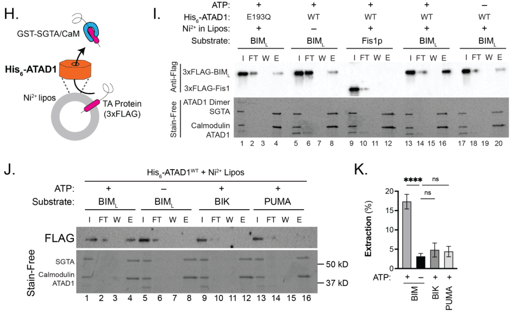
A key human-specific mechanistic finding is that ATAD1 directly extracts the pro-apoptotic BH3-only protein BIM from membranes/mitochondria. In a reconstituted liposome assay, extraction was ATP-dependent, required membrane anchoring, and showed substrate selectivity (BIM extracted, while related proteins such as PUMA/BIK were not, under the tested conditions). (winter2022collateraldeletionof pages 8-9, winter2022collateraldeletionof media 8a98eb16)
A 2024 bioRxiv preprint reports a quantitative reconstituted extraction assay and proposes that Msp1 recognizes substrates via hydrophobic mismatch between a substrateโs transmembrane domain (TMD) and the surrounding lipid bilayer; importantly, the authors conclude that TMD extraction from the bilayer is rate-limiting. While centered on yeast Msp1, the work explicitly frames relevance to the human homolog ATAD1 and provides a mechanistic model applicable to ATAD1-family substrate selectivity. (fresenius2024theaaa+protein pages 1-3, fresenius2024theaaa+protein pages 13-21)
A 2023 peer-reviewed review consolidates the โproofreadingโ model: Msp1/ATAD1 extracts mislocalized TA proteins from the OMM and supports their handoff to GET/TRC for ER targeting or channels them to degradation pathways, situating ATAD1 within a multilayer organelle targeting QC network. (matsumoto2023msp1mediatedproofreadingmechanism pages 3-4, matsumoto2023msp1mediatedproofreadingmechanism pages 1-2)
A 2023 PLOS Biology study of the fission yeast ATAD1 homolog (Yta4) reports a role in preventing excessive mitochondrial fission, including interaction with mitochondrial divisome components and ATPase/translocase-dependent delocalization of factors such as Fis1/Mdv1; while not human ATAD1 directly, it broadens mechanistic hypotheses about how ATAD1-family extractases can tune organelle dynamics beyond proteostasis. (he2023theaaaatpaseyta4atad1 pages 1-2)
A 2024 bioRxiv preprint using covalently linked dimers suggests a non-linear relationship between ATP hydrolysis rate and membrane extraction efficiency and proposes a minimum ATPase rate for effective TMH extraction; these results are mechanistically informative for ATAD1 by orthology, though not yet direct human ATAD1 measurements in the retrieved set. (fresenius2024theaaa+protein pages 1-3)
ATAD1 is near PTEN (10q23) and is frequently co-deleted with PTEN. The 2022 eLife study proposes a clinically actionable vulnerability: ATAD1 loss sensitizes cells to proteasome dysfunction through BIM-dependent apoptosis, and ATAD1-null cells/xenografts are hypersensitive to proteasome inhibitors. (winter2022collateraldeletionof pages 1-2, winter2022collateraldeletionof pages 11-13)
Quantitative/implementation-relevant details from the study and supporting excerpts include:
- PTEN deletion rates cited as >33% of metastatic prostate tumors and ~10% of melanoma and glioblastoma multiforme, contextualizing the prevalence of adjacent-locus deletions. (winter2022collateraldeletionof pages 1-2)
- Mouse xenografts: bortezomib dosing of 1 mg/kg reduced growth of ATAD1-deleted PC3 tumors (but not controls in the excerpted comparison). (winter2022collateraldeletionof pages 11-13)
- Tumorigenicity shift in a glioma model: EV-transduced SW1088 cells formed 0/17 tumors, while ATAD1-transduced formed 20/21 tumors in NOD/SCID mice (suggesting ATAD1 can promote tumor growth/fitness in that context). (winter2022collateraldeletionof pages 11-13)
Across the cited mCRPC context, median overall survival is reported as 77 months for tumors that are PTEN-null and ATAD1-null vs 37 months for PTEN-null or unaltered tumors. (winter2022collateraldeletionof pages 11-13, fresenius2023developmentofa pages 67-73)
In ZSD model systems, overexpression of ATAD1 (as a mitochondrial QC factor) was sufficient to rescue aspects of mitochondrial function, supporting an organelle-proteostasis โmodifierโ approach. (fresenius2023developmentofaa pages 19-23)
An authoritative 2020 Annual Review article synthesizes the field: ATAD1/Msp1 is a central mitochondrial proteostasis factor extracting mislocalized membrane proteins and import-stalled substrates; extracted clients are triaged to proteasomal degradation or rerouting, and the review highlights unresolved questions about substrate selection and how ATAD1โs specialization enables membrane extraction. (wang2020msp1atad1inprotein pages 1-2, wang2020msp1atad1inprotein pages 2-4)
The same review compiles biochemical and neuronal data indicating ATAD1 (Thorase) can regulate synaptic receptor trafficking: ATAD1 forms complexes with GluR2 and GRIP1; ATAD1 can disassemble GluR2โGRIP1 in an ATP-dependent manner; perturbing ATAD1 changes surface AMPAR levels and affects activity-dependent synaptic downscaling (e.g., bicuculline-induced AMPAR internalization). (wang2020msp1atad1inprotein pages 18-20)
A 2023 thesis-like source summarizes quantitative epidemiology and survival metrics related to ATAD1 loss in cancer:
- ATAD1 deletion frequency: 4.1% in gastric cancer; using global gastric cancer mortality (~800,000 deaths/year), the text estimates ~32,000 deaths/year associated with ATAD1-deficient gastric cancer. (fresenius2023developmentofac pages 67-73, fresenius2023developmentofa pages 67-73)
- Median overall survival in mCRPC: 77 vs 37 months (ATAD1/PTEN co-deleted vs PTEN-null/unaltered). (fresenius2023developmentofa pages 67-73, winter2022collateraldeletionof pages 11-13)
Figure evidence from Winter et al. 2022 directly supports ATP-dependent and selective extraction of BIM by ATAD1 in a reconstituted system. (winter2022collateraldeletionof media 8a98eb16, winter2022collateraldeletionof media eae7fce6)
Open Targets lists ATAD1 associations with terms including hereditary hyperekplexia / hyperekplexia 4 and broader neurodegenerative disease categories. These associations are useful for prioritizing follow-up but should be interpreted cautiously without direct primary-study confirmation in the same evidence bundle. (OpenTargets Search: -ATAD1)
ATAD1 is an OMM AAA+ ATPase extractase/translocase that uses ATP hydrolysis to remove membrane-embedded proteinsโespecially mislocalized tail-anchored proteins and stalled import substratesโfrom the mitochondrial outer membrane, promoting rerouting and/or degradation to maintain organelle proteostasis. (wang2020msp1atad1inprotein pages 1-2, wang2022conservedstructuralelements pages 1-2, chen2014msp1atad1maintainsmitochondrial pages 10-11)
The following table consolidates key findings, evidence types, and DOI URLs:
| Functional aspect | Key findings | Evidence type | Key sources with year + DOI URL |
|---|---|---|---|
| Gene/protein identity | Human ATAD1 = Thorase, ortholog of yeast Msp1; UniProt Q8NBU5. Literature consistently matches a membrane-anchored AAA+ ATPase/extractase rather than an unrelated gene symbol. Belongs to the MSP1 subfamily and functions in outer mitochondrial membrane protein quality control (wang2020msp1atad1inprotein pages 1-2, fresenius2023developmentofa pages 19-23, wang2022conservedstructuralelements pages 1-2) | Review, structure, database-aligned annotation | Wang & Walter 2020, doi: https://doi.org/10.1146/annurev-cellbio-031220-015840; Wang et al. 2022, doi: https://doi.org/10.7554/elife.73941 |
| Localization | ATAD1 is anchored by a single N-terminal transmembrane helix in the outer mitochondrial membrane (OMM) with the AAA domain exposed to the cytosol; several sources also note localization/function at peroxisomes for tail-anchored protein proofreading (wang2020msp1atad1inprotein pages 1-2, fresenius2023developmentofaa pages 19-23, fresenius2023developmentofab pages 19-23) | Review, cell biology, structural interpretation | Wang & Walter 2020, doi: https://doi.org/10.1146/annurev-cellbio-031220-015840; Chen et al. 2014, doi: https://doi.org/10.15252/embj.201487943 |
| Domain/oligomeric architecture | Human ATAD1 is a hexameric AAA+ ATPase that forms a right-handed spiral/lock-washer assembly during substrate engagement. Conserved pore loops and a C-terminal helix adapt it for membrane protein extraction (wang2022conservedstructuralelements pages 1-2, wang2022conservedstructuralelements pages 11-13, wang2022conservedstructuralelements pages 2-3) | Cryo-EM structure, mutagenesis | Wang et al. 2022, doi: https://doi.org/10.7554/elife.73941; Wang et al. 2020, doi: https://doi.org/10.7554/elife.54031 |
| Primary molecular activity | ATAD1 is an ATP-driven membrane protein extractase/translocase that removes mislocalized membrane proteins and proteins stalled in the mitochondrial import machinery; ATP hydrolysis is required for direct substrate removal from membranes (wang2020msp1atad1inprotein pages 1-2, winter2022collateraldeletionof pages 1-2, winter2022collateraldeletionof pages 8-9) | Review, biochemical reconstitution, cell biology | Wang & Walter 2020, doi: https://doi.org/10.1146/annurev-cellbio-031220-015840; Winter et al. 2022, doi: https://doi.org/10.7554/elife.82860 |
| Foundational substrate class: mistargeted tail-anchored proteins | The founding quality-control role is extraction of mislocalized tail-anchored (TA) proteins from mitochondria. In yeast, Pex15 and Gos1 are canonical substrates; mammalian conservation is supported by increased mitochondrial localization of PEX26 and GOS28 after ATAD1 depletion/knockdown (chen2014msp1atad1maintainsmitochondrial pages 6-7, chen2014msp1atad1maintainsmitochondrial pages 10-11, chen2014msp1atad1maintainsmitochondrial pages 9-10) | Genetics, substrate-trap biochemistry, microscopy | Chen et al. 2014, doi: https://doi.org/10.15252/embj.201487943 |
| Proofreading/rerouting pathway | Msp1/ATAD1-dependent extraction can reroute mislocalized TA proteins from mitochondria to the ER via the GET/TRC pathway, establishing an intracellular proofreading system rather than simple destruction in all cases (wang2020msp1atad1inprotein pages 2-4, matsumoto2023msp1mediatedproofreadingmechanism pages 5-6, matsumoto2023msp1mediatedproofreadingmechanism pages 1-2) | Time-lapse imaging, co-IP, review | Matsumoto et al. 2019, doi: https://doi.org/10.1016/j.molcel.2019.07.006; Matsumoto 2023, doi: https://doi.org/10.1093/jb/mvad025 |
| Degradation pathway coupling | For some substrates, extraction precedes ubiquitination and proteasomal degradation; evidence places Msp1/ATAD1 upstream of ubiquitin-dependent Cdc48/proteasome clearance for mislocalized TA substrates (matsumoto2023msp1mediatedproofreadingmechanism pages 3-4) | Genetics, inhibitor studies, biochemistry | Matsumoto 2023, doi: https://doi.org/10.1093/jb/mvad025 |
| Import-stress / translocase quality control | Beyond TA proteins, ATAD1/Msp1 extracts stuck import substrates from the mitochondrial outer membrane/translocase system, helping maintain mitochondrial protein import capacity and linking the protein to mitoCPR/import-stress pathways (wang2020msp1atad1inprotein pages 1-2, wang2022conservedstructuralelements pages 1-2, castanzo2020theaaa+atpase pages 1-2) | Review, genetics, mechanistic studies | Wang & Walter 2020, doi: https://doi.org/10.1146/annurev-cellbio-031220-015840; Castanzo et al. 2020, doi: https://doi.org/10.1073/pnas.1920109117 |
| Mechanism of substrate engagement | Structural work shows substrate threading through a hydrophobic/aromatic central pore. Human ATAD1 uses pore-loop residues including W166/Y167 to grip substrate; mutations impair activity or peptide binding by >100-fold in some assays (wang2022conservedstructuralelements pages 11-13, wang2022conservedstructuralelements pages 2-3) | Cryo-EM, mutagenesis, peptide-binding assays | Wang et al. 2022, doi: https://doi.org/10.7554/elife.73941 |
| Processive unfoldase/translocase behavior | Msp1/ATAD1-family enzymes act as processive protein translocases/unfoldases that thread substrates through the pore; ATPase activity depends on oligomeric state, supporting a mechanical extraction model for hydrophobic membrane proteins (castanzo2020theaaa+atpase pages 1-2) | Biochemical reconstitution, EM | Castanzo et al. 2020, doi: https://doi.org/10.1073/pnas.1920109117 |
| Human apoptotic substrate: BIM | A key human ATAD1-specific finding is direct, specific extraction of BIM from membranes/mitochondria to inactivate this pro-apoptotic factor. In liposome assays, extraction is ATP-dependent, requires membrane anchoring, and is lost with the catalytic E193Q mutant (winter2022collateraldeletionof pages 1-2, winter2022collateraldeletionof pages 8-9, winter2022collateraldeletionof media 8a98eb16) | Reconstituted biochemistry, co-IP, genetics, figure-level assay evidence | Winter et al. 2022, doi: https://doi.org/10.7554/elife.82860 |
| Substrate selectivity in apoptosis | ATAD1 extraction is selective, not universal for BH3-only proteins: in the reported reconstitution, ATAD1 extracted BIM but not BIK, PUMA, or yeast Fis1 under the same conditions (winter2022collateraldeletionof pages 8-9, winter2022collateraldeletionof media 8a98eb16) | Reconstituted biochemistry, figure quantification | Winter et al. 2022, doi: https://doi.org/10.7554/elife.82860 |
| Cancer relevance / collateral lethality | ATAD1 is adjacent to PTEN on 10q23 and is often co-deleted with PTEN in tumors. ATAD1 loss sensitizes cells and xenografts to proteasome inhibitors by increasing BIM-dependent apoptotic priming, suggesting a therapeutic vulnerability in ATAD1-null cancers (winter2022collateraldeletionof pages 1-2, winter2022collateraldeletionof pages 8-9) | Cancer genetics, cell biology, xenografts | Winter et al. 2022, doi: https://doi.org/10.7554/elife.82860 |
| Disease/application concept | Proposed application: exploit proteasome dysfunction/proteasome inhibitor sensitivity in cancers with ATAD1 loss; this is a preclinical therapeutic concept rather than an approved ATAD1-targeted therapy (winter2022collateraldeletionof pages 1-2) | Preclinical translational study | Winter et al. 2022, doi: https://doi.org/10.7554/elife.82860 |
| Mitochondrial dynamics (newer role) | A 2023 study of the homolog Yta4/ATAD1 identified a role in inhibiting mitochondrial fission by acting on divisome components (Fis1, Mdv1, Dnm1), expanding the conceptual landscape of ATAD1-family biology beyond proteostasis alone (he2023theaaaatpaseyta4atad1 pages 1-2) | Genetics, interaction assays, in vitro assembly assays | He et al. 2023, doi: https://doi.org/10.1371/journal.pbio.3002247 |
| 2023 conceptual update on proofreading | 2023 synthesis emphasized Msp1/ATAD1 as a proofreading system for TA protein localization, integrating extraction, GET-mediated rerouting, and selective degradation as a multilayer quality-control network (matsumoto2023msp1mediatedproofreadingmechanism pages 3-4, matsumoto2023msp1mediatedproofreadingmechanism pages 1-2) | Review | Matsumoto 2023, doi: https://doi.org/10.1093/jb/mvad025 |
| 2023-2024 reconstituted assay advance | New quantitative reconstituted assays using split-luciferase/defined proteoliposomes enabled controlled testing of substrate selectivity and membrane determinants of extraction, creating a more rigorous platform for ATAD1/Msp1 mechanism studies (fresenius2024theaaa+protein pages 1-3, fresenius2023developmentofac pages 110-116) | Method development, reconstitution | Fresenius et al. 2024, doi: https://doi.org/10.1101/2023.07.11.548587 |
| 2024 substrate-recognition model | 2024 work proposed that substrate recognition depends strongly on hydrophobic mismatch between the substrate transmembrane segment and the surrounding bilayer; extraction of a substrate TMD from the membrane appears to be the rate-limiting step (fresenius2024theaaa+protein pages 1-3, fresenius2024theaaa+protein pages 13-21) | Reconstituted biochemistry, membrane engineering | Fresenius et al. 2024, doi: https://doi.org/10.1101/2023.07.11.548587 |
| 2024 energetic/mechanistic direction | Emerging 2024 linked-dimer/energetic studies indicate a minimum ATP hydrolysis rate is needed for efficient TMH extraction and suggest mechanistic plasticity in subunit coordination during extraction; relevant to ATAD1 by homology but not yet a direct human ATAD1 paper in the retrieved evidence set (fresenius2024theaaa+protein pages 1-3) | Preprint mechanistic biochemistry (family-level inference) | Smith et al. 2024, doi: https://doi.org/10.1101/2024.09.23.614443 |
| Synaptic regulation | ATAD1/Thorase also has a noncanonical neuronal role in AMPA receptor trafficking. It forms complexes with GluR2 and GRIP1, can disassemble the GluR2โGRIP1 complex in an ATP-dependent manner, and is required for activity-dependent AMPAR internalization/downscaling (wang2020msp1atad1inprotein pages 1-2, wang2020msp1atad1inprotein pages 18-20) | Biochemistry, neuronal cell biology, mouse genetics | Wang & Walter 2020, doi: https://doi.org/10.1146/annurev-cellbio-031220-015840 |
| Neurological phenotypes | ATAD1 loss/function compromise is associated with seizures, impaired fear conditioning, worsened post-stroke deficits in mice, and severe human encephalopathy/stiffness/arthrogryposis in homozygous mutation cases; AMPAR antagonists reportedly ameliorated some defects (fresenius2023developmentofac pages 19-23, wang2020msp1atad1inprotein pages 18-20) | Mouse genetics, clinical association, review | Wang & Walter 2020, doi: https://doi.org/10.1146/annurev-cellbio-031220-015840 |
| Other disease associations / databases | Open Targets lists ATAD1 associations with hereditary hyperekplexia/hyperekplexia 4 and broader neurodegenerative phenotypes, but these database-level links should be interpreted as hypothesis-generating unless supported by primary studies (OpenTargets Search: -ATAD1) | Database | Open Targets platform query (context evidence) |
| Organelle-disease modifier role | In Zellweger-spectrum models, ATAD1 overexpression was reported to rescue aspects of mitochondrial dysfunction caused by mislocalized peroxisomal proteins, suggesting a disease-modifier role in organelle proteostasis (fresenius2023developmentofaa pages 19-23) | Cell biology, disease-model study | Nuebel et al. 2021, doi: https://doi.org/10.15252/embr.202051991 |
| Overall functional annotation | Best-supported primary annotation for human ATAD1 is: outer mitochondrial membrane AAA+ extractase/translocase that removes mislocalized TA proteins and stalled import substrates, coupling ATP hydrolysis to membrane protein extraction; additional metazoan-specialized roles include apoptosis control via BIM extraction and neuronal AMPAR trafficking (wang2020msp1atad1inprotein pages 1-2, winter2022collateraldeletionof pages 1-2, wang2022conservedstructuralelements pages 1-2, wang2020msp1atad1inprotein pages 18-20) | Integrated review of structure, biochemistry, genetics, disease evidence | Chen et al. 2014, doi: https://doi.org/10.15252/embj.201487943; Wang & Walter 2020, doi: https://doi.org/10.1146/annurev-cellbio-031220-015840; Wang et al. 2022, doi: https://doi.org/10.7554/elife.73941; Winter et al. 2022, doi: https://doi.org/10.7554/elife.82860 |
Table: This table summarizes the main functional annotation evidence for human ATAD1/Thorase (UniProt Q8NBU5), spanning localization, molecular mechanism, substrates, pathways, disease relevance, and translational implications. It highlights both foundational studies and newer 2023-2024 developments in substrate recognition and membrane-extraction mechanism.
Panels from Winter et al. 2022 eLife Figure 2 show ATP-dependent, selective extraction of BIM by ATAD1 in a reconstituted liposome assay. (winter2022collateraldeletionof media 8a98eb16, winter2022collateraldeletionof media eae7fce6)
References
(wang2020msp1atad1inprotein pages 1-2): Lan Wang and Peter Walter. Msp1/atad1 in protein quality control and regulation of synaptic activities. Annual Review of Cell and Developmental Biology, 36:141-164, Oct 2020. URL: https://doi.org/10.1146/annurev-cellbio-031220-015840, doi:10.1146/annurev-cellbio-031220-015840. This article has 62 citations and is from a domain leading peer-reviewed journal.
(wang2022conservedstructuralelements pages 1-2): Lan Wang, Hannah Toutkoushian, Vladislav Belyy, Claire Y Kokontis, and Peter Walter. Conserved structural elements specialize atad1 as a membrane protein extraction machine. May 2022. URL: https://doi.org/10.7554/elife.73941, doi:10.7554/elife.73941. This article has 27 citations and is from a domain leading peer-reviewed journal.
(winter2022collateraldeletionof pages 1-2): Jacob M Winter, Heidi L Fresenius, Corey N Cunningham, Peng Wei, Heather R Keys, Jordan Berg, Alex Bott, Tarun Yadav, Jeremy Ryan, Deepika Sirohi, Sheryl R Tripp, Paige Barta, Neeraj Agarwal, Anthony Letai, David M Sabatini, Matthew L Wohlever, and Jared Rutter. Collateral deletion of the mitochondrial aaa+ atpase atad1 sensitizes cancer cells to proteasome dysfunction. eLife, Nov 2022. URL: https://doi.org/10.7554/elife.82860, doi:10.7554/elife.82860. This article has 22 citations and is from a domain leading peer-reviewed journal.
(winter2022collateraldeletionof pages 8-9): Jacob M Winter, Heidi L Fresenius, Corey N Cunningham, Peng Wei, Heather R Keys, Jordan Berg, Alex Bott, Tarun Yadav, Jeremy Ryan, Deepika Sirohi, Sheryl R Tripp, Paige Barta, Neeraj Agarwal, Anthony Letai, David M Sabatini, Matthew L Wohlever, and Jared Rutter. Collateral deletion of the mitochondrial aaa+ atpase atad1 sensitizes cancer cells to proteasome dysfunction. eLife, Nov 2022. URL: https://doi.org/10.7554/elife.82860, doi:10.7554/elife.82860. This article has 22 citations and is from a domain leading peer-reviewed journal.
(wang2020msp1atad1inprotein pages 2-4): Lan Wang and Peter Walter. Msp1/atad1 in protein quality control and regulation of synaptic activities. Annual Review of Cell and Developmental Biology, 36:141-164, Oct 2020. URL: https://doi.org/10.1146/annurev-cellbio-031220-015840, doi:10.1146/annurev-cellbio-031220-015840. This article has 62 citations and is from a domain leading peer-reviewed journal.
(matsumoto2023msp1mediatedproofreadingmechanism pages 5-6): Shunsuke Matsumoto. Msp1-mediated proofreading mechanism for localization of tail-anchored membrane proteins. Journal of biochemistry, 174:13-20, Mar 2023. URL: https://doi.org/10.1093/jb/mvad025, doi:10.1093/jb/mvad025. This article has 4 citations and is from a peer-reviewed journal.
(matsumoto2023msp1mediatedproofreadingmechanism pages 1-2): Shunsuke Matsumoto. Msp1-mediated proofreading mechanism for localization of tail-anchored membrane proteins. Journal of biochemistry, 174:13-20, Mar 2023. URL: https://doi.org/10.1093/jb/mvad025, doi:10.1093/jb/mvad025. This article has 4 citations and is from a peer-reviewed journal.
(fresenius2023developmentofaa pages 19-23): H Fresenius. Development of a quantitative reconstituted system to study msp1/atad1 substrate selectivity and apoptotic regulation. Unknown journal, 2023.
(fresenius2023developmentofab pages 19-23): H Fresenius. Development of a quantitative reconstituted system to study msp1/atad1 substrate selectivity and apoptotic regulation. Unknown journal, 2023.
(wang2022conservedstructuralelements pages 2-3): Lan Wang, Hannah Toutkoushian, Vladislav Belyy, Claire Y Kokontis, and Peter Walter. Conserved structural elements specialize atad1 as a membrane protein extraction machine. May 2022. URL: https://doi.org/10.7554/elife.73941, doi:10.7554/elife.73941. This article has 27 citations and is from a domain leading peer-reviewed journal.
(wang2022conservedstructuralelements pages 11-13): Lan Wang, Hannah Toutkoushian, Vladislav Belyy, Claire Y Kokontis, and Peter Walter. Conserved structural elements specialize atad1 as a membrane protein extraction machine. May 2022. URL: https://doi.org/10.7554/elife.73941, doi:10.7554/elife.73941. This article has 27 citations and is from a domain leading peer-reviewed journal.
(chen2014msp1atad1maintainsmitochondrial pages 6-7): YuโChan Chen, George K E Umanah, Noah Dephoure, Shaida A Andrabi, Steven P Gygi, Ted M Dawson, Valina L Dawson, and Jared Rutter. Msp1/atad1 maintains mitochondrial function by facilitating the degradation of mislocalized tailโanchored proteins. The EMBO Journal, 33:1548-1564, Jul 2014. URL: https://doi.org/10.15252/embj.201487943, doi:10.15252/embj.201487943. This article has 267 citations.
(chen2014msp1atad1maintainsmitochondrial pages 10-11): YuโChan Chen, George K E Umanah, Noah Dephoure, Shaida A Andrabi, Steven P Gygi, Ted M Dawson, Valina L Dawson, and Jared Rutter. Msp1/atad1 maintains mitochondrial function by facilitating the degradation of mislocalized tailโanchored proteins. The EMBO Journal, 33:1548-1564, Jul 2014. URL: https://doi.org/10.15252/embj.201487943, doi:10.15252/embj.201487943. This article has 267 citations.
(chen2014msp1atad1maintainsmitochondrial pages 9-10): YuโChan Chen, George K E Umanah, Noah Dephoure, Shaida A Andrabi, Steven P Gygi, Ted M Dawson, Valina L Dawson, and Jared Rutter. Msp1/atad1 maintains mitochondrial function by facilitating the degradation of mislocalized tailโanchored proteins. The EMBO Journal, 33:1548-1564, Jul 2014. URL: https://doi.org/10.15252/embj.201487943, doi:10.15252/embj.201487943. This article has 267 citations.
(matsumoto2023msp1mediatedproofreadingmechanism pages 3-4): Shunsuke Matsumoto. Msp1-mediated proofreading mechanism for localization of tail-anchored membrane proteins. Journal of biochemistry, 174:13-20, Mar 2023. URL: https://doi.org/10.1093/jb/mvad025, doi:10.1093/jb/mvad025. This article has 4 citations and is from a peer-reviewed journal.
(winter2022collateraldeletionof media 8a98eb16): Jacob M Winter, Heidi L Fresenius, Corey N Cunningham, Peng Wei, Heather R Keys, Jordan Berg, Alex Bott, Tarun Yadav, Jeremy Ryan, Deepika Sirohi, Sheryl R Tripp, Paige Barta, Neeraj Agarwal, Anthony Letai, David M Sabatini, Matthew L Wohlever, and Jared Rutter. Collateral deletion of the mitochondrial aaa+ atpase atad1 sensitizes cancer cells to proteasome dysfunction. eLife, Nov 2022. URL: https://doi.org/10.7554/elife.82860, doi:10.7554/elife.82860. This article has 22 citations and is from a domain leading peer-reviewed journal.
(fresenius2024theaaa+protein pages 1-3): Heidi L. Fresenius, Deepika Gaur, Baylee Smith, Brian Acquaviva, and Matthew L. Wohlever. The aaa+ protein msp1 recognizes substrates by a hydrophobic mismatch. bioRxiv, Jul 2024. URL: https://doi.org/10.1101/2023.07.11.548587, doi:10.1101/2023.07.11.548587. This article has 5 citations.
(fresenius2024theaaa+protein pages 13-21): Heidi L. Fresenius, Deepika Gaur, Baylee Smith, Brian Acquaviva, and Matthew L. Wohlever. The aaa+ protein msp1 recognizes substrates by a hydrophobic mismatch. bioRxiv, Jul 2024. URL: https://doi.org/10.1101/2023.07.11.548587, doi:10.1101/2023.07.11.548587. This article has 5 citations.
(he2023theaaaatpaseyta4atad1 pages 1-2): Jiajia He, Ke Liu, Yifan Wu, Chenhui Zhao, Shuaijie Yan, Jia-Hui Chen, Lizhu Hu, Dongmei Wang, Fan Zheng, Wenfan Wei, Chao Xu, Chengdong Huang, Xing Liu, Xuebiao Yao, Lijun Ding, Zhiyou Fang, Ai-Hui Tang, and Chuanhai Fu. The aaa-atpase yta4/atad1 interacts with the mitochondrial divisome to inhibit mitochondrial fission. PLOS Biology, 21:e3002247, Aug 2023. URL: https://doi.org/10.1371/journal.pbio.3002247, doi:10.1371/journal.pbio.3002247. This article has 11 citations and is from a highest quality peer-reviewed journal.
(winter2022collateraldeletionof pages 11-13): Jacob M Winter, Heidi L Fresenius, Corey N Cunningham, Peng Wei, Heather R Keys, Jordan Berg, Alex Bott, Tarun Yadav, Jeremy Ryan, Deepika Sirohi, Sheryl R Tripp, Paige Barta, Neeraj Agarwal, Anthony Letai, David M Sabatini, Matthew L Wohlever, and Jared Rutter. Collateral deletion of the mitochondrial aaa+ atpase atad1 sensitizes cancer cells to proteasome dysfunction. eLife, Nov 2022. URL: https://doi.org/10.7554/elife.82860, doi:10.7554/elife.82860. This article has 22 citations and is from a domain leading peer-reviewed journal.
(fresenius2023developmentofa pages 67-73): H Fresenius. Development of a quantitative reconstituted system to study msp1/atad1 substrate selectivity and apoptotic regulation. Unknown journal, 2023.
(wang2020msp1atad1inprotein pages 18-20): Lan Wang and Peter Walter. Msp1/atad1 in protein quality control and regulation of synaptic activities. Annual Review of Cell and Developmental Biology, 36:141-164, Oct 2020. URL: https://doi.org/10.1146/annurev-cellbio-031220-015840, doi:10.1146/annurev-cellbio-031220-015840. This article has 62 citations and is from a domain leading peer-reviewed journal.
(fresenius2023developmentofac pages 67-73): H Fresenius. Development of a quantitative reconstituted system to study msp1/atad1 substrate selectivity and apoptotic regulation. Unknown journal, 2023.
(winter2022collateraldeletionof media eae7fce6): Jacob M Winter, Heidi L Fresenius, Corey N Cunningham, Peng Wei, Heather R Keys, Jordan Berg, Alex Bott, Tarun Yadav, Jeremy Ryan, Deepika Sirohi, Sheryl R Tripp, Paige Barta, Neeraj Agarwal, Anthony Letai, David M Sabatini, Matthew L Wohlever, and Jared Rutter. Collateral deletion of the mitochondrial aaa+ atpase atad1 sensitizes cancer cells to proteasome dysfunction. eLife, Nov 2022. URL: https://doi.org/10.7554/elife.82860, doi:10.7554/elife.82860. This article has 22 citations and is from a domain leading peer-reviewed journal.
(OpenTargets Search: -ATAD1): Open Targets Query (-ATAD1, 5 results). Buniello, A. et al. (2025). Open Targets Platform: facilitating therapeutic hypotheses building in drug discovery. Nucleic Acids Research.
(fresenius2023developmentofa pages 19-23): H Fresenius. Development of a quantitative reconstituted system to study msp1/atad1 substrate selectivity and apoptotic regulation. Unknown journal, 2023.
(castanzo2020theaaa+atpase pages 1-2): Dominic T. Castanzo, Benjamin LaFrance, and Andreas Martin. The aaa+ atpase msp1 is a processive protein translocase with robust unfoldase activity. Proceedings of the National Academy of Sciences, 117:14970-14977, Jun 2020. URL: https://doi.org/10.1073/pnas.1920109117, doi:10.1073/pnas.1920109117. This article has 41 citations and is from a highest quality peer-reviewed journal.
(fresenius2023developmentofac pages 110-116): H Fresenius. Development of a quantitative reconstituted system to study msp1/atad1 substrate selectivity and apoptotic regulation. Unknown journal, 2023.
(fresenius2023developmentofac pages 19-23): H Fresenius. Development of a quantitative reconstituted system to study msp1/atad1 substrate selectivity and apoptotic regulation. Unknown journal, 2023.
(wang2020structureofthe pages 1-2): Lan Wang, Alexander Myasnikov, Xingjie Pan, and Peter Walter. Structure of the aaa protein msp1 reveals mechanism of mislocalized membrane protein extraction. eLife, Jan 2020. URL: https://doi.org/10.7554/elife.54031, doi:10.7554/elife.54031. This article has 72 citations and is from a domain leading peer-reviewed journal.
Falcon deep research was attempted with the required perplexity-lite fallback.
Falcon timed out after 600 seconds and the fallback failed with a Perplexity API
quota error, so this review uses cached primary evidence, UniProt, Reactome, and
the local PN projection reports rather than a generated Falcon research artifact.
ATAD1 is a strong fit for the mitochondrial proteostasis batch, but the safest
core term remains the existing specific process GO:0140570 extraction of
mislocalized protein from mitochondrial outer membrane. The direct ATAD1/Msp1
paper says human ATAD1 limits mitochondrial mislocalization of PEX26 and GOS28
and proposes ATAD1/Msp1 as mitochondrial protein quality-control factors that
promote extraction and degradation of mislocalized tail-anchored proteins
[PMID:24843043 "human ATAD1 limits the mitochondrial mislocalization of PEX26
and GOS28"; PMID:24843043 "promote the extraction and degradation of
mislocalized TA proteins"].
The PN projection report proposes GO:0035694 mitochondrial protein catabolic
process for ATAD1 from
Mitochondrial proteostasis|Organelle-specific protein degradation|mitoCPR
pathway. This is a class-level propagation, and the mapping audit flags this
source as requiring manual gene-level review before changing a review. I accepted
the projected term only as a broad NEW candidate because PMID:24843043 supports
degradation as the downstream outcome, not because the PN source alone is
sufficient. I used TAS rather than IMP for this candidate because the cached
paper text supports a traceable degradation claim, while the explicit
protein-level knockout evidence available in the cached abstract is from mouse
tissue. It should not replace GO:0140570, which is the sharper mechanism.
Peroxisomal membrane localization is retained as non-core. UniProt reports
peroxisome membrane localization and Reactome lists ATAD1 as a class I
peroxisomal membrane protein bound by PEX19, but the current evidence does not
establish peroxisomal substrate extraction as ATAD1's primary function
[file:human/ATAD1/ATAD1-uniprot.txt "Peroxisome membrane";
Reactome:R-HSA-9603804 "ATAD1 (Liu et al. 2016)"].
The synaptic/behavioral block is treated conservatively. UniProt summarizes
mouse-derived AMPAR trafficking, synaptic plasticity, learning, and memory
biology by similarity, and human ATAD1 encephalopathy provides disease context
[file:human/ATAD1/ATAD1-uniprot.txt "Required for NMDA-stimulated AMPAR
internalization"; PMID:29659736 "ATAD1 encephalopathy and stiff baby syndrome"].
I kept postsynaptic/receptor-internalization terms as non-core and marked the
high-level learning/memory annotations as over-annotations.
A Falcon (Edison Scientific) deep research report was generated successfully (the
earlier 2026-06-03 run had failed). It largely CONFIRMS the existing review but
adds several mechanistic/translational findings that were not previously cited.
Key synthesis (CONFIRMS / NEW / PROVISIONAL labels are relative to the prior
review):
CONFIRMS core function. ATAD1/Msp1 is a hexameric OMM AAA+ extractase that
uses ATP hydrolysis to thread membrane-embedded substrates (especially
mislocalized tail-anchored proteins, e.g. PEX26/GOS28, and stalled import
substrates) through its central pore for rerouting or degradation
PMID:24843043. This is already the accepted core (GO:0140567, GO:0140570).
NEW (human, direct, primary): ATAD1 directly and selectively extracts the
pro-apoptotic BH3-only protein BIM from mitochondrial membranes to inactivate
it; extraction is ATP-dependent, requires membrane anchoring, and is selective
(BIM extracted but not BIK/PUMA/Fis1 under the same reconstituted conditions;
lost in the catalytic E193Q mutant). This connects ATAD1 to apoptotic priming
and to a cancer "collateral lethality" with PTEN (10q23 co-deletion):
ATAD1-null cells/xenografts are hypersensitized to proteasome inhibitors via
BIM-dependent apoptosis [PMID:36409067 Winter et al. 2022 eLife, doi:10.7554/eLife.82860
"ATAD1 directly and specifically extracts the pro-apoptotic protein BIM from
mitochondria to inactivate it"]. This is a genuinely new molecular substrate
axis and a new disease/translational link not in the prior review.
NEW (human, structural/mechanistic, primary): Cryo-EM of human ATAD1 bound to
a peptide substrate shows phylogenetically conserved pore-loop aromatics
(pore-loop 1 W166/Y167) are required to grip hydrophobic substrate and cannot
be replaced by aliphatic residues; a C-terminal alpha-helix promotes
oligomerization, specializing ATAD1 among AAA proteins [PMID:35550246 Wang et
al. 2022 eLife, doi:10.7554/eLife.73941 "both aromatic amino acids in pore-loop 1
are required for ATAD1's function"]. Mechanistic support for the dislocase /
ATP-hydrolysis core via yeast Msp1 structure [PMID:31999255 Wang et al. 2020
eLife, doi:10.7554/eLife.54031] and processive bidirectional translocase /
unfoldase activity, dependent on hexamerization and inhibited by Pex3
[PMID:32541053 Castanzo et al. 2020 PNAS, doi:10.1073/pnas.1920109117].
NEW/CONFIRMS (pathway coupling): Extracted TA proteins are not obligatorily
degraded; they can be rerouted to the ER via the GET/TRC (Get3) pathway as an
intracellular "proofreading" system, or handed to ubiquitin-proteasome
(Cdc48) clearance [Matsumoto 2023 J Biochem doi:10.1093/jb/mvad025;
Matsumoto et al. 2019 Mol Cell doi:10.1016/j.molcel.2019.07.006 โ yeast Msp1].
The 2019 Mol Cell DOI did not resolve to a confirmed PMID via PubMed and is
yeast-focused, so I am NOT adding it to references; kept in notes only.
PROVISIONAL (homolog, not human): A fission-yeast ATAD1 ortholog (Yta4) was
reported to inhibit excessive mitochondrial fission by acting on divisome
components Fis1/Mdv1/Dnm1 โ a role beyond proteostasis [He et al. 2023 PLOS
Biology, PMID:37590302, doi:10.1371/journal.pbio.3002247]. Because this is the
Schizosaccharomyces homolog rather than human ATAD1, I treat it as
hypothesis-generating only and do NOT use it to add/modify human annotations.
PROVISIONAL (preprint / database): A 2024 bioRxiv preprint proposes Msp1
substrate recognition by hydrophobic mismatch with TMD extraction as the
rate-limiting step [Fresenius et al. 2024 bioRxiv doi:10.1101/2023.07.11.548587];
Open Targets lists ATAD1 with hyperekplexia 4 / neurodegenerative terms. These
are preprint/database-level and explicitly NOT used to change annotations.
CONFIRMS (neuronal axis): The Wang & Walter 2020 review compiles the
noncanonical neuronal role โ ATAD1/Thorase forms complexes with GluR2 and
GRIP1 and disassembles GluR2-GRIP1 in an ATP-dependent manner, regulating
surface AMPAR levels and activity-dependent synaptic downscaling
[Wang & Walter 2020 Annu Rev Cell Dev Biol doi:10.1146/annurev-cellbio-031220-015840].
This supports keeping the existing AMPAR/postsynaptic annotations as non-core
(no change warranted).
Conservative actions taken in the YAML: added the four human-relevant primary
references (PMID:36409067, PMID:35550246, PMID:31999255, PMID:32541053) as
statement-only (full_text_unavailable, no supporting_text since none are cached
locally); added one suggested question (BIM/apoptosis axis) and one suggested
experiment (selective BIM extraction). No existing annotation actions were
changed โ all new findings reinforce rather than contradict the current calls.
Mitochondrial proteostasis|Organelle-specific protein degradation|mitoCPR pathway ; PN-node mapping: group(mitoCPR)=no_mapping; class(Organelle-specific protein degradation)=mapped/ok GO:0035694 (mitochondrial protein catabolic process, new_to_goa); branch=no_mapping.This file is generated from the current PROTEOSTASIS phase-1 dossier and local gene-review artifacts. Edit the source review, PN mapping, or dossier rather than this generated note when correcting the underlying curation.
id: Q8NBU5
gene_symbol: ATAD1
product_type: PROTEIN
status: COMPLETE
taxon:
id: NCBITaxon:9606
label: Homo sapiens
description: >-
ATAD1 is a conserved single-pass AAA+ ATPase anchored mainly in the
mitochondrial outer membrane, with additional evidence for peroxisomal membrane
localization. It functions as an ATP-dependent membrane protein dislocase that
extracts mistargeted tail-anchored membrane proteins from the mitochondrial
outer membrane so that they can be cleared, thereby protecting mitochondrial
integrity. In neurons, ATAD1/Thorase has a separate disease-relevant role in
AMPA receptor complex disassembly and postsynaptic receptor trafficking, but
the conserved molecular activity remains ATP-driven membrane protein
extraction.
alternative_products:
- name: '1'
id: Q8NBU5-1
- name: '2'
id: Q8NBU5-2
sequence_note: VSP_037304
existing_annotations:
- term:
id: GO:0140570
label: extraction of mislocalized protein from mitochondrial outer membrane
evidence_type: IBA
original_reference_id: GO_REF:0000033
qualifier: involved_in
review:
summary: >-
This is the most specific existing biological-process annotation for the
conserved ATAD1/Msp1 pathway.
action: ACCEPT
reason: >-
The IBA call matches direct human/yeast evidence that ATAD1/Msp1 limits
accumulation of mistargeted tail-anchored proteins on mitochondria and
promotes their extraction and degradation. This should be retained as a
core function and is more precise than the PN-projected parent process.
supported_by:
- reference_id: PMID:24843043
supporting_text: >-
human ATAD1 limits the mitochondrial mislocalization of PEX26 and
GOS28
- reference_id: PMID:24843043
supporting_text: >-
conserved members of the mitochondrial protein quality control system
that might promote the extraction and degradation of mislocalized TA
proteins
- reference_id: PMID:35550246
supporting_text: >-
removes mislocalized membrane proteins, as well as stuck import
substrates from the mitochondrial outer membrane, facilitating their
re-insertion into their cognate organelles and maintaining
mitochondria's protein import capacity. In doing so, it helps to
maintain proteostasis in mitochondria
- term:
id: GO:0005741
label: mitochondrial outer membrane
evidence_type: IBA
original_reference_id: GO_REF:0000033
qualifier: is_active_in
review:
summary: >-
ATAD1 is an outer-mitochondrial-membrane anchored AAA+ dislocase.
action: ACCEPT
reason: >-
The mitochondrial outer membrane is the active location for ATAD1-mediated
removal of mistargeted tail-anchored proteins.
supported_by:
- reference_id: file:human/ATAD1/ATAD1-uniprot.txt
supporting_text: 'SUBCELLULAR LOCATION: Mitochondrion outer membrane'
- reference_id: PMID:24843043
supporting_text: >-
Msp1 limits the accumulation of mislocalized TA proteins on
mitochondria
- term:
id: GO:0005524
label: ATP binding
evidence_type: IEA
original_reference_id: GO_REF:0000002
qualifier: enables
review:
summary: >-
ATAD1 has a canonical AAA ATPase domain and predicted ATP-binding
residues.
action: KEEP_AS_NON_CORE
reason: >-
ATP binding is accurate but less informative than ATP hydrolysis activity
and membrane protein dislocase activity, which capture the functional
mechanism.
supported_by:
- reference_id: file:human/ATAD1/ATAD1-uniprot.txt
supporting_text: /ligand="ATP"
- term:
id: GO:0005741
label: mitochondrial outer membrane
evidence_type: IEA
original_reference_id: GO_REF:0000044
qualifier: located_in
review:
summary: >-
UniProt subcellular-location mapping correctly places ATAD1 in the
mitochondrial outer membrane.
action: ACCEPT
reason: >-
This is the core membrane location for the dislocase function.
supported_by:
- reference_id: file:human/ATAD1/ATAD1-uniprot.txt
supporting_text: 'SUBCELLULAR LOCATION: Mitochondrion outer membrane'
- term:
id: GO:0005778
label: peroxisomal membrane
evidence_type: IEA
original_reference_id: GO_REF:0000044
qualifier: located_in
review:
summary: >-
UniProt reports peroxisomal membrane localization, but the reviewed core
function is mitochondrial outer-membrane protein extraction.
action: KEEP_AS_NON_CORE
reason: >-
Keep this localization as supported non-core context. Current evidence
does not establish peroxisomal membrane extraction as ATAD1's main
conserved function.
supported_by:
- reference_id: file:human/ATAD1/ATAD1-uniprot.txt
supporting_text: 'Peroxisome membrane {ECO:0000269|PubMed:24843043}'
- term:
id: GO:0016020
label: membrane
evidence_type: IEA
original_reference_id: GO_REF:0000117
qualifier: located_in
review:
summary: >-
ATAD1 is a membrane protein, but the generic term loses the informative
mitochondrial outer-membrane and peroxisomal-membrane localizations.
action: MODIFY
reason: >-
Replace the generic membrane annotation with the specific experimentally
supported membrane locations.
proposed_replacement_terms:
- id: GO:0005741
label: mitochondrial outer membrane
- id: GO:0005778
label: peroxisomal membrane
supported_by:
- reference_id: file:human/ATAD1/ATAD1-uniprot.txt
supporting_text: 'Mitochondrion outer membrane'
- reference_id: file:human/ATAD1/ATAD1-uniprot.txt
supporting_text: 'Peroxisome membrane'
- term:
id: GO:0016887
label: ATP hydrolysis activity
evidence_type: IEA
original_reference_id: GO_REF:0000120
qualifier: enables
review:
summary: >-
ATP hydrolysis is the enzymatic activity that powers ATAD1 dislocase
function. The cryo-EM structures of human ATAD1 (PDB 7UPR with ATP/Mg;
7UPT with ADP+ATP/Mg) capture the hexameric AAA+ assembly engaging a
peptide substrate, consistent with ATP-hydrolysis-driven substrate
translocation through the central pore.
action: ACCEPT
reason: >-
Retain this molecular-function annotation as a core activity of the AAA+
ATPase.
supported_by:
- reference_id: file:human/ATAD1/ATAD1-uniprot.txt
supporting_text: 'ATAD1-catalyzed ATP hydrolysis'
- reference_id: PMID:35550246
supporting_text: >-
extract hydrophobic membrane proteins from the lipid
bilayer...utilization of multiple aromatic amino acids to firmly
grip the substrate in the central pore
- term:
id: GO:0045211
label: postsynaptic membrane
evidence_type: IEA
original_reference_id: GO_REF:0000120
qualifier: located_in
review:
summary: >-
Postsynaptic localization is transferred from mouse Thorase/Atad1
biology and is relevant to the AMPA receptor trafficking phenotype.
action: KEEP_AS_NON_CORE
reason: >-
Keep as non-core neuronal context. The conserved core activity is
membrane protein extraction at the mitochondrial outer membrane.
supported_by:
- reference_id: file:human/ATAD1/ATAD1-uniprot.txt
supporting_text: 'Postsynaptic cell membrane'
- reference_id: PMID:29659736
supporting_text: 'ATAD1 encephalopathy and stiff baby syndrome'
- term:
id: GO:0140567
label: membrane protein dislocase activity
evidence_type: IEA
original_reference_id: GO_REF:0000116
qualifier: enables
review:
summary: >-
Membrane protein dislocase activity captures the core molecular function
of ATAD1 more informatively than ATP binding alone. The cryo-EM
structures of human ATAD1 (PDB 7UPR/7UPT) bound to a peptide substrate
show a hexameric AAA+ spiral that grips the substrate in its central pore
via conserved aromatic pore-loop 1 residues, directly visualizing the
extraction/dislocase mechanism.
action: ACCEPT
reason: >-
UniProt describes ATAD1 as a dislocase that mediates ATP-dependent
extraction of mistargeted tail-anchored transmembrane proteins; the Rhea
mapping is therefore biologically appropriate.
supported_by:
- reference_id: file:human/ATAD1/ATAD1-uniprot.txt
supporting_text: >-
acts as a dislocase that mediates the ATP-dependent extraction of
mistargeted tail-anchored transmembrane proteins
- reference_id: PMID:35550246
supporting_text: >-
removes mislocalized membrane proteins, as well as stuck import
substrates from the mitochondrial outer membrane...utilization of
multiple aromatic amino acids to firmly grip the substrate in the
central pore...both aromatic amino acids in pore-loop 1 are required
for ATAD1's function and cannot be substituted by aliphatic amino
acids
- term:
id: GO:0002092
label: positive regulation of receptor internalization
evidence_type: IEA
original_reference_id: GO_REF:0000107
qualifier: involved_in
review:
summary: >-
This automated transfer reflects AMPA receptor internalization biology in
the Thorase/Atad1 literature.
action: KEEP_AS_NON_CORE
reason: >-
Retain as a non-core neuronal receptor-trafficking process. It is not the
primary conserved proteostasis function emphasized by the direct ATAD1
mitochondrial evidence.
supported_by:
- reference_id: file:human/ATAD1/ATAD1-uniprot.txt
supporting_text: 'Required for NMDA-stimulated AMPAR internalization'
- term:
id: GO:0007612
label: learning
evidence_type: IEA
original_reference_id: GO_REF:0000107
qualifier: involved_in
review:
summary: >-
Learning is a high-level organismal phenotype transferred from mouse
Thorase/Atad1 studies.
action: MARK_AS_OVER_ANNOTATED
reason: >-
The term is too far downstream for a human ATAD1 gene-function review.
The mechanistic neuronal annotations should be retained instead of
treating learning as a core ATAD1 function.
supported_by:
- reference_id: file:human/ATAD1/ATAD1-uniprot.txt
supporting_text: 'thereby regulating synaptic plasticity and learning'
- term:
id: GO:0007613
label: memory
evidence_type: IEA
original_reference_id: GO_REF:0000107
qualifier: involved_in
review:
summary: >-
Memory is a high-level behavioral consequence inferred from mouse
Thorase/Atad1 studies.
action: MARK_AS_OVER_ANNOTATED
reason: >-
This phenotype-level term is too indirect for the core human annotation
set. AMPAR receptor trafficking terms are more mechanistically useful.
supported_by:
- reference_id: file:human/ATAD1/ATAD1-uniprot.txt
supporting_text: 'learning and memory (By similarity)'
- term:
id: GO:0051967
label: negative regulation of synaptic transmission, glutamatergic
evidence_type: IEA
original_reference_id: GO_REF:0000107
qualifier: involved_in
review:
summary: >-
This term summarizes the inferred synaptic consequence of ATAD1-dependent
AMPAR trafficking.
action: KEEP_AS_NON_CORE
reason: >-
The annotation is plausible for neuronal ATAD1/Thorase biology but is
secondary to the conserved mitochondrial dislocase/protein-quality-control
function.
supported_by:
- reference_id: file:human/ATAD1/ATAD1-uniprot.txt
supporting_text: 'regulating synaptic plasticity'
- term:
id: GO:0098794
label: postsynapse
evidence_type: IEA
original_reference_id: GO_REF:0000107
qualifier: is_active_in
review:
summary: >-
Postsynapse is a transferred neuronal location consistent with the
AMPAR-trafficking model.
action: KEEP_AS_NON_CORE
reason: >-
Keep as non-core neuronal context; it should not displace the
mitochondrial outer membrane as the principal active location.
supported_by:
- reference_id: file:human/ATAD1/ATAD1-uniprot.txt
supporting_text: 'Postsynaptic cell membrane'
- term:
id: GO:0098978
label: glutamatergic synapse
evidence_type: IEA
original_reference_id: GO_REF:0000107
qualifier: is_active_in
review:
summary: >-
Glutamatergic synapse is a transferred neuronal location for the
AMPAR-trafficking role.
action: KEEP_AS_NON_CORE
reason: >-
Keep as non-core context because the mechanistic evidence is by
similarity and disease context rather than direct human localization.
supported_by:
- reference_id: file:human/ATAD1/ATAD1-uniprot.txt
supporting_text: 'Required for NMDA-stimulated AMPAR internalization'
- term:
id: GO:0099149
label: regulation of postsynaptic neurotransmitter receptor internalization
evidence_type: IEA
original_reference_id: GO_REF:0000107
qualifier: involved_in
review:
summary: >-
This is the most specific of the transferred receptor-internalization
annotations.
action: KEEP_AS_NON_CORE
reason: >-
Retain as non-core neuronal context. It is mechanistically more
appropriate than learning or memory but remains secondary to ATAD1's
conserved membrane protein dislocase role.
supported_by:
- reference_id: file:human/ATAD1/ATAD1-uniprot.txt
supporting_text: 'Required for NMDA-stimulated AMPAR internalization'
- term:
id: GO:0045211
label: postsynaptic membrane
evidence_type: ISS
original_reference_id: GO_REF:0000024
qualifier: located_in
review:
summary: >-
The manual transfer from mouse supports a neuronal postsynaptic membrane
context.
action: KEEP_AS_NON_CORE
reason: >-
Keep as non-core because ATAD1's best-supported conserved location is the
mitochondrial outer membrane.
supported_by:
- reference_id: file:human/ATAD1/ATAD1-uniprot.txt
supporting_text: 'Postsynaptic cell membrane'
- term:
id: GO:0005739
label: mitochondrion
evidence_type: HTP
original_reference_id: PMID:34800366
qualifier: located_in
review:
summary: >-
The MitoCoP proteomics study supports mitochondrial assignment, but the
more precise location for ATAD1 is the mitochondrial outer membrane.
action: MODIFY
reason: >-
Replace the broad mitochondrion term with mitochondrial outer membrane
when representing ATAD1's active localization.
proposed_replacement_terms:
- id: GO:0005741
label: mitochondrial outer membrane
supported_by:
- reference_id: PMID:34800366
supporting_text: >-
mitochondrial high-confidence proteome of >1,100 proteins
- reference_id: file:human/ATAD1/ATAD1-uniprot.txt
supporting_text: 'Mitochondrion outer membrane'
- term:
id: GO:0016887
label: ATP hydrolysis activity
evidence_type: ISS
original_reference_id: GO_REF:0000024
qualifier: enables
review:
summary: >-
ATP hydrolysis activity is conserved across ATAD1/Msp1 orthologs and
powers dislocation/extraction.
action: ACCEPT
reason: >-
Retain as a core molecular function of the AAA+ ATPase.
supported_by:
- reference_id: file:human/ATAD1/ATAD1-uniprot.txt
supporting_text: 'ATAD1-catalyzed ATP hydrolysis'
- term:
id: GO:0005741
label: mitochondrial outer membrane
evidence_type: IDA
original_reference_id: PMID:24843043
qualifier: located_in
review:
summary: >-
Direct ATAD1 work supports mitochondrial localization for the protein
quality-control function.
action: ACCEPT
reason: >-
The mitochondrial outer membrane is the site from which ATAD1 extracts
mistargeted tail-anchored proteins.
supported_by:
- reference_id: PMID:24843043
supporting_text: >-
Msp1 limits the accumulation of mislocalized TA proteins on
mitochondria
- reference_id: file:human/ATAD1/ATAD1-uniprot.txt
supporting_text: 'Mitochondrion outer membrane'
- term:
id: GO:0005778
label: peroxisomal membrane
evidence_type: IDA
original_reference_id: PMID:24843043
qualifier: located_in
review:
summary: >-
Peroxisomal membrane localization is supported, but the direct functional
evidence in this paper centers on mitochondrial mislocalization and
mitochondrial quality control.
action: KEEP_AS_NON_CORE
reason: >-
Keep the location as supported non-core context. Do not infer an
equivalent peroxisomal extraction function without direct evidence.
supported_by:
- reference_id: file:human/ATAD1/ATAD1-uniprot.txt
supporting_text: 'Peroxisome membrane {ECO:0000269|PubMed:24843043}'
- term:
id: GO:0140570
label: extraction of mislocalized protein from mitochondrial outer membrane
evidence_type: IDA
original_reference_id: PMID:24843043
qualifier: involved_in
review:
summary: >-
Direct ATAD1/Msp1 evidence supports extraction of mislocalized
tail-anchored proteins from mitochondria.
action: ACCEPT
reason: >-
This term captures the direct, specific ATAD1 biological process and is
the best annotation for the PN-relevant mitochondrial quality-control
role.
supported_by:
- reference_id: PMID:24843043
supporting_text: >-
human ATAD1 limits the mitochondrial mislocalization of PEX26 and
GOS28
- reference_id: PMID:24843043
supporting_text: >-
promote the extraction and degradation of mislocalized TA proteins
- term:
id: GO:0005778
label: peroxisomal membrane
evidence_type: TAS
original_reference_id: Reactome:R-HSA-9603775
qualifier: located_in
review:
summary: >-
Reactome treats ATAD1 as a class I peroxisomal membrane protein in the
PEX19/PEX3 import pathway.
action: KEEP_AS_NON_CORE
reason: >-
Keep as non-core localization/pathway context. This does not change the
core function from mitochondrial outer-membrane dislocation.
additional_reference_ids:
- Reactome:R-HSA-9603804
supported_by:
- reference_id: Reactome:R-HSA-9603804
supporting_text: >-
Human class I peroxisomal membrane proteins that are bound by PEX19
include
- reference_id: Reactome:R-HSA-9603804
supporting_text: 'ATAD1 (Liu et al. 2016)'
- term:
id: GO:0005829
label: cytosol
evidence_type: TAS
original_reference_id: Reactome:R-HSA-9603775
qualifier: located_in
review:
summary: >-
The cytosol localization appears to come from the PEX19 cytosolic step in
peroxisomal membrane protein import rather than ATAD1 itself.
action: REMOVE
reason: >-
ATAD1 is a single-pass membrane protein with mitochondrial outer membrane,
peroxisomal membrane, and postsynaptic membrane annotations; cytosol is
not an appropriate cellular-component annotation for the gene product.
additional_reference_ids:
- Reactome:R-HSA-9603804
supported_by:
- reference_id: Reactome:R-HSA-9603804
supporting_text: >-
In the cytosol, PEX19 binds newly synthesized class I peroxisomal
membrane proteins
- reference_id: file:human/ATAD1/ATAD1-uniprot.txt
supporting_text: 'Single-pass membrane protein'
- term:
id: GO:0005829
label: cytosol
evidence_type: TAS
original_reference_id: Reactome:R-HSA-9603784
qualifier: located_in
review:
summary: >-
This Reactome cytosol annotation reflects cytosolic PEX19-cargo handling,
not a soluble ATAD1 pool.
action: REMOVE
reason: >-
Cytosol is inappropriate for ATAD1 because the protein is membrane
anchored. The reaction can remain pathway context, but not as an ATAD1
cellular-component annotation.
additional_reference_ids:
- Reactome:R-HSA-9603804
supported_by:
- reference_id: Reactome:R-HSA-9603784
supporting_text: 'Cytosolic PEX19 bound to a peroxisomal membrane protein'
- reference_id: file:human/ATAD1/ATAD1-uniprot.txt
supporting_text: 'Single-pass membrane protein'
- term:
id: GO:0005829
label: cytosol
evidence_type: TAS
original_reference_id: Reactome:R-HSA-9603804
qualifier: located_in
review:
summary: >-
Reactome explicitly places PEX19 in the cytosol; ATAD1 is one of the
membrane protein cargos listed in the same pathway context.
action: REMOVE
reason: >-
The cytosolic reaction context should not be propagated as ATAD1
cytosolic localization.
supported_by:
- reference_id: Reactome:R-HSA-9603804
supporting_text: >-
In the cytosol, PEX19 binds newly synthesized class I peroxisomal
membrane proteins
- reference_id: Reactome:R-HSA-9603804
supporting_text: 'ATAD1 (Liu et al. 2016)'
- term:
id: GO:0016020
label: membrane
evidence_type: HDA
original_reference_id: PMID:19946888
qualifier: located_in
review:
summary: >-
The high-throughput NK-cell membrane proteome annotation supports ATAD1
as membrane-associated but is less specific than curated subcellular
locations.
action: MODIFY
reason: >-
Replace the broad membrane term with mitochondrial outer membrane and
peroxisomal membrane where relevant.
proposed_replacement_terms:
- id: GO:0005741
label: mitochondrial outer membrane
- id: GO:0005778
label: peroxisomal membrane
supported_by:
- reference_id: PMID:19946888
supporting_text: >-
approximately 40% of the identified proteins were predicted as
plausible membrane proteins
- reference_id: file:human/ATAD1/ATAD1-uniprot.txt
supporting_text: 'Mitochondrion outer membrane'
- term:
id: GO:0005778
label: peroxisomal membrane
evidence_type: HDA
original_reference_id: PMID:21525035
qualifier: located_in
review:
summary: >-
The peroxisomal proteomics/co-complex study is compatible with ATAD1
peroxisomal membrane localization, but it does not define the main ATAD1
function.
action: KEEP_AS_NON_CORE
reason: >-
Keep as non-core localization. The peroxisomal evidence is useful but
weaker for functional inference than the direct mitochondrial
quality-control evidence.
supported_by:
- reference_id: PMID:21525035
supporting_text: >-
Using mass spectrometric analysis, almost all known human peroxins
involved in protein import were identified
- reference_id: file:human/ATAD1/ATAD1-uniprot.txt
supporting_text: 'Peroxisome membrane'
- term:
id: GO:0002092
label: positive regulation of receptor internalization
evidence_type: ISS
original_reference_id: GO_REF:0000024
qualifier: involved_in
review:
summary: >-
Manual transfer from mouse captures ATAD1/Thorase-dependent AMPAR
internalization.
action: KEEP_AS_NON_CORE
reason: >-
Keep as a secondary neuronal function. The more specific postsynaptic
neurotransmitter receptor internalization term is preferable when
representing this axis.
supported_by:
- reference_id: file:human/ATAD1/ATAD1-uniprot.txt
supporting_text: 'Required for NMDA-stimulated AMPAR internalization'
- term:
id: GO:0007612
label: learning
evidence_type: ISS
original_reference_id: GO_REF:0000024
qualifier: involved_in
review:
summary: >-
Learning is a downstream phenotype from transferred mouse evidence.
action: MARK_AS_OVER_ANNOTATED
reason: >-
Do not retain behavioral phenotype terms as core human ATAD1 function.
Receptor trafficking and synaptic transmission annotations better capture
the mechanistic neuronal axis.
supported_by:
- reference_id: file:human/ATAD1/ATAD1-uniprot.txt
supporting_text: 'thereby regulating synaptic plasticity and learning'
- term:
id: GO:0007613
label: memory
evidence_type: ISS
original_reference_id: GO_REF:0000024
qualifier: involved_in
review:
summary: >-
Memory is a downstream behavioral phenotype from transferred mouse
evidence.
action: MARK_AS_OVER_ANNOTATED
reason: >-
The term is too high-level and indirect for ATAD1's gene-function core.
supported_by:
- reference_id: file:human/ATAD1/ATAD1-uniprot.txt
supporting_text: 'learning and memory (By similarity)'
- term:
id: GO:0045211
label: postsynaptic membrane
evidence_type: ISS
original_reference_id: GO_REF:0000024
qualifier: located_in
review:
summary: >-
Manual orthology transfer supports a postsynaptic membrane context.
action: KEEP_AS_NON_CORE
reason: >-
Retain as secondary neuronal localization; mitochondrial outer membrane
remains the principal location for the core dislocase function.
supported_by:
- reference_id: file:human/ATAD1/ATAD1-uniprot.txt
supporting_text: 'Postsynaptic cell membrane'
- term:
id: GO:0051967
label: negative regulation of synaptic transmission, glutamatergic
evidence_type: ISS
original_reference_id: GO_REF:0000024
qualifier: involved_in
review:
summary: >-
This GOA row was missing from the initial seeded review YAML and was
added manually from ATAD1-goa.tsv to complete review coverage. The term
is a transferred synaptic consequence of ATAD1/Thorase AMPAR trafficking.
action: KEEP_AS_NON_CORE
reason: >-
Retain as non-core neuronal context rather than as a defining ATAD1
function. It is more mechanistic than learning/memory but still secondary
to the conserved mitochondrial protein-quality-control role.
supported_by:
- reference_id: file:human/ATAD1/ATAD1-uniprot.txt
supporting_text: 'regulating synaptic plasticity'
- term:
id: GO:0035694
label: mitochondrial protein catabolic process
evidence_type: TAS
original_reference_id: PMID:24843043
qualifier: involved_in
review:
summary: >-
The PN projection proposed mitochondrial protein catabolic process for
ATAD1 from the class-level organelle-specific protein degradation bucket.
Direct ATAD1 evidence supports this as a broad downstream process because
ATAD1 facilitates clearance of mislocalized mitochondrial outer-membrane
tail-anchored proteins.
action: NEW
reason: >-
Add conservatively as a broad PN-relevant candidate, supported by the
traceable author statement and abstract-level evidence in the direct
ATAD1 degradation paper. TAS is used rather than IMP because the cached
evidence supports the process as a reported downstream outcome, while
the stricter perturbation evidence for protein-level accumulation is
from ATAD1(-/-) mouse tissue. This should not replace the more specific
existing GO:0140570 extraction annotation, which remains the preferred
core mechanistic process.
additional_reference_ids:
- file:projects/PROTEOSTASIS/reports/pn_projection/pn_projected_candidate_additions.tsv
- file:projects/PROTEOSTASIS/reports/pn_mapping_audit/current_mapping_scrutiny.tsv
supported_by:
- reference_id: PMID:24843043
supporting_text: >-
facilitating the degradation of mislocalized tail-anchored proteins
- reference_id: PMID:24843043
supporting_text: >-
GOS28 protein level is also increased in ATAD1(-/-) mouse tissues
references:
- id: GO_REF:0000002
title: Gene Ontology annotation through association of InterPro records with GO terms
findings:
- statement: >-
InterPro AAA ATPase domains support the generic ATP-binding annotation,
but more informative ATP hydrolysis and dislocase terms are available.
- id: GO_REF:0000024
title: Manual transfer of experimentally-verified manual GO annotation data to orthologs by curator judgment of sequence similarity
findings:
- statement: >-
Manual transfer from mouse supports the AMPAR/postsynaptic annotation
block, which is retained as non-core neuronal context.
- id: GO_REF:0000033
title: Annotation inferences using phylogenetic trees
findings:
- statement: >-
The IBA mitochondrial outer-membrane extraction and localization
annotations align with direct ATAD1/Msp1 evidence.
- id: GO_REF:0000044
title: >-
Gene Ontology annotation based on UniProtKB/Swiss-Prot Subcellular
Location vocabulary mapping, accompanied by conservative changes to GO
terms applied by UniProt
findings:
- statement: >-
UniProt location mapping correctly captures mitochondrial outer
membrane and peroxisomal membrane localizations.
- id: GO_REF:0000107
title: Automatic transfer of experimentally verified manual GO annotation data to orthologs using Ensembl Compara
findings:
- statement: >-
Ensembl Compara transfers mouse ATAD1/Thorase postsynaptic and
receptor-trafficking annotations; these are not the conserved core
proteostasis role.
- id: GO_REF:0000116
title: Automatic Gene Ontology annotation based on Rhea mapping
findings:
- statement: >-
Rhea mapping supports the translocase/dislocase reaction driven by ATP
hydrolysis.
- id: GO_REF:0000117
title: Electronic Gene Ontology annotations created by ARBA machine learning models
findings:
- statement: >-
The generic membrane annotation should be replaced by specific membrane
locations.
- id: GO_REF:0000120
title: Combined Automated Annotation using Multiple IEA Methods
findings:
- statement: >-
Combined automated ATPase and postsynaptic annotations are broadly
compatible with UniProt but require core/non-core separation.
- id: PMID:24843043
title: Msp1/ATAD1 maintains mitochondrial function by facilitating the degradation of mislocalized tail-anchored proteins.
full_text_unavailable: true
findings:
- statement: >-
Human ATAD1 limits mitochondrial mislocalization of PEX26 and GOS28 and
is proposed as a conserved mitochondrial protein quality-control factor.
supporting_text: >-
human ATAD1 limits the mitochondrial mislocalization of PEX26 and GOS28
- statement: >-
ATAD1/Msp1 promotes extraction and degradation of mislocalized
tail-anchored proteins.
supporting_text: >-
promote the extraction and degradation of mislocalized TA proteins
- id: PMID:19946888
title: Defining the membrane proteome of NK cells.
full_text_unavailable: true
findings:
- statement: >-
High-throughput membrane proteomics supports a broad membrane
annotation but not a specific ATAD1 active compartment.
supporting_text: >-
approximately 40% of the identified proteins were predicted as plausible
membrane proteins
- id: PMID:21525035
title: PEX14 is required for microtubule-based peroxisome motility in human cells.
full_text_unavailable: true
findings:
- statement: >-
Peroxisomal proteomics provides supporting context for peroxisomal
membrane localization but not ATAD1 core function.
supporting_text: >-
Using mass spectrometric analysis, almost all known human peroxins
involved in protein import were identified
- id: PMID:34800366
title: Quantitative high-confidence human mitochondrial proteome and its dynamics in cellular context.
findings:
- statement: >-
MitoCoP supports ATAD1 as part of the high-confidence human
mitochondrial proteome.
supporting_text: >-
mitochondrial high-confidence proteome of >1,100 proteins
- id: PMID:29659736
title: 'ATAD1 encephalopathy and stiff baby syndrome: a recognizable clinical presentation.'
full_text_unavailable: true
findings:
- statement: >-
Human ATAD1 disease provides context for neuronal/post-synaptic
relevance, although this short article is not the primary mechanistic
AMPAR trafficking paper.
supporting_text: 'ATAD1 encephalopathy and stiff baby syndrome'
- id: PMID:36409067
title: >-
Collateral deletion of the mitochondrial AAA+ ATPase ATAD1 sensitizes
cancer cells to proteasome dysfunction.
full_text_unavailable: true
findings:
- statement: >-
Human ATAD1 directly and selectively extracts the pro-apoptotic
BH3-only protein BIM from mitochondria to inactivate it; extraction is
ATP-dependent, requires membrane anchoring, and is lost in the
catalytic E193Q mutant, supporting the dislocase/extractase activity.
- statement: >-
ATAD1 lies adjacent to PTEN on chromosome 10q23 and is frequently
co-deleted; ATAD1 loss sensitizes cells and xenografts to proteasome
inhibitors via BIM-dependent apoptosis, a candidate therapeutic
vulnerability rather than a core annotation.
- id: PMID:35550246
title: >-
Conserved structural elements specialize ATAD1 as a membrane protein
extraction machine.
full_text_unavailable: true
reference_review:
relevance: HIGH
correctness: VERIFIED
review_notes: >-
PubMed-verified cryo-EM structure of the human ATAD1 hexamer bound to a
peptide substrate (PDB 7UPR/7UPT) supporting ATAD1's AAA+
membrane-protein-extraction function via conserved aromatic pore-loop
residues.
findings:
- statement: >-
Cryo-EM of human ATAD1 bound to a peptide substrate shows it forms a
hexameric AAA+ spiral that threads substrate through a central pore;
pore-loop 1 aromatic residues are required to grip hydrophobic
substrate and a C-terminal helix promotes oligomerization, specializing
ATAD1 for membrane protein extraction.
- id: PMID:31999255
title: >-
Structure of the AAA protein Msp1 reveals mechanism of mislocalized
membrane protein extraction.
full_text_unavailable: true
findings:
- statement: >-
Cryo-EM structures of the ATAD1 ortholog Msp1 in complex with substrate
establish that it forms hexameric spirals translocating substrate
through a central aromatic pore, coupling ATP hydrolysis to membrane
protein extraction.
- id: PMID:32541053
title: >-
The AAA+ ATPase Msp1 is a processive protein translocase with robust
unfoldase activity.
full_text_unavailable: true
findings:
- statement: >-
The ATAD1 ortholog Msp1 is a processive, bidirectional protein
translocase with unfoldase activity that threads substrates through its
central pore; activity depends on the hexameric state and is inhibited
by Pex3.
- id: Reactome:R-HSA-9603775
title: PEX3:PEX19:class I PMP dissociates
findings:
- statement: >-
Reactome records a PEX19/PEX3 class I peroxisomal membrane protein
import step.
supporting_text: >-
The PEX19:PEX3:peroxisomal membrane protein complex dissociates
- id: Reactome:R-HSA-9603784
title: PEX19:class I PMP binds PEX3
findings:
- statement: >-
Reactome places cytosolic PEX19 in this pathway step, explaining why
cytosol should not be propagated as ATAD1 localization.
supporting_text: 'Cytosolic PEX19 bound to a peroxisomal membrane protein'
- id: Reactome:R-HSA-9603804
title: PEX19 binds class I peroxisomal membrane proteins
findings:
- statement: >-
Reactome lists ATAD1 among class I peroxisomal membrane proteins bound
by PEX19.
supporting_text: 'ATAD1 (Liu et al. 2016)'
- id: file:human/ATAD1/ATAD1-uniprot.txt
title: UniProtKB ATAD1_HUMAN record
findings:
- statement: >-
UniProt summarizes ATAD1 as an outer mitochondrial transmembrane helix
translocase with ATP-dependent dislocase activity.
supporting_text: >-
acts as a dislocase that mediates the ATP-dependent extraction of
mistargeted tail-anchored transmembrane proteins
- statement: >-
UniProt records mitochondrial outer membrane, peroxisome membrane, and
postsynaptic cell membrane localizations.
supporting_text: 'SUBCELLULAR LOCATION: Mitochondrion outer membrane'
- id: file:projects/PROTEOSTASIS/reports/pn_projection/pn_projected_candidate_additions.tsv
title: PN projected candidate additions report
findings:
- statement: >-
The PN projection flags ATAD1 as a new-to-GOA candidate for
mitochondrial protein catabolic process from the organelle-specific
protein degradation class.
- id: file:projects/PROTEOSTASIS/reports/pn_mapping_audit/current_mapping_scrutiny.tsv
title: PN mapping scrutiny report
findings:
- statement: >-
The mitochondrial protein catabolic process mapping is class-level and
requires manual gene-level review before changing a gene review.
core_functions:
- molecular_function:
id: GO:0140567
label: membrane protein dislocase activity
description: >-
ATAD1 is an ATP-dependent membrane protein dislocase that extracts
mistargeted tail-anchored proteins from the mitochondrial outer membrane.
This is the main conserved molecular role of the protein.
directly_involved_in:
- id: GO:0140570
label: extraction of mislocalized protein from mitochondrial outer membrane
- id: GO:0035694
label: mitochondrial protein catabolic process
locations:
- id: GO:0005741
label: mitochondrial outer membrane
supported_by:
- reference_id: PMID:24843043
supporting_text: >-
human ATAD1 limits the mitochondrial mislocalization of PEX26 and GOS28
- reference_id: file:human/ATAD1/ATAD1-uniprot.txt
supporting_text: >-
acts as a dislocase that mediates the ATP-dependent extraction of
mistargeted tail-anchored transmembrane proteins
- molecular_function:
id: GO:0016887
label: ATP hydrolysis activity
description: >-
ATAD1 hydrolyzes ATP through its AAA+ ATPase domain to power extraction of
membrane protein substrates and disassembly of selected protein complexes.
directly_involved_in:
- id: GO:0140570
label: extraction of mislocalized protein from mitochondrial outer membrane
locations:
- id: GO:0005741
label: mitochondrial outer membrane
supported_by:
- reference_id: file:human/ATAD1/ATAD1-uniprot.txt
supporting_text: 'ATAD1-catalyzed ATP hydrolysis'
proposed_new_terms: []
suggested_questions:
- question: >-
What is the direct molecular function of ATAD1 at the peroxisomal membrane,
and does it extract or remodel peroxisomal membrane protein substrates in
vivo?
- question: >-
For human ATAD1 disease, how much of the neurologic phenotype is caused by
AMPAR trafficking defects versus mitochondrial protein quality-control
defects?
- question: >-
Does ATAD1-mediated extraction of the pro-apoptotic protein BIM constitute
a distinct, dedicated apoptotic-regulation function warranting its own GO
annotation, or is it best represented as one substrate of the general
membrane protein dislocase activity (GO:0140567)?
suggested_experiments:
- description: >-
Reconstitute human ATAD1 with candidate mitochondrial and peroxisomal
tail-anchored substrates and assay ATP-dependent extraction, substrate
turnover, and downstream proteasomal dependence.
hypothesis: >-
ATAD1 directly extracts mistargeted mitochondrial outer-membrane substrates
and may have a narrower or substrate-specific peroxisomal dislocase role.
- description: >-
In human neurons carrying ATAD1 loss-of-function or ATPase-defective
variants, jointly assay AMPAR internalization, mitochondrial
tail-anchored-protein accumulation, and mitochondrial health.
hypothesis: >-
ATAD1 neurologic disease reflects both postsynaptic receptor trafficking
defects and mitochondrial outer-membrane protein quality-control failure.
- description: >-
Use a reconstituted proteoliposome extraction assay to test whether human
ATAD1 selectively extracts BIM but not other BH3-only proteins (e.g.,
BIK, PUMA), comparing wild-type ATAD1 against a Walker-B/catalytic mutant.
hypothesis: >-
ATAD1 exerts substrate-selective, ATP-dependent extraction of the
pro-apoptotic protein BIM from the mitochondrial outer membrane, linking
its dislocase activity to regulation of apoptotic priming.
tags:
- proteostasis-pn
- mitochondrial-proteostasis