EDEM1 (ER degradation-enhancing alpha-mannosidase-like protein 1) is a single-pass type II endoplasmic reticulum membrane protein of glycoside hydrolase family 47 (GH47), one of three mammalian Htm1/Mns1 homologues (EDEM1, EDEM2, EDEM3) acting in the ER-associated degradation of glycoproteins (gpERAD). EDEM1 extracts terminally misfolded glycoproteins, but not proteins undergoing productive folding, from the calnexin/calreticulin folding cycle and accelerates their clearance, delivering aberrant substrates to the SEL1L/HRD1 dislocation and ubiquitination machinery and, via Derlin-2/-3, to the p97/VCP retrotranslocation system. It recognizes non-native protein structure in a glycan-independent manner (acting in part as a lectin/holdase-like factor), binding both glycosylated and nonglycosylated misfolded substrates, and it requires its mannosidase-like domain for association with SEL1L. EDEM1 possesses low alpha-1,2-mannosidase activity, contributing to the second mannose-trimming step (Man8GlcNAc2 to Man7GlcNAc2) that exposes the alpha-1,6-mannose recognized by downstream lectins (OS-9/XTP3-B); its catalytic activity is weak relative to its recognition/delivery role and was historically debated. EDEM1 is induced by the IRE1-XBP1 branch of the unfolded protein response, resides in the ER membrane and concentrates in the ER-derived quality control compartment (ERQC), and promotes ER-to-cytosol retrotranslocation of substrates including the ricin A chain.
| GO Term | Evidence | Action | Reason |
|---|---|---|---|
|
GO:0005509
calcium ion binding
|
IEA
GO_REF:0000002 |
KEEP AS NON CORE |
Summary: GH47-family mannosidases use a calcium ion in the active site; EDEM1 retains this fold and binds calcium as a structural/catalytic cofactor. This is subsidiary to its recognition and ERAD functions, especially given its weak mannosidase activity.
Reason: Accurate structural cofactor attribute of the GH47 mannosidase-like domain, but not a standalone core function; the informative functions are misfolded protein recognition and ERAD.
Supporting Evidence:
file:human/EDEM1/EDEM1-uniprot.txt
Belongs to the glycosyl hydrolase 47 family
|
|
GO:0005783
endoplasmic reticulum
|
IEA
GO_REF:0000117 |
ACCEPT |
Summary: EDEM1 is an ER-resident protein; electronic (ARBA) assignment of ER localization is consistent with experimental evidence.
Reason: Correct site of action; redundant with IDA ER and ERQC annotations.
Supporting Evidence:
file:human/EDEM1/EDEM1-uniprot.txt
SUBCELLULAR LOCATION: Endoplasmic reticulum membrane
|
|
GO:0005789
endoplasmic reticulum membrane
|
IEA
GO_REF:0000120 |
ACCEPT |
Summary: EDEM1 is a single-pass type II ER membrane protein; electronic transfer of ER membrane localization is correct.
Reason: Correct compartment; redundant with the ISS ER membrane annotation and UniProt subcellular location.
Supporting Evidence:
file:human/EDEM1/EDEM1-uniprot.txt
Single-pass type II membrane protein
|
|
GO:0005975
carbohydrate metabolic process
|
IEA
GO_REF:0000002 |
MARK AS OVER ANNOTATED |
Summary: Generic carbohydrate metabolic process from InterPro; far less informative than the specific ER mannose trimming and ERAD processes EDEM1 participates in.
Reason: Over-general parent; the specific ER mannose trimming (GO:1904380) and ERAD (GO:0036503) terms better capture the biology.
Supporting Evidence:
file:human/EDEM1/EDEM1-uniprot.txt
Belongs to the glycosyl hydrolase 47 family
|
|
GO:0016020
membrane
|
IEA
GO_REF:0000002 |
MARK AS OVER ANNOTATED |
Summary: Generic membrane localization from InterPro, superseded by the specific ER membrane annotation.
Reason: Uninformative parent; EDEM1 is specifically an ER membrane protein.
Proposed replacements:
endoplasmic reticulum membrane
Supporting Evidence:
file:human/EDEM1/EDEM1-uniprot.txt
SUBCELLULAR LOCATION: Endoplasmic reticulum membrane
|
|
GO:0036503
ERAD pathway
|
IEA
GO_REF:0000117 |
ACCEPT |
Summary: Electronic (ARBA) assignment of the ERAD pathway, consistent with extensive experimental evidence that EDEM1 accelerates ERAD of misfolded glycoproteins.
Reason: Correct core biological process; redundant with IMP/ISS evidence.
Supporting Evidence:
file:human/EDEM1/EDEM1-uniprot.txt
It is directly involved in endoplasmic reticulum-associated degradation (ERAD)
|
|
GO:1904380
endoplasmic reticulum mannose trimming
|
IEA
GO_REF:0000120 |
ACCEPT |
Summary: EDEM1 contributes to ER mannose trimming (the Man8 to Man7 step); electronic assignment is consistent with the IMP evidence.
Reason: Correct biological process; redundant with the IMP annotation from endogenous knockout analysis.
Supporting Evidence:
PMID:25092655
Mannose trimming from Man8GlcNAc2 to Man7GlcNAc2 is performed mainly by EDEM3 and to a lesser extent by EDEM1
|
|
GO:1904154
positive regulation of retrograde protein transport, ER to cytosol
|
IEA
GO_REF:0000120 |
KEEP AS NON CORE |
Summary: EDEM1 promotes retrotranslocation of ERAD substrates from the ER to the cytosol; electronic assignment is consistent with the experimental ricin retrotranslocation data.
Reason: Real, specific aspect of EDEM1 function (substrate dislocation) but subordinate to the core recognition/ERAD role; redundant with the IMP/IGI annotations.
Supporting Evidence:
PMID:24200403
This transport is promoted by EDEM1
|
|
GO:0004571
mannosyl-oligosaccharide 1,2-alpha-mannosidase activity
|
TAS
Reactome:R-HSA-6782685 |
ACCEPT |
Summary: Reactome curation of EDEM1 (with EDEM3) hydrolysing Man8 to Man5 glycans. EDEM1 has genuine but low alpha-1,2-mannosidase activity contributing to the second trimming step.
Reason: Correct molecular function (weak but real, demonstrated by endogenous knockout); kept as supporting rather than the dominant function.
Supporting Evidence:
PMID:25092655
all endogenous EDEMs possess mannosidase activity
|
|
GO:0005783
endoplasmic reticulum
|
IDA
GO_REF:0000052 |
ACCEPT |
Summary: Direct immunofluorescence (HPA) evidence for ER localization, consistent with EDEM1's site of action.
Reason: Correct compartment with direct experimental support.
Supporting Evidence:
file:human/EDEM1/EDEM1-uniprot.txt
SUBCELLULAR LOCATION: Endoplasmic reticulum membrane
|
|
GO:1904380
endoplasmic reticulum mannose trimming
|
IMP
PMID:25092655 EDEM2 initiates mammalian glycoprotein ERAD by catalyzing th... |
ACCEPT |
Summary: Endogenous EDEM1 knockout in human and chicken cells increased Man8B levels, showing EDEM1 contributes (with EDEM3) to the second ER mannose-trimming step from Man8GlcNAc2 to Man7GlcNAc2.
Reason: Core biological process with direct experimental (IMP) support from endogenous gene knockout.
Supporting Evidence:
PMID:25092655
Mannose trimming from Man8GlcNAc2 to Man7GlcNAc2 is performed mainly by EDEM3 and to a lesser extent by EDEM1
|
|
GO:0006511
ubiquitin-dependent protein catabolic process
|
IMP
PMID:25092655 EDEM2 initiates mammalian glycoprotein ERAD by catalyzing th... |
KEEP AS NON CORE |
Summary: EDEM1 knockout delayed degradation of the gpERAD substrate ATF6alpha, which is degraded by the ubiquitin-proteasome system; supports a role in ubiquitin-dependent catabolism via ERAD.
Reason: Correct but generic parent process; the more specific ERAD pathway (GO:0036503) better captures EDEM1's role.
Supporting Evidence:
PMID:25092655
deletion of hEDEM1 significantly delayed the degradation of hATF6
|
|
GO:0036503
ERAD pathway
|
IMP
PMID:21062743 Mannose trimming is required for delivery of a glycoprotein ... |
ACCEPT |
Summary: EDEM1 functions in ERAD by handing off substrate glycoproteins, after mannose trimming, to the late ERAD lectin XTP3-B and downstream E3 ligases.
Reason: Core biological process; experimentally links EDEM1 to the downstream ERAD machinery.
Supporting Evidence:
PMID:21062743
mannose trimming enables delivery of a substrate glycoprotein from EDEM1 to late ERAD steps through association with XTP3-B
|
|
GO:0036503
ERAD pathway
|
IMP
PMID:25092655 EDEM2 initiates mammalian glycoprotein ERAD by catalyzing th... |
ACCEPT |
Summary: Endogenous EDEM1 knockout delayed gpERAD of ATF6alpha, directly demonstrating EDEM1's role in the ERAD pathway.
Reason: Core biological process with direct experimental (IMP) support.
Supporting Evidence:
PMID:25092655
deletion of hEDEM1 significantly delayed the degradation of hATF6
|
|
GO:1904382
mannose trimming involved in glycoprotein ERAD pathway
|
TAS
Reactome:R-HSA-6782685 |
ACCEPT |
Summary: Reactome curation of EDEM1 mannose trimming within the glycoprotein ERAD pathway; an accurate, specific refinement of EDEM1's trimming contribution to ERAD.
Reason: Correct specific biological process linking the trimming activity to ERAD.
Supporting Evidence:
PMID:25092655
M8B is trimmed by EDEM1 and EDEM3 to Man7-5GlcNAc2, which are recognized by lectin OS-9
|
|
GO:0044322
endoplasmic reticulum quality control compartment
|
IDA
PMID:23233672 A shared endoplasmic reticulum-associated degradation pathwa... |
ACCEPT |
Summary: On proteasome inhibition, EDEM1 and other ERAD machinery accumulate with substrates in the ER-derived quality control compartment (ERQC), supporting an ERQC localization.
Reason: Genuine, functionally relevant localization with direct experimental support.
Supporting Evidence:
PMID:23233672
accumulation of the nonglycosylated proteins and ERAD machinery in the endoplasmic reticulum-derived quality control compartment
|
|
GO:0045047
protein targeting to ER
|
IMP
PMID:23233672 A shared endoplasmic reticulum-associated degradation pathwa... |
MARK AS OVER ANNOTATED |
Summary: This annotation derives from work showing EDEM1 targets misfolded substrates to the ER membrane dislocation/ERAD machinery, not co-translational targeting of nascent proteins to the ER. The GO term protein targeting to ER (import into the ER) poorly captures the demonstrated delivery-to-ERAD biology.
Reason: The term denotes targeting of proteins into the ER (e.g. co-translational import), which is not what EDEM1 does; the experimental result is substrate delivery to the ER-membrane ERAD/dislocation complex. Retained (experimental basis) rather than removed, but the term is a poor fit.
Supporting Evidence:
PMID:19524542
target aberrant proteins to the ER membrane dislocation and ubiquitination complex containing SEL1L
|
|
GO:0051787
misfolded protein binding
|
IMP
PMID:23233672 A shared endoplasmic reticulum-associated degradation pathwa... |
ACCEPT |
Summary: EDEM1 binds misfolded glycosylated and nonglycosylated substrates, associating with nonglycosylated proteins through a region outside its mannosidase-like domain; recognition of non-native protein structure is a core EDEM1 function.
Reason: Core molecular function; EDEM1's defining role is recognition of misfolded/non-native proteins for ERAD.
Supporting Evidence:
PMID:23233672
EDEM1 associates through a region outside of its mannosidase-like domain with the nonglycosylated proteins
|
|
GO:0004571
mannosyl-oligosaccharide 1,2-alpha-mannosidase activity
|
IMP
PMID:25092655 EDEM2 initiates mammalian glycoprotein ERAD by catalyzing th... |
ACCEPT |
Summary: Endogenous gene knockout established that EDEM1 possesses (weak) alpha-1,2-mannosidase activity, contributing to the second trimming step Man8B to Man7. This resolved the long-standing mannosidase-versus-lectin controversy in favor of genuine but low catalytic activity.
Reason: Molecular function supported by endogenous knockout (IMP); EDEM1 has the weakest mannosidase activity of the three EDEMs, so this is retained as a supporting rather than dominant function.
Supporting Evidence:
PMID:25092655
all endogenous EDEMs possess mannosidase activity
|
|
GO:1904154
positive regulation of retrograde protein transport, ER to cytosol
|
IMP
PMID:24200403 The role of EDEM2 compared with EDEM1 in ricin transport fro... |
KEEP AS NON CORE |
Summary: EDEM1 promotes ER-to-cytosol retrotranslocation of the ricin A chain, a model retrotranslocation substrate handled like a misfolded ER protein.
Reason: Specific, experimentally supported aspect of EDEM1's ERAD/dislocation activity, but subordinate to the core recognition/ERAD role.
Supporting Evidence:
PMID:24200403
This transport is promoted by EDEM1
|
|
GO:1904154
positive regulation of retrograde protein transport, ER to cytosol
|
IGI
PMID:24200403 The role of EDEM2 compared with EDEM1 in ricin transport fro... |
KEEP AS NON CORE |
Summary: Genetic-interaction evidence (with EDEM2, UniProtKB:Q9BV94) that EDEM1 promotes ricin A-chain retrotranslocation from the ER to the cytosol.
Reason: Consistent with the IMP retrotranslocation annotation; a specific aspect of the dislocation function rather than the core role.
Supporting Evidence:
PMID:24200403
more ricin can interact with EDEM2 in comparison with EDEM1
|
|
GO:0044322
endoplasmic reticulum quality control compartment
|
TAS
Reactome:R-HSA-1791155 |
ACCEPT |
Summary: Reactome curation of EDEM1 ERQC localization (EDEM expression context); consistent with direct IDA evidence.
Reason: Correct compartment; redundant with the IDA ERQC annotation.
Supporting Evidence:
PMID:23233672
endoplasmic reticulum-derived quality control compartment
|
|
GO:0044322
endoplasmic reticulum quality control compartment
|
TAS
Reactome:R-HSA-6782685 |
ACCEPT |
Summary: Reactome curation of EDEM1 ERQC localization in the mannose-trimming reaction context.
Reason: Correct compartment; redundant with the IDA ERQC annotation.
Supporting Evidence:
PMID:23233672
endoplasmic reticulum-derived quality control compartment
|
|
GO:0005783
endoplasmic reticulum
|
IMP
PMID:21062743 Mannose trimming is required for delivery of a glycoprotein ... |
ACCEPT |
Summary: ER localization in the context of EDEM1-dependent ERAD substrate handoff.
Reason: Correct site of action; consistent with experimental ER/ERQC evidence.
Supporting Evidence:
file:human/EDEM1/EDEM1-uniprot.txt
SUBCELLULAR LOCATION: Endoplasmic reticulum membrane
|
|
GO:0005783
endoplasmic reticulum
|
IDA
PMID:24200403 The role of EDEM2 compared with EDEM1 in ricin transport fro... |
ACCEPT |
Summary: Direct evidence for ER localization of EDEM1 in the ricin retrotranslocation study.
Reason: Correct compartment with direct experimental support.
Supporting Evidence:
file:human/EDEM1/EDEM1-uniprot.txt
SUBCELLULAR LOCATION: Endoplasmic reticulum membrane
|
|
GO:0004571
mannosyl-oligosaccharide 1,2-alpha-mannosidase activity
|
IDA
NOT
PMID:12610306 Role of EDEM in the release of misfolded glycoproteins from ... |
KEEP AS NON CORE |
Summary: A negated (NOT) experimental annotation reflecting the original proposal that EDEM1 lacks alpha-1,2-mannosidase activity, based on overexpression biochemistry. Later endogenous-knockout analysis (PMID:25092655) demonstrated that EDEM1 does possess weak mannosidase activity, so this negation is superseded but retained as the curated record of the early finding.
Reason: Genuine historical experimental (IDA) annotation that conflicts with later endogenous-KO evidence; per guidelines an experimental annotation is not removed on weak grounds. Flagged as superseded by PMID:25092655.
Supporting Evidence:
PMID:25092655
it had originally been proposed that EDEM1 has no Ξ±1,2-mannosidase activity
|
|
GO:0005515
protein binding
|
IPI
PMID:19524542 EDEM1 recognition and delivery of misfolded proteins to the ... |
KEEP AS NON CORE |
Summary: Physical interaction with SEL1L (UniProtKB:Q9UBV2), the HRD1-complex adaptor through which EDEM1 delivers substrates to the dislocation machinery. The bare protein binding term is uninformative; the SEL1L interaction is captured by the misfolded protein binding and ERAD annotations.
Reason: Records a real interaction (SEL1L) but bare protein binding is uninformative per curation guidelines; the functional consequence is reflected in the ERAD/recognition terms.
Supporting Evidence:
PMID:19524542
its association with the ER membrane adaptor protein SEL1L
|
|
GO:0005515
protein binding
|
IPI
PMID:19934218 A dual role for EDEM1 in the processing of rod opsin. |
KEEP AS NON CORE |
Summary: Physical interaction with rod opsin (UniProtKB:P08100), an EDEM1 client glycoprotein. The bare protein binding term is uninformative.
Reason: Real client interaction (rod opsin) but bare protein binding is uninformative; EDEM1's recognition function is captured by misfolded protein binding.
Supporting Evidence:
PMID:19934218
rod opsin co-immunoprecipitated with EDEM1
|
|
GO:0051787
misfolded protein binding
|
IDA
PMID:19524542 EDEM1 recognition and delivery of misfolded proteins to the ... |
ACCEPT |
Summary: EDEM1 directly and specifically binds non-native proteins in a glycan-independent manner, demonstrating recognition of misfolded protein structure independent of N-glycan trimming.
Reason: Core molecular function; direct demonstration of misfolded/non-native protein recognition.
Supporting Evidence:
PMID:19524542
EDEM1 specifically binds nonnative proteins in a glycan-independent manner
|
|
GO:0005515
protein binding
|
IPI
PMID:16449189 Derlin-2 and Derlin-3 are regulated by the mammalian unfolde... |
KEEP AS NON CORE |
Summary: Physical interactions with Derlin-2 (UniProtKB:Q9GZP9) and Derlin-3 (UniProtKB:Q96Q80), which link EDEM to p97/VCP for substrate extraction. The bare protein binding term is uninformative; the functional role is reflected in the ERAD annotations.
Reason: Records real Derlin-2/-3 interactions providing the EDEM-to-p97 link, but bare protein binding is uninformative per guidelines.
Supporting Evidence:
PMID:16449189
Derlin-2 and -3 are associated with EDEM and p97
|
|
GO:0005789
endoplasmic reticulum membrane
|
ISS
GO_REF:0000024 |
ACCEPT |
Summary: Sequence-similarity transfer (from mouse Q925U4) of ER membrane localization; EDEM1 is a single-pass type II ER membrane protein.
Reason: Correct compartment; consistent with UniProt subcellular location and IDA evidence.
Supporting Evidence:
file:human/EDEM1/EDEM1-uniprot.txt
Single-pass type II membrane protein
|
|
GO:0036503
ERAD pathway
|
ISS
GO_REF:0000024 |
ACCEPT |
Summary: Sequence-similarity transfer of the ERAD pathway role from the mouse ortholog; consistent with extensive human experimental evidence.
Reason: Correct core biological process; redundant with IMP evidence.
Supporting Evidence:
file:human/EDEM1/EDEM1-uniprot.txt
It is directly involved in endoplasmic reticulum-associated degradation (ERAD)
|
Q: To what extent does EDEM1 function in vivo as a catalytic mannosidase versus a lectin/holdase-like recognition factor, and how is the balance set by expression level and ER stress?
Q: What structural features outside the mannosidase-like domain mediate EDEM1's glycan-independent recognition of non-native protein structure?
Experiment: Reconstitute substrate handoff with purified EDEM1, ERManI, EDEM2/3 and SEL1L on defined misfolded glycoprotein substrates to quantify the relative contributions of EDEM1 catalysis versus recognition/holdase activity to ERAD commitment.
Experiment: Domain-swap and point-mutation analysis of the EDEM1 mannosidase-like domain in endogenous knock-in cells to separate catalytic, SEL1L-binding, and non-native-protein-binding activities and test their individual requirements for ERAD of glycosylated and nonglycosylated substrates.
The research report should be a detailed narrative explaining the function, biological processes, and localization of the gene product. Citations should be given for all claims.
You should prioritize authoritative reviews and primary scientific literature when conducting research. You can supplement
this with annotations you find in gene/protein databases, but these can be outdated or inaccurate.
We are specifically interested in the primary function of the gene - for enzymes, what reaction is catalyzed, and what is the substrate specificity? For transporters, what is the substrate? For structural proteins or adapters, what is the broader structural role? For signaling molecules, what is the role in the pathway.
We are interested in where in or outside the cell the gene product carries out its function.
We are also interested in the signaling or biochemical pathways in which the gene functions. We are less interested in broad pleiotropic effects, except where these elucidate the precise role.
Include evidence where possible. We are interested in both experimental evidence as well as inference from structure, evolution, or bioinformatic analysis. Precise studies should be prioritized over high-throughput, where available.
The gene symbol EDEM1 in human refers to βER degradation-enhancing alpha-mannosidase-like protein 1β, an ER quality-control factor implicated in glycoprotein surveillance and ER-associated degradation (ERAD), consistent with the UniProt entry Q92611 (human) and with the functional framing in mechanistic studies of ERAD and glycoprotein demannosylation. (chiritoiu2020edem1drivesmisfolded pages 1-3, shenkman2018mannosidaseactivityof pages 1-2)
ERAD is a conserved proteostasis pathway in which misfolded proteins in the ER are recognized and triaged for retrotranslocation, ubiquitination, and proteasomal degradation. In mammalian ERAD for luminal glycoproteins (ERAD-L), glycan processing and lectin recognition help distinguish terminally misfolded proteins from folding intermediates. (chiritoiu2020edem1drivesmisfolded pages 3-5, fasana2024ertolysosomeassociateddegradationacts pages 7-8)
EDEM1 is classically described as a factor that helps extract terminally misfolded glycoproteins from the calnexin folding cycle and promote their delivery to downstream ERAD machinery (e.g., HRD1βSEL1L), including ER luminal lectins such as OS-9/XTP3-B. (chiritoiu2020edem1drivesmisfolded pages 1-3, chiritoiu2020edem1drivesmisfolded pages 3-5)
A central concept in glycoprotein ERAD is that progressive demannosylation of N-glycans (removal of specific Ξ±1,2-mannose residues) can generate a degradation signal that increases affinity for ERAD lectins and supports commitment to disposal. EDEM1 belongs to the glycoside hydrolase family 47 (GH47) mannosidase-like proteins that participate in this process. (shenkman2018mannosidaseactivityof pages 1-2, manica2021edem3domainscooperate pages 1-2)
Direct in vitro evidence indicates that EDEM1 has bona fide mannosidase activity, i.e., it trims Ξ±1,2-linked mannose residues from N-linked high-mannose glycans on glycoproteins. (shenkman2018mannosidaseactivityof pages 1-2, shenkman2018mannosidaseactivityof pages 5-6)
A key mechanistic result is that EDEM1βs mannosidase activity is strongly dependent on the conformational state of the substrate glycoprotein: activity is modest on free glycans or native glycoproteins but increases substantially on denatured/unfolded glycoproteins, resolving how slow background trimming can become selective for misfolded species. (shenkman2018mannosidaseactivityof pages 4-4, shenkman2018mannosidaseactivityof pages 1-2)
Experimentally, EDEM1 was observed to trim oligomannose structures consistent with steps from M8 toward shorter M7βM5 species, with cellular data supporting prominent contributions to M8βM7 and M6βM5 trimming steps. (shenkman2018mannosidaseactivityof pages 4-5, shenkman2018mannosidaseactivityof pages 5-6)
Mutation of a conserved catalytic residue (EDEM1 E488Q) abrogates mannosidase activity and yields a dominant-negative functional effect on ERAD substrate degradation, supporting that catalytic trimming can be functionally important in ERAD routing for at least some clients. (shenkman2018mannosidaseactivityof pages 4-4)
EDEM1 also engages substrates through proteinβprotein interactions beyond pure glycan recognition. For example, knockdown of EDEM1 markedly decreased interaction of an ERAD substrate with the ERAD lectin OS-9 to ~20% of control (β80% reduction), consistent with EDEM1 contributing to formation/exposure of lectin-binding glycan signals and/or stabilizing substrate handoff. (shenkman2018mannosidaseactivityof pages 4-4, shenkman2018mannosidaseactivityof pages 1-2)
EDEM1 functions in the endoplasmic reticulum within ER quality-control/ERAD-associated complexes. (chiritoiu2020edem1drivesmisfolded pages 1-3, chiritoiu2020edem1drivesmisfolded pages 3-5)
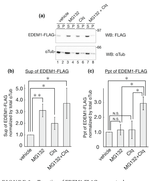
A 2024 study focused on EDEM1βs own turnover provides biochemical evidence that EDEM1 can exist in both soluble and membrane-associated (including a type II transmembrane) forms, consistent with post-translational processing that yields distinct topologies. Fractionation data supported substantial membrane association (example soluble/pellet distributions reported as ~20/80 or 35/65 depending on conditions/cofactors). (katsuki2024turnoverofedem1 pages 1-2, katsuki2024turnoverofedem1 pages 10-11)
Microscopy evidence from the same study shows ER localization under mannosidase inhibition (kifunensine) and relocalization to ubiquitin-positive aggresome-like structures upon proteasome inhibition. (katsuki2024turnoverofedem1 media 21e83ca5)
EDEM1 physically associates with multiple components of ER quality-control and ERAD machinery, including SEL1L, HRD1, OS-9, XTP3-B, and ER chaperones/co-chaperones (e.g., ERdj proteins), supporting a role as an organizing/processing factor that helps route clients through the ERAD-L pipeline. (chiritoiu2020edem1drivesmisfolded pages 3-5)
EDEM1 is not only an ERAD factor but also becomes a substrate of ER proteostasis pathways. Perturbations to ERAD (e.g., inhibition of mannose trimming) can shift complex composition/distribution and affect EDEM1 stability. (chiritoiu2020edem1drivesmisfolded pages 3-5)
Katsuki et al. (Genes to Cells, Apr 2024, https://doi.org/10.1111/gtc.13117) report that EDEM1, itself an ERAD-accelerating factor, is turned over rapidly in cells (cycloheximide chase half-life ~3 h) and can be degraded via both ERAD/proteasome and autophagy, with distinct folded/aggregated states contributing to route choice. (katsuki2024turnoverofedem1 pages 2-3, katsuki2024turnoverofedem1 media 21e83ca5)
Mechanistically, the study identifies ERAD components that contribute to EDEM1 clearance, including SEL1L/Hrd1, the deubiquitinase YOD1, and other factors (e.g., XTP3B, ERdj3, VIMP, BAG6, JB12), while OS9 binds EDEM1 but did not drive turnover in their overexpression assays. (katsuki2024turnoverofedem1 pages 1-2, katsuki2024turnoverofedem1 pages 6-7)
Fasana et al. (EMBO Reports, May 2024, https://doi.org/10.1038/s44319-024-00165-y) demonstrate that genetic or pharmacologic inhibition of ERAD componentsβincluding silencing EDEM1 or inhibiting luminal Ξ±1,2-mannosidases with kifunensine (KIF)βcan redirect canonical ERAD clients such as NHK and BACE457Ξ to LAMP1-positive endolysosomes via ER-to-lysosome-associated degradation (ERLAD). (fasana2024ertolysosomeassociateddegradationacts pages 3-4, fasana2024ertolysosomeassociateddegradationacts pages 9-10)
Importantly, this compensatory route requires the ER-phagy receptor FAM134B and its LC3-interacting region (LIR), and depends on LC3 lipidation machinery (e.g., ATG7) but is reported to be independent of classical autophagosome biogenesis factors (e.g., ATG13) in their assays. (fasana2024ertolysosomeassociateddegradationacts pages 7-8, fasana2024ertolysosomeassociateddegradationacts pages 6-7)
A recent high-authority review (Christianson et al., Nature Reviews Molecular Cell Biology, Aug 2023, https://doi.org/10.1038/s41580-023-00633-8) situates EDEM1 among the luminal processing factors that cooperate with chaperones and lectins to deliver misfolded proteins to HRD1βSEL1L-dependent ERAD, reinforcing consensus pathway placement and mechanistic framing. (fasana2024ertolysosomeassociateddegradationacts pages 10-11)
In practice, EDEM1 is widely used as:
- A node/marker of ER proteostasis pathways (ER stress/UPRβERAD axis) in perturbation experiments; and
- A mechanistic handle to shift the fate of misfolded glycoproteins between proteasomal ERAD and lysosomal ERLAD/ER-phagy-like disposal when ERAD is compromised. (katsuki2024turnoverofedem1 pages 1-2, fasana2024ertolysosomeassociateddegradationacts pages 7-8)
Although this report did not identify established clinical EDEM1-targeting therapies in the retrieved sources, EDEM1 is directly relevant to disease-linked proteostasis by controlling disposal of misfolded glycoproteins and by engaging compensatory lysosomal routes under ERAD impairment. (fasana2024ertolysosomeassociateddegradationacts pages 3-4, fasana2024ertolysosomeassociateddegradationacts pages 6-7)
Overall, EDEM1 is best understood as a misfolding-selective GH47 mannosidase-like factor that helps commit certain ER clients to degradation by coupling (i) preferential action on unfolded glycoprotein substrates with (ii) lectin/chaperone-mediated handoff to core ERAD machinery. The recent 2024 literature adds an important systems-level perspective: EDEM1 is tightly regulated at the protein level by the same pathways it supports (ERAD and autophagy), and when EDEM1-dependent ERAD processes are impaired, cells can compensate by engaging FAM134B/LC3-dependent ERLAD to deliver ERAD clients to lysosomes. (shenkman2018mannosidaseactivityof pages 4-4, katsuki2024turnoverofedem1 pages 1-2, fasana2024ertolysosomeassociateddegradationacts pages 7-8)
| Aspect | Key findings (1-3 bullets) | Evidence & quantitative details | Key sources (first author year, journal) | URL |
|---|---|---|---|---|
| Definition / target verification | β’ Human EDEM1 corresponds to ER degradation-enhancing alpha-mannosidase-like protein 1 (UniProt Q92611). β’ Member of glycosyl hydrolase family 47 / mannosidase-like ER quality-control proteins. β’ Functions in ER glycoprotein quality control and ER-associated degradation (ERAD). |
Experimental/review context consistently places EDEM1 in mammalian ERAD as a GH47 mannosidase-like factor acting on misfolded glycoproteins rather than an unrelated protein; family assignment and ERAD role are concordant across mechanistic studies and reviews (chiritoiu2020edem1drivesmisfolded pages 1-3, shenkman2018mannosidaseactivityof pages 1-2, manica2021edem3domainscooperate pages 1-2). | Shenkman 2018, Communications Biology; Chiritoiu 2020, IJMS | https://doi.org/10.1038/s42003-018-0174-8 ; https://doi.org/10.3390/ijms21103468 |
| Primary molecular function / enzymatic activity | β’ Bona fide mannosidase activity demonstrated in vitro for human EDEM1. β’ Activity is much stronger on unfolded/denatured glycoproteins than on free glycans or native glycoproteins. β’ Catalytic residue E488 is required for activity and ERAD support. |
EDEM1 shows only modest trimming on free N-glycans/native glycoproteins but >3-fold higher activity on denatured glycoproteins; trims oligomannose species from M8 toward M5, contributing especially to M8βM7 and M6βM5 steps. E488Q mutant loses mannosidase activity and acts dominant-negatively on ERAD substrate degradation (shenkman2018mannosidaseactivityof pages 4-4, shenkman2018mannosidaseactivityof pages 4-5, shenkman2018mannosidaseactivityof pages 1-2, shenkman2018mannosidaseactivityof pages 5-6). | Shenkman 2018, Communications Biology; Lamriben 2018, JBC | https://doi.org/10.1038/s42003-018-0174-8 ; https://doi.org/10.1074/jbc.ra118.004183 |
| Substrate specificity / recognition mode | β’ Prefers misfolded or unfolded glycoproteins rather than properly folded substrates. β’ Recognition is not purely glycan-based: EDEM1 also uses proteinβprotein interactions and redox-sensitive contacts. β’ N- and C-terminal intrinsically disordered regions (IDRs) contribute to substrate/partner binding. |
EDEM1 mannosidase activity rises sharply when glycoprotein substrate is denatured; knockdown reduces OS-9 association of ERAD substrate H2a to ~20% of control (~80% reduction). The mannosidase-like domain can bind ERAD clients in a thiol/redox-sensitive manner; IDRs are required for interaction with ERAD factor ERdj5 and for efficient client binding/degradation (shenkman2018mannosidaseactivityof pages 4-4, shenkman2018mannosidaseactivityof pages 1-2, manica2021edem3domainscooperate pages 1-2). | Shenkman 2018, Communications Biology; Lamriben 2018, JBC; Chiritoiu 2020, IJMS | https://doi.org/10.1038/s42003-018-0174-8 ; https://doi.org/10.1074/jbc.ra118.004183 ; https://doi.org/10.3390/ijms21103468 |
| Localization / topology | β’ EDEM1 is an ER-resident quality-control factor. β’ 2024 work indicates both soluble and membrane-associated / transmembrane forms can exist. β’ Membrane-associated form appears more aggregation-prone and can be selectively handled by some turnover factors. |
Katsuki et al. report EDEM1 has five N-glycans, undergoes post-translational signal-sequence cleavage, and yields soluble and type II transmembrane forms; alkaline extraction/fractionation showed substantial membrane association (S/P ~20/80 or 35/65 depending on condition/cofactors). Figure-based evidence shows ER localization under KIF and aggresome relocalization with proteasome inhibition (katsuki2024turnoverofedem1 pages 1-2, katsuki2024turnoverofedem1 pages 10-11, katsuki2024turnoverofedem1 media 21e83ca5). | Katsuki 2024, Genes to Cells; Chiritoiu 2020, IJMS | https://doi.org/10.1111/gtc.13117 ; https://doi.org/10.3390/ijms21103468 |
| Pathway role in ER quality control / ERAD | β’ EDEM1 helps extract terminally misfolded proteins from the calnexin cycle and route them to HRD1βSEL1L ERAD. β’ Associates with ERAD lectins/chaperones including OS-9, XTP3-B, SEL1L, HRD1, ERdj proteins. β’ Can contribute to degradation of some nonglycosylated misfolded proteins via protein-based recognition. |
Co-complexing with SEL1L, OS-9, XTP3-B, HRD1, PSMC6, ERdj4/5, calnexin, UGGTs supports placement in luminal ERAD handoff complexes; catalytic and noncatalytic substrate engagement both contribute to routing. Earlier work also supports shared ERAD machinery for glycosylated and nonglycosylated substrates involving EDEM1 (chiritoiu2020edem1drivesmisfolded pages 1-3, chiritoiu2020edem1drivesmisfolded pages 3-5, manica2021edem3domainscooperate pages 1-2). | Chiritoiu 2020, IJMS; Christianson 2023, Nat Rev Mol Cell Biol; Shenkman 2013, JBC | https://doi.org/10.3390/ijms21103468 ; https://doi.org/10.1038/s41580-023-00633-8 ; https://doi.org/10.1074/jbc.m112.438275 |
| Regulation by ER stress / UPR | β’ UPR/ER stress induces EDEM1 expression. β’ EDEM1 itself is also tightly controlled post-translationally by degradation pathways. β’ Perturbing ERAD can modestly raise EDEM1 and BiP levels. |
Katsuki 2024 states EDEM1 gene expression is upregulated by ER stress/UPR; protein turnover is fast (half-life ~3 h by CHX chase). Chiritoiu 2020 found kifunensine and SEL1L depletion increase EDEM1 stability/abundance, with mild BiP upregulation during ERAD perturbation (katsuki2024turnoverofedem1 pages 1-2, katsuki2024turnoverofedem1 pages 2-3, chiritoiu2020edem1drivesmisfolded pages 3-5, katsuki2024turnoverofedem1 media 21e83ca5). | Katsuki 2024, Genes to Cells; Chiritoiu 2020, IJMS | https://doi.org/10.1111/gtc.13117 ; https://doi.org/10.3390/ijms21103468 |
| Turnover and autoregulation (2024 emphasis) | β’ EDEM1 is itself degraded by ERAD and autophagy. β’ SEL1L/Hrd1, YOD1, XTP3B, ERdj3, VIMP, BAG6, JB12 participate in EDEM1 turnover. β’ OS9 binds EDEM1 but did not measurably drive its turnover in the 2024 study. |
CHX chase: ~3 h half-life. KIF or MG132 stabilizes EDEM1. SEL1L knockout upregulates EDEM1; Hrd1 C329S impairs turnover; XTP3B overexpression lowers EDEM1 in a KIF-sensitive manner; inactive YOD1 C160S increases ubiquitinated, detergent-insoluble EDEM1. Statistical analyses reported from 3β4 independent experiments, with significance including P < 0.05 and P < 0.01 (katsuki2024turnoverofedem1 pages 1-2, katsuki2024turnoverofedem1 pages 10-11, katsuki2024turnoverofedem1 pages 6-7, katsuki2024turnoverofedem1 pages 2-3, katsuki2024turnoverofedem1 media 21e83ca5). | Katsuki 2024, Genes to Cells | https://doi.org/10.1111/gtc.13117 |
| Backup lysosomal disposal / ER-phagy-ERLAD | β’ When ERAD is impaired, EDEM1-linked pathways connect to ER-phagy / ER-to-lysosome-associated degradation (ERLAD) as a failsafe. β’ FAM134B and LC3 lipidation are required for rerouting canonical ERAD clients. β’ Supports a model in which EDEM1 loss/inhibition does not fully block disposal but shifts it to lysosomes. |
Fasana 2024: EDEM1 silencing redirects NHK to degradative endolysosomes; pharmacologic ERAD inhibition (KIF, PS341) reroutes NHK and BACE457Ξ to LAMP1+ compartments. Delivery requires FAM134B and its LIR motif; ATG7 deletion blocks delivery, whereas ATG13 deletion does not. Example pulse-chase retentions at 120 min under ERAD inhibition: NHK mock 37β49% vs KIF 53β80%, PS341 52β73%, and even higher with lysosome block; BACE457Ξ mock 36β54% vs KIF 72β75%, PS341 70β84%. LysoQuant analyses used multiple cell counts and N=2β3 experiments; several comparisons reached **P < 0.0001 (fasana2024ertolysosomeassociateddegradationacts pages 1-2, fasana2024ertolysosomeassociateddegradationacts pages 7-8, fasana2024ertolysosomeassociateddegradationacts pages 3-4, fasana2024ertolysosomeassociateddegradationacts pages 9-10, fasana2024ertolysosomeassociateddegradationacts pages 6-7). | Fasana 2024, EMBO Reports; Chiritoiu 2020, IJMS | https://doi.org/10.1038/s44319-024-00165-y ; https://doi.org/10.3390/ijms21103468 |
| Real-world / disease-linked applications | β’ EDEM1 level or activity is being used mainly as a proteostasis/ER stress marker in cell biology and disease models. β’ Manipulating EDEM1 can alter fate of disease-relevant clients such as APP and viral proteins. β’ No approved EDEM1-targeted therapy was identified; current use is mechanistic and preclinical. |
In human cell models, EDEM1 overproduction reduces APP levels and decreases AΞ²40/AΞ²42 secretion, supporting relevance to Alzheimer-related proteostasis; recent literature also cites EDEM1 in viral exploitation and stress-pathway studies, but translation remains preclinical. The strongest current βimplementationβ is use of EDEM1 as a pathway node/marker in ERAD- and UPR-focused experiments rather than a clinical biomarker or drug target (chiritoiu2020edem1drivesmisfolded pages 1-3, katsuki2024turnoverofedem1 pages 1-2). | Nowakowska-GoΕacka 2021, IJMS; Katsuki 2024, Genes to Cells | https://doi.org/10.3390/ijms23010117 ; https://doi.org/10.1111/gtc.13117 |
Table: This table summarizes core evidence for the identity, function, localization, regulation, pathway role, and applications of human EDEM1 (UniProt Q92611). It emphasizes the most informative mechanistic studies, especially Katsuki 2024 on EDEM1 turnover and Fasana 2024 on ERLAD compensation when ERAD is impaired.
References
(chiritoiu2020edem1drivesmisfolded pages 1-3): Marioara Chiritoiu, Gabriela N. Chiritoiu, Cristian V. A. Munteanu, Florin Pastrama, N. Erwin Ivessa, and Stefana M. Petrescu. Edem1 drives misfolded protein degradation via erad and exploits er-phagy as back-up mechanism when erad is impaired. International Journal of Molecular Sciences, 21:3468, May 2020. URL: https://doi.org/10.3390/ijms21103468, doi:10.3390/ijms21103468. This article has 38 citations.
(shenkman2018mannosidaseactivityof pages 1-2): Marina Shenkman, Efrat Ron, Rivka Yehuda, Ron Benyair, Isam Khalaila, and Gerardo Z. Lederkremer. Mannosidase activity of edem1 and edem2 depends on an unfolded state of their glycoprotein substrates. Communications Biology, Oct 2018. URL: https://doi.org/10.1038/s42003-018-0174-8, doi:10.1038/s42003-018-0174-8. This article has 71 citations and is from a peer-reviewed journal.
(chiritoiu2020edem1drivesmisfolded pages 3-5): Marioara Chiritoiu, Gabriela N. Chiritoiu, Cristian V. A. Munteanu, Florin Pastrama, N. Erwin Ivessa, and Stefana M. Petrescu. Edem1 drives misfolded protein degradation via erad and exploits er-phagy as back-up mechanism when erad is impaired. International Journal of Molecular Sciences, 21:3468, May 2020. URL: https://doi.org/10.3390/ijms21103468, doi:10.3390/ijms21103468. This article has 38 citations.
(fasana2024ertolysosomeassociateddegradationacts pages 7-8): Elisa Fasana, Ilaria Fregno, Carmela Galli, Tatiana SoldΓ , and Maurizio Molinari. Er-to-lysosome-associated degradation acts as failsafe mechanism upon erad dysfunction. EMBO Reports, 25:2773-2785, May 2024. URL: https://doi.org/10.1038/s44319-024-00165-y, doi:10.1038/s44319-024-00165-y. This article has 37 citations and is from a highest quality peer-reviewed journal.
(manica2021edem3domainscooperate pages 1-2): Georgiana Manica, Simona Ghenea, Cristian V. A. Munteanu, Eliza C. Martin, Cristian Butnaru, Marius Surleac, Gabriela N. Chiritoiu, Petruta R. Alexandru, Andrei-Jose Petrescu, and Stefana M. Petrescu. Edem3 domains cooperate to perform its overall cell functioning. International Journal of Molecular Sciences, 22:2172, Feb 2021. URL: https://doi.org/10.3390/ijms22042172, doi:10.3390/ijms22042172. This article has 17 citations.
(shenkman2018mannosidaseactivityof pages 5-6): Marina Shenkman, Efrat Ron, Rivka Yehuda, Ron Benyair, Isam Khalaila, and Gerardo Z. Lederkremer. Mannosidase activity of edem1 and edem2 depends on an unfolded state of their glycoprotein substrates. Communications Biology, Oct 2018. URL: https://doi.org/10.1038/s42003-018-0174-8, doi:10.1038/s42003-018-0174-8. This article has 71 citations and is from a peer-reviewed journal.
(shenkman2018mannosidaseactivityof pages 4-4): Marina Shenkman, Efrat Ron, Rivka Yehuda, Ron Benyair, Isam Khalaila, and Gerardo Z. Lederkremer. Mannosidase activity of edem1 and edem2 depends on an unfolded state of their glycoprotein substrates. Communications Biology, Oct 2018. URL: https://doi.org/10.1038/s42003-018-0174-8, doi:10.1038/s42003-018-0174-8. This article has 71 citations and is from a peer-reviewed journal.
(shenkman2018mannosidaseactivityof pages 4-5): Marina Shenkman, Efrat Ron, Rivka Yehuda, Ron Benyair, Isam Khalaila, and Gerardo Z. Lederkremer. Mannosidase activity of edem1 and edem2 depends on an unfolded state of their glycoprotein substrates. Communications Biology, Oct 2018. URL: https://doi.org/10.1038/s42003-018-0174-8, doi:10.1038/s42003-018-0174-8. This article has 71 citations and is from a peer-reviewed journal.
(katsuki2024turnoverofedem1 pages 1-2): Riko Katsuki, Mai Kanuka, Ren Ohta, Shusei Yoshida, and Taku Tamura. Turnover of edem1, an eradβenhancing factor, is mediated by multiple degradation routes. Genes to Cells, 29:486-502, Apr 2024. URL: https://doi.org/10.1111/gtc.13117, doi:10.1111/gtc.13117. This article has 5 citations and is from a peer-reviewed journal.
(katsuki2024turnoverofedem1 pages 10-11): Riko Katsuki, Mai Kanuka, Ren Ohta, Shusei Yoshida, and Taku Tamura. Turnover of edem1, an eradβenhancing factor, is mediated by multiple degradation routes. Genes to Cells, 29:486-502, Apr 2024. URL: https://doi.org/10.1111/gtc.13117, doi:10.1111/gtc.13117. This article has 5 citations and is from a peer-reviewed journal.
(katsuki2024turnoverofedem1 media 21e83ca5): Riko Katsuki, Mai Kanuka, Ren Ohta, Shusei Yoshida, and Taku Tamura. Turnover of edem1, an eradβenhancing factor, is mediated by multiple degradation routes. Genes to Cells, 29:486-502, Apr 2024. URL: https://doi.org/10.1111/gtc.13117, doi:10.1111/gtc.13117. This article has 5 citations and is from a peer-reviewed journal.
(katsuki2024turnoverofedem1 pages 2-3): Riko Katsuki, Mai Kanuka, Ren Ohta, Shusei Yoshida, and Taku Tamura. Turnover of edem1, an eradβenhancing factor, is mediated by multiple degradation routes. Genes to Cells, 29:486-502, Apr 2024. URL: https://doi.org/10.1111/gtc.13117, doi:10.1111/gtc.13117. This article has 5 citations and is from a peer-reviewed journal.
(katsuki2024turnoverofedem1 pages 6-7): Riko Katsuki, Mai Kanuka, Ren Ohta, Shusei Yoshida, and Taku Tamura. Turnover of edem1, an eradβenhancing factor, is mediated by multiple degradation routes. Genes to Cells, 29:486-502, Apr 2024. URL: https://doi.org/10.1111/gtc.13117, doi:10.1111/gtc.13117. This article has 5 citations and is from a peer-reviewed journal.
(fasana2024ertolysosomeassociateddegradationacts pages 3-4): Elisa Fasana, Ilaria Fregno, Carmela Galli, Tatiana SoldΓ , and Maurizio Molinari. Er-to-lysosome-associated degradation acts as failsafe mechanism upon erad dysfunction. EMBO Reports, 25:2773-2785, May 2024. URL: https://doi.org/10.1038/s44319-024-00165-y, doi:10.1038/s44319-024-00165-y. This article has 37 citations and is from a highest quality peer-reviewed journal.
(fasana2024ertolysosomeassociateddegradationacts pages 9-10): Elisa Fasana, Ilaria Fregno, Carmela Galli, Tatiana SoldΓ , and Maurizio Molinari. Er-to-lysosome-associated degradation acts as failsafe mechanism upon erad dysfunction. EMBO Reports, 25:2773-2785, May 2024. URL: https://doi.org/10.1038/s44319-024-00165-y, doi:10.1038/s44319-024-00165-y. This article has 37 citations and is from a highest quality peer-reviewed journal.
(fasana2024ertolysosomeassociateddegradationacts pages 6-7): Elisa Fasana, Ilaria Fregno, Carmela Galli, Tatiana SoldΓ , and Maurizio Molinari. Er-to-lysosome-associated degradation acts as failsafe mechanism upon erad dysfunction. EMBO Reports, 25:2773-2785, May 2024. URL: https://doi.org/10.1038/s44319-024-00165-y, doi:10.1038/s44319-024-00165-y. This article has 37 citations and is from a highest quality peer-reviewed journal.
(fasana2024ertolysosomeassociateddegradationacts pages 10-11): Elisa Fasana, Ilaria Fregno, Carmela Galli, Tatiana SoldΓ , and Maurizio Molinari. Er-to-lysosome-associated degradation acts as failsafe mechanism upon erad dysfunction. EMBO Reports, 25:2773-2785, May 2024. URL: https://doi.org/10.1038/s44319-024-00165-y, doi:10.1038/s44319-024-00165-y. This article has 37 citations and is from a highest quality peer-reviewed journal.
(fasana2024ertolysosomeassociateddegradationacts pages 1-2): Elisa Fasana, Ilaria Fregno, Carmela Galli, Tatiana SoldΓ , and Maurizio Molinari. Er-to-lysosome-associated degradation acts as failsafe mechanism upon erad dysfunction. EMBO Reports, 25:2773-2785, May 2024. URL: https://doi.org/10.1038/s44319-024-00165-y, doi:10.1038/s44319-024-00165-y. This article has 37 citations and is from a highest quality peer-reviewed journal.
already_in_goa_exact (TAS Reactome + matches review). EDEM1's contribution is already captured (GO:1904380, GO:1904382, GO:0036503, GO:0004571 IMP). No NEW term needed; review proposes none. Already captured.ER proteostasis|Glycoproteostasis|N-glycosylation system|N-glycan processing|Mannose trimming ; PN-node mapping: subtype "Mannose trimming"=mapped/ok GO:1904382 (mannose trimming in glycoprotein ERAD, already_in_goa_exact); group "N-glycosylation system"=mapped/ok GO:0006487 protein N-linked glycosylation (new_to_goa); intermediate type/class/branch=no_mapping.already_in_goa_exact (TAS Reactome + matches review). EDEM1's contribution is already captured (GO:1904380, GO:1904382, GO:0036503, GO:0004571 IMP). No NEW term needed; review proposes none. Already captured.This file is generated from the current PROTEOSTASIS phase-1 dossier and local gene-review artifacts. Edit the source review, PN mapping, or dossier rather than this generated note when correcting the underlying curation.
id: Q92611
gene_symbol: EDEM1
product_type: PROTEIN
status: COMPLETE
taxon:
id: NCBITaxon:9606
label: Homo sapiens
description: EDEM1 (ER degradation-enhancing alpha-mannosidase-like protein 1) is a single-pass type II endoplasmic reticulum membrane protein of glycoside hydrolase family 47 (GH47), one of three mammalian Htm1/Mns1 homologues (EDEM1, EDEM2, EDEM3) acting in the ER-associated degradation of glycoproteins (gpERAD). EDEM1 extracts terminally misfolded glycoproteins, but not proteins undergoing productive folding, from the calnexin/calreticulin folding cycle and accelerates their clearance, delivering aberrant substrates to the SEL1L/HRD1 dislocation and ubiquitination machinery and, via Derlin-2/-3, to the p97/VCP retrotranslocation system. It recognizes non-native protein structure in a glycan-independent manner (acting in part as a lectin/holdase-like factor), binding both glycosylated and nonglycosylated misfolded substrates, and it requires its mannosidase-like domain for association with SEL1L. EDEM1 possesses low alpha-1,2-mannosidase activity, contributing to the second mannose-trimming step (Man8GlcNAc2 to Man7GlcNAc2) that exposes the alpha-1,6-mannose recognized by downstream lectins (OS-9/XTP3-B); its catalytic activity is weak relative to its recognition/delivery role and was historically debated. EDEM1 is induced by the IRE1-XBP1 branch of the unfolded protein response, resides in the ER membrane and concentrates in the ER-derived quality control compartment (ERQC), and promotes ER-to-cytosol retrotranslocation of substrates including the ricin A chain.
alternative_products:
- name: '1'
id: Q92611-1
- name: '2'
id: Q92611-2
sequence_note: VSP_056703, VSP_056704
existing_annotations:
- term:
id: GO:0005509
label: calcium ion binding
evidence_type: IEA
original_reference_id: GO_REF:0000002
qualifier: enables
review:
summary: GH47-family mannosidases use a calcium ion in the active site; EDEM1 retains this fold and binds calcium as a structural/catalytic cofactor. This is subsidiary to its recognition and ERAD functions, especially given its weak mannosidase activity.
action: KEEP_AS_NON_CORE
reason: Accurate structural cofactor attribute of the GH47 mannosidase-like domain, but not a standalone core function; the informative functions are misfolded protein recognition and ERAD.
supported_by:
- reference_id: file:human/EDEM1/EDEM1-uniprot.txt
supporting_text: Belongs to the glycosyl hydrolase 47 family
- term:
id: GO:0005783
label: endoplasmic reticulum
evidence_type: IEA
original_reference_id: GO_REF:0000117
qualifier: located_in
review:
summary: EDEM1 is an ER-resident protein; electronic (ARBA) assignment of ER localization is consistent with experimental evidence.
action: ACCEPT
reason: Correct site of action; redundant with IDA ER and ERQC annotations.
supported_by:
- reference_id: file:human/EDEM1/EDEM1-uniprot.txt
supporting_text: 'SUBCELLULAR LOCATION: Endoplasmic reticulum membrane'
- term:
id: GO:0005789
label: endoplasmic reticulum membrane
evidence_type: IEA
original_reference_id: GO_REF:0000120
qualifier: located_in
review:
summary: EDEM1 is a single-pass type II ER membrane protein; electronic transfer of ER membrane localization is correct.
action: ACCEPT
reason: Correct compartment; redundant with the ISS ER membrane annotation and UniProt subcellular location.
supported_by:
- reference_id: file:human/EDEM1/EDEM1-uniprot.txt
supporting_text: Single-pass type II membrane protein
- term:
id: GO:0005975
label: carbohydrate metabolic process
evidence_type: IEA
original_reference_id: GO_REF:0000002
qualifier: involved_in
review:
summary: Generic carbohydrate metabolic process from InterPro; far less informative than the specific ER mannose trimming and ERAD processes EDEM1 participates in.
action: MARK_AS_OVER_ANNOTATED
reason: Over-general parent; the specific ER mannose trimming (GO:1904380) and ERAD (GO:0036503) terms better capture the biology.
supported_by:
- reference_id: file:human/EDEM1/EDEM1-uniprot.txt
supporting_text: Belongs to the glycosyl hydrolase 47 family
- term:
id: GO:0016020
label: membrane
evidence_type: IEA
original_reference_id: GO_REF:0000002
qualifier: located_in
review:
summary: Generic membrane localization from InterPro, superseded by the specific ER membrane annotation.
action: MARK_AS_OVER_ANNOTATED
reason: Uninformative parent; EDEM1 is specifically an ER membrane protein.
proposed_replacement_terms:
- id: GO:0005789
label: endoplasmic reticulum membrane
supported_by:
- reference_id: file:human/EDEM1/EDEM1-uniprot.txt
supporting_text: 'SUBCELLULAR LOCATION: Endoplasmic reticulum membrane'
- term:
id: GO:0036503
label: ERAD pathway
evidence_type: IEA
original_reference_id: GO_REF:0000117
qualifier: involved_in
review:
summary: Electronic (ARBA) assignment of the ERAD pathway, consistent with extensive experimental evidence that EDEM1 accelerates ERAD of misfolded glycoproteins.
action: ACCEPT
reason: Correct core biological process; redundant with IMP/ISS evidence.
supported_by:
- reference_id: file:human/EDEM1/EDEM1-uniprot.txt
supporting_text: It is directly involved in endoplasmic reticulum-associated degradation (ERAD)
- term:
id: GO:1904380
label: endoplasmic reticulum mannose trimming
evidence_type: IEA
original_reference_id: GO_REF:0000120
qualifier: involved_in
review:
summary: EDEM1 contributes to ER mannose trimming (the Man8 to Man7 step); electronic assignment is consistent with the IMP evidence.
action: ACCEPT
reason: Correct biological process; redundant with the IMP annotation from endogenous knockout analysis.
supported_by:
- reference_id: PMID:25092655
supporting_text: Mannose trimming from Man8GlcNAc2 to Man7GlcNAc2 is performed mainly by EDEM3 and to a lesser extent by EDEM1
- term:
id: GO:1904154
label: positive regulation of retrograde protein transport, ER to cytosol
evidence_type: IEA
original_reference_id: GO_REF:0000120
qualifier: involved_in
review:
summary: EDEM1 promotes retrotranslocation of ERAD substrates from the ER to the cytosol; electronic assignment is consistent with the experimental ricin retrotranslocation data.
action: KEEP_AS_NON_CORE
reason: Real, specific aspect of EDEM1 function (substrate dislocation) but subordinate to the core recognition/ERAD role; redundant with the IMP/IGI annotations.
supported_by:
- reference_id: PMID:24200403
supporting_text: This transport is promoted by EDEM1
- term:
id: GO:0004571
label: mannosyl-oligosaccharide 1,2-alpha-mannosidase activity
evidence_type: TAS
original_reference_id: Reactome:R-HSA-6782685
qualifier: enables
review:
summary: Reactome curation of EDEM1 (with EDEM3) hydrolysing Man8 to Man5 glycans. EDEM1 has genuine but low alpha-1,2-mannosidase activity contributing to the second trimming step.
action: ACCEPT
reason: Correct molecular function (weak but real, demonstrated by endogenous knockout); kept as supporting rather than the dominant function.
supported_by:
- reference_id: PMID:25092655
supporting_text: all endogenous EDEMs possess mannosidase activity
- term:
id: GO:0005783
label: endoplasmic reticulum
evidence_type: IDA
original_reference_id: GO_REF:0000052
qualifier: located_in
review:
summary: Direct immunofluorescence (HPA) evidence for ER localization, consistent with EDEM1's site of action.
action: ACCEPT
reason: Correct compartment with direct experimental support.
supported_by:
- reference_id: file:human/EDEM1/EDEM1-uniprot.txt
supporting_text: 'SUBCELLULAR LOCATION: Endoplasmic reticulum membrane'
- term:
id: GO:1904380
label: endoplasmic reticulum mannose trimming
evidence_type: IMP
original_reference_id: PMID:25092655
qualifier: involved_in
review:
summary: Endogenous EDEM1 knockout in human and chicken cells increased Man8B levels, showing EDEM1 contributes (with EDEM3) to the second ER mannose-trimming step from Man8GlcNAc2 to Man7GlcNAc2.
action: ACCEPT
reason: Core biological process with direct experimental (IMP) support from endogenous gene knockout.
supported_by:
- reference_id: PMID:25092655
supporting_text: Mannose trimming from Man8GlcNAc2 to Man7GlcNAc2 is performed mainly by EDEM3 and to a lesser extent by EDEM1
- term:
id: GO:0006511
label: ubiquitin-dependent protein catabolic process
evidence_type: IMP
original_reference_id: PMID:25092655
qualifier: involved_in
review:
summary: EDEM1 knockout delayed degradation of the gpERAD substrate ATF6alpha, which is degraded by the ubiquitin-proteasome system; supports a role in ubiquitin-dependent catabolism via ERAD.
action: KEEP_AS_NON_CORE
reason: Correct but generic parent process; the more specific ERAD pathway (GO:0036503) better captures EDEM1's role.
supported_by:
- reference_id: PMID:25092655
supporting_text: deletion of hEDEM1 significantly delayed the degradation of hATF6
- term:
id: GO:0036503
label: ERAD pathway
evidence_type: IMP
original_reference_id: PMID:21062743
qualifier: involved_in
review:
summary: EDEM1 functions in ERAD by handing off substrate glycoproteins, after mannose trimming, to the late ERAD lectin XTP3-B and downstream E3 ligases.
action: ACCEPT
reason: Core biological process; experimentally links EDEM1 to the downstream ERAD machinery.
supported_by:
- reference_id: PMID:21062743
supporting_text: mannose trimming enables delivery of a substrate glycoprotein from EDEM1 to late ERAD steps through association with XTP3-B
- term:
id: GO:0036503
label: ERAD pathway
evidence_type: IMP
original_reference_id: PMID:25092655
qualifier: involved_in
review:
summary: Endogenous EDEM1 knockout delayed gpERAD of ATF6alpha, directly demonstrating EDEM1's role in the ERAD pathway.
action: ACCEPT
reason: Core biological process with direct experimental (IMP) support.
supported_by:
- reference_id: PMID:25092655
supporting_text: deletion of hEDEM1 significantly delayed the degradation of hATF6
- term:
id: GO:1904382
label: mannose trimming involved in glycoprotein ERAD pathway
evidence_type: TAS
original_reference_id: Reactome:R-HSA-6782685
qualifier: involved_in
review:
summary: Reactome curation of EDEM1 mannose trimming within the glycoprotein ERAD pathway; an accurate, specific refinement of EDEM1's trimming contribution to ERAD.
action: ACCEPT
reason: Correct specific biological process linking the trimming activity to ERAD.
supported_by:
- reference_id: PMID:25092655
supporting_text: M8B is trimmed by EDEM1 and EDEM3 to Man7-5GlcNAc2, which are recognized by lectin OS-9
- term:
id: GO:0044322
label: endoplasmic reticulum quality control compartment
evidence_type: IDA
original_reference_id: PMID:23233672
qualifier: located_in
review:
summary: On proteasome inhibition, EDEM1 and other ERAD machinery accumulate with substrates in the ER-derived quality control compartment (ERQC), supporting an ERQC localization.
action: ACCEPT
reason: Genuine, functionally relevant localization with direct experimental support.
supported_by:
- reference_id: PMID:23233672
supporting_text: accumulation of the nonglycosylated proteins and ERAD machinery in the endoplasmic reticulum-derived quality control compartment
- term:
id: GO:0045047
label: protein targeting to ER
evidence_type: IMP
original_reference_id: PMID:23233672
qualifier: involved_in
review:
summary: This annotation derives from work showing EDEM1 targets misfolded substrates to the ER membrane dislocation/ERAD machinery, not co-translational targeting of nascent proteins to the ER. The GO term protein targeting to ER (import into the ER) poorly captures the demonstrated delivery-to-ERAD biology.
action: MARK_AS_OVER_ANNOTATED
reason: The term denotes targeting of proteins into the ER (e.g. co-translational import), which is not what EDEM1 does; the experimental result is substrate delivery to the ER-membrane ERAD/dislocation complex. Retained (experimental basis) rather than removed, but the term is a poor fit.
supported_by:
- reference_id: PMID:19524542
supporting_text: target aberrant proteins to the ER membrane dislocation and ubiquitination complex containing SEL1L
- term:
id: GO:0051787
label: misfolded protein binding
evidence_type: IMP
original_reference_id: PMID:23233672
qualifier: enables
review:
summary: EDEM1 binds misfolded glycosylated and nonglycosylated substrates, associating with nonglycosylated proteins through a region outside its mannosidase-like domain; recognition of non-native protein structure is a core EDEM1 function.
action: ACCEPT
reason: Core molecular function; EDEM1's defining role is recognition of misfolded/non-native proteins for ERAD.
supported_by:
- reference_id: PMID:23233672
supporting_text: EDEM1 associates through a region outside of its mannosidase-like domain with the nonglycosylated proteins
- term:
id: GO:0004571
label: mannosyl-oligosaccharide 1,2-alpha-mannosidase activity
evidence_type: IMP
original_reference_id: PMID:25092655
qualifier: enables
review:
summary: Endogenous gene knockout established that EDEM1 possesses (weak) alpha-1,2-mannosidase activity, contributing to the second trimming step Man8B to Man7. This resolved the long-standing mannosidase-versus-lectin controversy in favor of genuine but low catalytic activity.
action: ACCEPT
reason: Molecular function supported by endogenous knockout (IMP); EDEM1 has the weakest mannosidase activity of the three EDEMs, so this is retained as a supporting rather than dominant function.
supported_by:
- reference_id: PMID:25092655
supporting_text: all endogenous EDEMs possess mannosidase activity
- term:
id: GO:1904154
label: positive regulation of retrograde protein transport, ER to cytosol
evidence_type: IMP
original_reference_id: PMID:24200403
qualifier: involved_in
review:
summary: EDEM1 promotes ER-to-cytosol retrotranslocation of the ricin A chain, a model retrotranslocation substrate handled like a misfolded ER protein.
action: KEEP_AS_NON_CORE
reason: Specific, experimentally supported aspect of EDEM1's ERAD/dislocation activity, but subordinate to the core recognition/ERAD role.
supported_by:
- reference_id: PMID:24200403
supporting_text: This transport is promoted by EDEM1
- term:
id: GO:1904154
label: positive regulation of retrograde protein transport, ER to cytosol
evidence_type: IGI
original_reference_id: PMID:24200403
qualifier: involved_in
review:
summary: Genetic-interaction evidence (with EDEM2, UniProtKB:Q9BV94) that EDEM1 promotes ricin A-chain retrotranslocation from the ER to the cytosol.
action: KEEP_AS_NON_CORE
reason: Consistent with the IMP retrotranslocation annotation; a specific aspect of the dislocation function rather than the core role.
supported_by:
- reference_id: PMID:24200403
supporting_text: more ricin can interact with EDEM2 in comparison with EDEM1
- term:
id: GO:0044322
label: endoplasmic reticulum quality control compartment
evidence_type: TAS
original_reference_id: Reactome:R-HSA-1791155
qualifier: located_in
review:
summary: Reactome curation of EDEM1 ERQC localization (EDEM expression context); consistent with direct IDA evidence.
action: ACCEPT
reason: Correct compartment; redundant with the IDA ERQC annotation.
supported_by:
- reference_id: PMID:23233672
supporting_text: endoplasmic reticulum-derived quality control compartment
- term:
id: GO:0044322
label: endoplasmic reticulum quality control compartment
evidence_type: TAS
original_reference_id: Reactome:R-HSA-6782685
qualifier: located_in
review:
summary: Reactome curation of EDEM1 ERQC localization in the mannose-trimming reaction context.
action: ACCEPT
reason: Correct compartment; redundant with the IDA ERQC annotation.
supported_by:
- reference_id: PMID:23233672
supporting_text: endoplasmic reticulum-derived quality control compartment
- term:
id: GO:0005783
label: endoplasmic reticulum
evidence_type: IMP
original_reference_id: PMID:21062743
qualifier: located_in
review:
summary: ER localization in the context of EDEM1-dependent ERAD substrate handoff.
action: ACCEPT
reason: Correct site of action; consistent with experimental ER/ERQC evidence.
supported_by:
- reference_id: file:human/EDEM1/EDEM1-uniprot.txt
supporting_text: 'SUBCELLULAR LOCATION: Endoplasmic reticulum membrane'
- term:
id: GO:0005783
label: endoplasmic reticulum
evidence_type: IDA
original_reference_id: PMID:24200403
qualifier: located_in
review:
summary: Direct evidence for ER localization of EDEM1 in the ricin retrotranslocation study.
action: ACCEPT
reason: Correct compartment with direct experimental support.
supported_by:
- reference_id: file:human/EDEM1/EDEM1-uniprot.txt
supporting_text: 'SUBCELLULAR LOCATION: Endoplasmic reticulum membrane'
- term:
id: GO:0004571
label: mannosyl-oligosaccharide 1,2-alpha-mannosidase activity
evidence_type: IDA
original_reference_id: PMID:12610306
qualifier: enables
negated: true
review:
summary: A negated (NOT) experimental annotation reflecting the original proposal that EDEM1 lacks alpha-1,2-mannosidase activity, based on overexpression biochemistry. Later endogenous-knockout analysis (PMID:25092655) demonstrated that EDEM1 does possess weak mannosidase activity, so this negation is superseded but retained as the curated record of the early finding.
action: KEEP_AS_NON_CORE
reason: Genuine historical experimental (IDA) annotation that conflicts with later endogenous-KO evidence; per guidelines an experimental annotation is not removed on weak grounds. Flagged as superseded by PMID:25092655.
supported_by:
- reference_id: PMID:25092655
supporting_text: it had originally been proposed that EDEM1 has no Ξ±1,2-mannosidase
activity
- term:
id: GO:0005515
label: protein binding
evidence_type: IPI
original_reference_id: PMID:19524542
qualifier: enables
review:
summary: Physical interaction with SEL1L (UniProtKB:Q9UBV2), the HRD1-complex adaptor through which EDEM1 delivers substrates to the dislocation machinery. The bare protein binding term is uninformative; the SEL1L interaction is captured by the misfolded protein binding and ERAD annotations.
action: KEEP_AS_NON_CORE
reason: Records a real interaction (SEL1L) but bare protein binding is uninformative per curation guidelines; the functional consequence is reflected in the ERAD/recognition terms.
supported_by:
- reference_id: PMID:19524542
supporting_text: its association with the ER membrane adaptor protein SEL1L
- term:
id: GO:0005515
label: protein binding
evidence_type: IPI
original_reference_id: PMID:19934218
qualifier: enables
review:
summary: Physical interaction with rod opsin (UniProtKB:P08100), an EDEM1 client glycoprotein. The bare protein binding term is uninformative.
action: KEEP_AS_NON_CORE
reason: Real client interaction (rod opsin) but bare protein binding is uninformative; EDEM1's recognition function is captured by misfolded protein binding.
supported_by:
- reference_id: PMID:19934218
supporting_text: rod opsin co-immunoprecipitated with EDEM1
- term:
id: GO:0051787
label: misfolded protein binding
evidence_type: IDA
original_reference_id: PMID:19524542
qualifier: enables
review:
summary: EDEM1 directly and specifically binds non-native proteins in a glycan-independent manner, demonstrating recognition of misfolded protein structure independent of N-glycan trimming.
action: ACCEPT
reason: Core molecular function; direct demonstration of misfolded/non-native protein recognition.
supported_by:
- reference_id: PMID:19524542
supporting_text: EDEM1 specifically binds nonnative proteins in a glycan-independent manner
- term:
id: GO:0005515
label: protein binding
evidence_type: IPI
original_reference_id: PMID:16449189
qualifier: enables
review:
summary: Physical interactions with Derlin-2 (UniProtKB:Q9GZP9) and Derlin-3 (UniProtKB:Q96Q80), which link EDEM to p97/VCP for substrate extraction. The bare protein binding term is uninformative; the functional role is reflected in the ERAD annotations.
action: KEEP_AS_NON_CORE
reason: Records real Derlin-2/-3 interactions providing the EDEM-to-p97 link, but bare protein binding is uninformative per guidelines.
supported_by:
- reference_id: PMID:16449189
supporting_text: Derlin-2 and -3 are associated with EDEM and p97
- term:
id: GO:0005789
label: endoplasmic reticulum membrane
evidence_type: ISS
original_reference_id: GO_REF:0000024
qualifier: located_in
review:
summary: Sequence-similarity transfer (from mouse Q925U4) of ER membrane localization; EDEM1 is a single-pass type II ER membrane protein.
action: ACCEPT
reason: Correct compartment; consistent with UniProt subcellular location and IDA evidence.
supported_by:
- reference_id: file:human/EDEM1/EDEM1-uniprot.txt
supporting_text: Single-pass type II membrane protein
- term:
id: GO:0036503
label: ERAD pathway
evidence_type: ISS
original_reference_id: GO_REF:0000024
qualifier: involved_in
review:
summary: Sequence-similarity transfer of the ERAD pathway role from the mouse ortholog; consistent with extensive human experimental evidence.
action: ACCEPT
reason: Correct core biological process; redundant with IMP evidence.
supported_by:
- reference_id: file:human/EDEM1/EDEM1-uniprot.txt
supporting_text: It is directly involved in endoplasmic reticulum-associated degradation (ERAD)
references:
- id: GO_REF:0000002
title: Gene Ontology annotation through association of InterPro records with GO terms
findings: []
- id: GO_REF:0000024
title: Manual transfer of experimentally-verified manual GO annotation data to orthologs by curator judgment of sequence similarity
findings: []
- id: GO_REF:0000052
title: Gene Ontology annotation based on curation of immunofluorescence data
findings: []
- id: GO_REF:0000117
title: Electronic Gene Ontology annotations created by ARBA machine learning models
findings: []
- id: GO_REF:0000120
title: Combined Automated Annotation using Multiple IEA Methods
findings: []
- id: PMID:12610306
title: Role of EDEM in the release of misfolded glycoproteins from the calnexin cycle.
findings:
- statement: EDEM extracts misfolded glycoproteins, but not glycoproteins undergoing productive folding, from the calnexin cycle; overexpression accelerates release and ERAD.
reference_section_type: ABSTRACT
reference_review:
relevance: HIGH
correctness: VERIFIED
review_notes: Foundational demonstration of EDEM1's role in extracting misfolded glycoproteins from the calnexin cycle. Also the source of the NOT mannosidase IDA (early no-activity view, later superseded by PMID:25092655).
- id: PMID:16449189
title: Derlin-2 and Derlin-3 are regulated by the mammalian unfolded protein response and are required for ER-associated degradation.
findings:
- statement: Derlin-2 and -3 are associated with EDEM and p97 and provide the missing link between EDEM and p97 in degradation of misfolded glycoproteins.
reference_section_type: ABSTRACT
reference_review:
relevance: MEDIUM
correctness: VERIFIED
review_notes: Source of the EDEM1-DERL2/DERL3 protein binding annotations; establishes the EDEM-to-p97 link.
- id: PMID:19524542
title: EDEM1 recognition and delivery of misfolded proteins to the SEL1L-containing ERAD complex.
findings:
- statement: EDEM1 specifically binds non-native proteins in a glycan-independent manner; its mannosidase-like domain is required for SEL1L association but not for substrate binding.
reference_section_type: ABSTRACT
reference_review:
relevance: HIGH
correctness: VERIFIED
review_notes: Key evidence for glycan-independent misfolded protein recognition and the SEL1L delivery model; supports the misfolded protein binding core function.
- id: PMID:19934218
title: A dual role for EDEM1 in the processing of rod opsin.
findings:
- statement: EDEM1 is a chaperone of rod opsin; it promotes degradation of misfolded P23H rod opsin and reduces aggregation, with binding independent of mannose trimming.
reference_section_type: ABSTRACT
reference_review:
relevance: MEDIUM
correctness: VERIFIED
review_notes: Source of the rod opsin (P08100) interaction; supports trimming-independent client recognition and ER membrane localization.
- id: PMID:21062743
title: Mannose trimming is required for delivery of a glycoprotein from EDEM1 to XTP3-B and to late endoplasmic reticulum-associated degradation steps.
findings:
- statement: Mannose trimming enables handoff of a substrate glycoprotein from EDEM1 to the late ERAD lectin XTP3-B and downstream E3 ligases; EDEM1 binding itself is trimming-independent.
reference_section_type: ABSTRACT
reference_review:
relevance: HIGH
correctness: VERIFIED
review_notes: Places EDEM1 upstream of XTP3-B/HRD1 in the ERAD pathway; supports the ERAD and ER localization annotations.
- id: PMID:23233672
title: A shared endoplasmic reticulum-associated degradation pathway involving the EDEM1 protein for glycosylated and nonglycosylated proteins.
findings:
- statement: EDEM1 is used by both glycosylated and nonglycosylated misfolded substrates, binding nonglycosylated proteins through a region outside its mannosidase-like domain; ERAD machinery accumulates in the ERQC on proteasome inhibition.
reference_section_type: ABSTRACT
reference_review:
relevance: HIGH
correctness: VERIFIED
review_notes: Supports misfolded protein binding (glycan-independent), ERQC localization, and the shared ERAD pathway.
- id: PMID:24200403
title: The role of EDEM2 compared with EDEM1 in ricin transport from the endoplasmic reticulum to the cytosol.
findings:
- statement: EDEM1 (and EDEM2) promote retrotranslocation of the ricin A chain from the ER to the cytosol, modeling ERAD-substrate dislocation.
reference_section_type: ABSTRACT
reference_review:
relevance: MEDIUM
correctness: VERIFIED
review_notes: Supports the positive regulation of retrograde protein transport (ER to cytosol) annotations and ER localization.
- id: PMID:25092655
title: EDEM2 initiates mammalian glycoprotein ERAD by catalyzing the first mannose trimming step.
findings:
- statement: All endogenous EDEMs possess mannosidase activity; EDEM3 and to a lesser extent EDEM1 perform the second trimming step (Man8B to Man7), resolving the mannosidase-versus-lectin controversy.
reference_section_type: ABSTRACT
- statement: EDEM1 has the weakest mannosidase activity of the three EDEMs and can also enhance gpERAD independently of catalysis (lectin-like) when overexpressed.
reference_section_type: RESULTS
reference_review:
relevance: HIGH
correctness: VERIFIED
review_notes: Definitive endogenous-knockout study; establishes EDEM1's weak mannosidase activity and its role in the second trimming step and gpERAD.
- id: PMID:30374462
title: Mannosidase activity of EDEM1 and EDEM2 depends on an unfolded state of their glycoprotein substrates.
findings:
- statement: In vitro, EDEM1 (and EDEM2) mannosidase activity is modest on free oligosaccharides and native glycoproteins but significantly higher on a denatured glycoprotein, explaining how slow background trimming becomes selective for misfolded substrates; the EDEMs associate with oxidoreductases including PDI and TXNDC11.
reference_section_type: ABSTRACT
reference_review:
relevance: HIGH
correctness: VERIFIED
review_notes: PubMed-verified (PMID:30374462, doi:10.1038/s42003-018-0174-8). Demonstrates EDEM1 has bona fide in vitro mannosidase activity that is folding-state dependent (preferential on unfolded/denatured glycoproteins), complementing the endogenous-KO evidence (PMID:25092655). Not cached; reference added without verbatim supporting_text.
- id: PMID:32423001
title: EDEM1 Drives Misfolded Protein Degradation via ERAD and Exploits ER-Phagy as Back-Up Mechanism When ERAD Is Impaired.
findings:
- statement: EDEM1 is found in auto-regulatory complexes with ERAD components; its N-terminal disordered region mediates interaction with misfolded proteins, and EDEM1 overexpression can drive client degradation via autophagy/ER-phagy when proteasomal degradation or dislocation is impaired.
reference_section_type: ABSTRACT
reference_review:
relevance: HIGH
correctness: VERIFIED
review_notes: PubMed-verified (PMID:32423001, doi:10.3390/ijms21103468). Adds the N-terminal intrinsically disordered region as the misfolded-protein-interaction module and shows EDEM1 engages autophagy/ER-phagy as a backup ERAD route. Not cached; reference added without verbatim supporting_text.
- id: PMID:38682256
title: Turnover of EDEM1, an ERAD-enhancing factor, is mediated by multiple degradation routes.
findings:
- statement: EDEM1 is itself turned over by both ERAD (SEL1L/Hrd1, YOD1, XTP3B, ERdj3, VIMP, BAG6, JB12) and autophagy in folded-state-dependent manner; OS9 binds EDEM1 but did not drive its turnover. EDEM1 has a fast half-life (~3 h) and exists in soluble and membrane-associated forms.
reference_section_type: ABSTRACT
reference_review:
relevance: MEDIUM
correctness: VERIFIED
review_notes: PubMed-verified (PMID:38682256, doi:10.1111/gtc.13117). Establishes that EDEM1, an ERAD accelerator, is itself an ERAD/autophagy substrate (autoregulation). Not cached; reference added without verbatim supporting_text.
- id: PMID:38773321
title: ER-to-lysosome-associated degradation acts as failsafe mechanism upon ERAD dysfunction.
findings:
- statement: Pharmacologic or genetic inhibition of ERAD components, including silencing EDEM1, reroutes canonical ERAD clients (NHK, BACE457delta) to degradative endolysosomes via the ER-phagy receptor FAM134B and the LC3 lipidation machinery.
reference_section_type: ABSTRACT
reference_review:
relevance: MEDIUM
correctness: VERIFIED
review_notes: PubMed-verified (PMID:38773321, doi:10.1038/s44319-024-00165-y). Shows EDEM1 loss/inhibition activates a compensatory FAM134B-dependent ERLAD route. Not cached; reference added without verbatim supporting_text.
- id: PMID:37528230
title: Mechanisms of substrate processing during ER-associated protein degradation.
findings:
- statement: Authoritative review placing EDEM-family mannosidases among the luminal processing factors that cooperate with chaperones and lectins to deliver misfolded glycoproteins to HRD1-SEL1L-dependent ERAD.
reference_section_type: OTHER
reference_review:
relevance: LOW
correctness: VERIFIED
review_notes: PubMed-verified (PMID:37528230, doi:10.1038/s41580-023-00633-8). Recent Nat Rev MCB review providing consensus ERAD pathway framing for EDEM1. Background/contextual.
- id: Reactome:R-HSA-1791155
title: Expression of EDEM
findings: []
- id: Reactome:R-HSA-6782685
title: EDEM1,3 hydrolyse (GlcNAc)2 (Man)8b to (GlcNAc)2 (Man)5
findings: []
- id: file:human/EDEM1/EDEM1-uniprot.txt
title: UniProt entry Q92611 (EDEM1_HUMAN), ER degradation-enhancing alpha-mannosidase-like protein 1
findings:
- statement: Single-pass type II ER membrane GH47 protein that extracts misfolded glycoproteins from the calnexin cycle and targets them for ERAD (N-glycan-independent, via SEL1L); has low mannosidase activity (Man8GlcNAc2 to Man7GlcNAc2); interacts with SEL1L, DERL2, DERL3.
reference_section_type: OTHER
core_functions:
- description: Recognizes terminally misfolded/non-native glycoproteins (and some nonglycosylated proteins) and extracts them from the calnexin/calreticulin folding cycle, delivering them to the SEL1L/HRD1 dislocation and ubiquitination machinery for ER-associated degradation.
molecular_function:
id: GO:0051787
label: misfolded protein binding
locations:
- id: GO:0005789
label: endoplasmic reticulum membrane
- id: GO:0044322
label: endoplasmic reticulum quality control compartment
supported_by:
- reference_id: PMID:19524542
supporting_text: EDEM1 specifically binds nonnative proteins in a glycan-independent manner
- reference_id: PMID:12610306
supporting_text: EDEM was shown to extract misfolded glycoproteins, but not glycoproteins undergoing productive folding, from the calnexin cycle
directly_involved_in:
- id: GO:0036503
label: ERAD pathway
- description: Possesses low alpha-1,2-mannosidase activity contributing to the second ER mannose-trimming step (Man8GlcNAc2 to Man7GlcNAc2), generating the alpha-1,6-mannose-exposed glycan recognized by downstream lectins (OS-9/XTP3-B) in glycoprotein ERAD.
molecular_function:
id: GO:0004571
label: mannosyl-oligosaccharide 1,2-alpha-mannosidase activity
locations:
- id: GO:0005789
label: endoplasmic reticulum membrane
supported_by:
- reference_id: PMID:25092655
supporting_text: Mannose trimming from Man8GlcNAc2 to Man7GlcNAc2 is performed mainly by EDEM3 and to a lesser extent by EDEM1
- reference_id: PMID:30374462
directly_involved_in:
- id: GO:1904380
label: endoplasmic reticulum mannose trimming
proposed_new_terms: []
suggested_questions:
- question: To what extent does EDEM1 function in vivo as a catalytic mannosidase versus a lectin/holdase-like recognition factor, and how is the balance set by expression level and ER stress?
- question: What structural features outside the mannosidase-like domain mediate EDEM1's glycan-independent recognition of non-native protein structure?
suggested_experiments:
- description: Reconstitute substrate handoff with purified EDEM1, ERManI, EDEM2/3 and SEL1L on defined misfolded glycoprotein substrates to quantify the relative contributions of EDEM1 catalysis versus recognition/holdase activity to ERAD commitment.
- description: Domain-swap and point-mutation analysis of the EDEM1 mannosidase-like domain in endogenous knock-in cells to separate catalytic, SEL1L-binding, and non-native-protein-binding activities and test their individual requirements for ERAD of glycosylated and nonglycosylated substrates.